. 2000 Sep 4;150(5):1209-14.
doi: 10.1083/jcb.150.5.1209.

The dystrophin complex forms a mechanically strong link between the sarcolemma and costameric actin

Affiliations

The dystrophin complex forms a mechanically strong link between the sarcolemma and costameric actin

I N Rybakova et al. J Cell Biol. .

Abstract

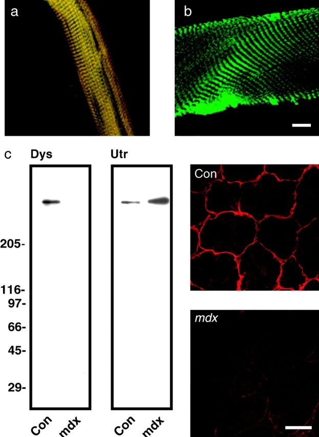
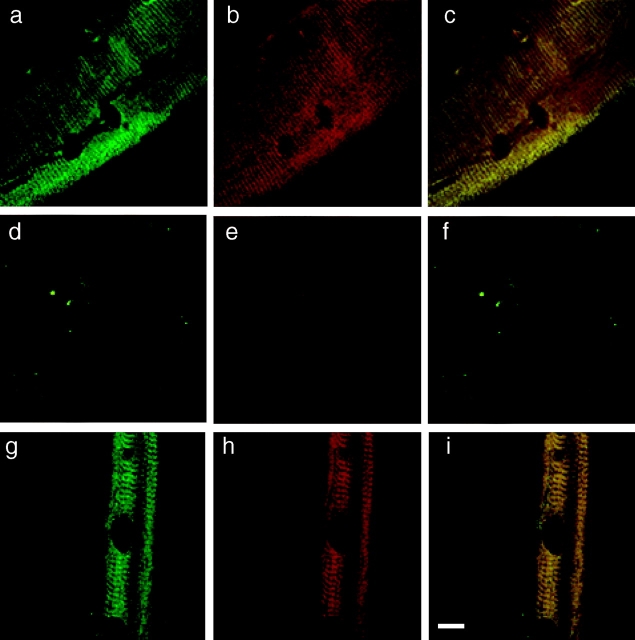
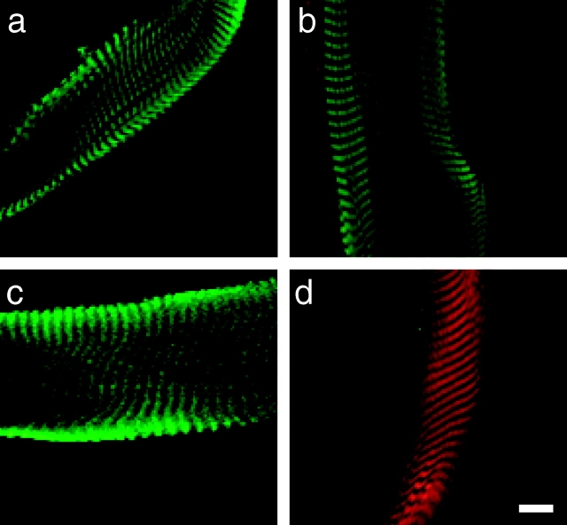
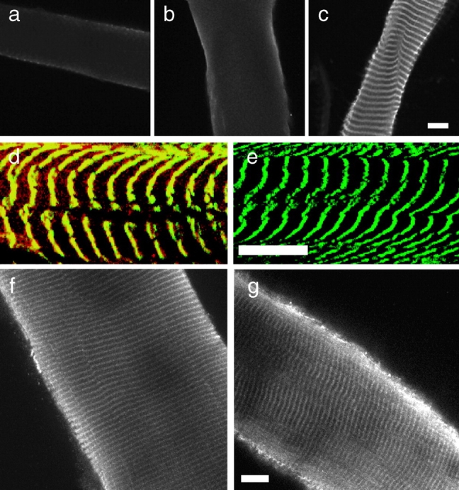
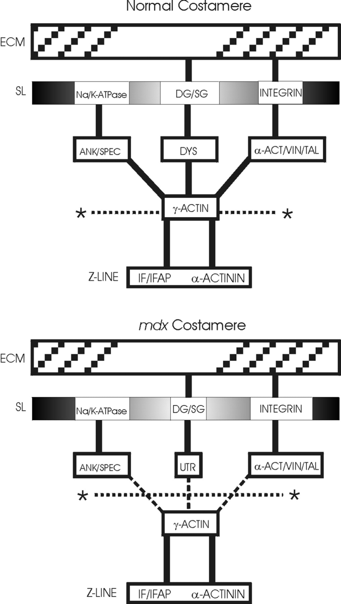
The absence of dystrophin complex leads to disorganization of the force-transmitting costameric cytoskeleton and disruption of sarcolemmal membrane integrity in skeletal muscle. However, it has not been determined whether the dystrophin complex can form a mechanically strong bond with any costameric protein. We performed confocal immunofluorescence analysis of isolated sarcolemma that were mechanically peeled from skeletal fibers of mouse hindlimb muscle. A population of gamma-actin filaments was stably associated with sarcolemma isolated from normal muscle and displayed a costameric pattern that precisely overlapped with dystrophin. However, costameric actin was absent from all sarcolemma isolated from dystrophin-deficient mdx mouse muscle even though it was localized to costameres in situ. Vinculin, alpha-actinin, beta-dystroglycan and utrophin were all retained on mdx sarcolemma, indicating that the loss of costameric actin was not due to generalized membrane instability. Our data demonstrate that the dystrophin complex forms a mechanically strong link between the sarcolemma and the costameric cytoskeleton through interaction with gamma-actin filaments. Destabilization of costameric actin filaments may also be an important precursor to the costamere disarray observed in dystrophin-deficient muscle. Finally, these methods will be broadly useful in assessing the mechanical integrity of the membrane cytoskeleton in dystrophic animal models lacking other costameric proteins.


Figures

Figure 1
Figure 1
Dystrophin and F-actin colocalize on mechanically isolated sarcolemma in a costameric pattern. Shown is a mechanically isolated sarcolemma (a), or a skinned myofiber (b) both stained with Alexa488-phalloidin (green) and rabbit 2 antiserum to dystrophin (red). Red and green channels were collected simultaneously and areas of coincidence appear yellow. Shown on the left in (c) are immunoblots containing WGA-Sepharose eluates from detergent solubilized control and mdx skeletal muscle membranes stained with rabbit 2 antiserum to dystrophin (Dys), or rabbit 56 antiserum to utrophin (Utr). Shown on the right in c are transverse cryosections of control and mdx skeletal muscle stained with rabbit 2 antiserum to dystrophin. Bars: (b) 10 μm; (c) 50 μm.
Figure 2
Figure 2
Costameric actin is absent from dystrophin-deficient mdx sarcolemma. Shown are representative images of sarcolemma isolated from control (a–c), dystrophin-deficient mdx (d–f), or laminin-2–deficient dy/dy (g–i) muscle stained with rabbit 2 antiserum to dystrophin (green) and monoclonal antibody C4 to actin (red). Green (a, d, and g) and red (b, e, and h) channels were collected separately, or simultaneously (c, f, and i) where areas of coincidence appear yellow. Dark ovoid shaped areas are nuclear lacunae, which mark the location of peripheral myonuclei before skinning (Straub et al. 1992). Bar, 10 μm.
Figure 3
Figure 3
Retention of vinculin, β-dystroglycan and utrophin on mdx Sarcolemma. Shown are images of mechanically isolated sarcolemma from mdx (a–c) or control muscle (d) stained with Alexa568-phalloidin (a–d) in red, and antibodies to vinculin (a), β-dystroglycan (b), or utrophin (c and d) in green. Bar, 10 μm.
Figure 4
Figure 4
Costameric γ-actin is appropriately expressed and localized in mdx muscle in situ. Shown in a–c are images of sarcolemma from normal mouse muscle stained with antibodies specific to α-sarcomeric (a), β-nonmuscle (b), or γ-actin (c). Shown in d and e are images of control (d), or mdx sarcolemma (e) double stained with monoclonal antibodies to α-actinin (green) and polyclonal antibodies to γ-actin (red). Areas of coincidence between the two probes appear yellow. Shown in f and g are images of fixed and permeabilized myofibers from control (f) and mdx (g) muscle stained with antibodies specific for γ-actin. Bars, 10 μm.
Figure 5
Figure 5
Costameric actin as a common nexus for mechanical coupling between the contractile apparatus and extracellular matrix of striated muscle. Shown is a schematic model depicting the molecular organization of costameres in normal (top) and dystrophin-deficient mdx mouse muscle (bottom). Based on evidence presented in the current report, the fracture plane effected by mechanical peeling is also indicated (*……….*).

References

    1. Amann K.J., Guo W.X.A., Ervasti J.M. Utrophin lacks the rod domain actin binding activity of dystrophin. J. Biol. Chem. 1999;274:35375–35380. - PubMed
    1. Amann K.J., Renley B.A., Ervasti J.M. A cluster of basic repeats in the dystrophin rod domain binds F-actin through an electrostatic interaction. J. Biol. Chem. 1998;273:28419–28423. - PubMed
    1. Andra K., Lassman H., Bittner R., Shorny S., Fassler R., Propst F., Wiche G. Targeted inactivation of plectin reveals essential function in maintaining the integrity of skin, muscle, and heart cytoarchitecture. Genes Dev. 1997;11:3143–3156. - PMC - PubMed
    1. Campbell K.P. Three muscular dystrophiesLoss of cytoskeleton-extracellular matrix linkage. Cell. 1995;80:675–679. - PubMed
    1. Chamberlain J.S., Corrado K., Rafael J.A., Cox G.A., Hauser M., Lumeng C. Interactions between dystrophin and the sarcolemma membrane. In: Froehner S.C., Bennett V., editors. Cytoskeletal Regulation of Membrane Function. The Rockefeller University Press; New York: 1997. pp. 19–29. - PubMed

Publication types