. 2000 Sep 4;150(5):1215-21.
doi: 10.1083/jcb.150.5.1215.

Regulation of programmed cell death by basement membranes in embryonic development

Affiliations

Regulation of programmed cell death by basement membranes in embryonic development

P Murray et al. J Cell Biol. .

Abstract

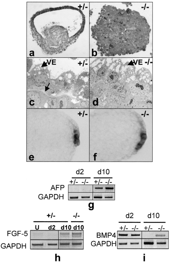
The formation of the proamniotic cavity in the mammalian embryo is the earliest of many instances throughout development in which programmed cell death and the formation of epithelia play fundamental roles (Coucouvanis, E., and G.R. Martin. 1995. Cell. 83:279-287). To determine the role of the basement membrane (BM) in cavitation, we use embryoid bodies derived from mouse embryonic stem cells in which the LAMC1 genes have been inactivated to prevent BM deposition (Smyth, N., H.S. Vatansever, P. Murray, M. Meyer, C. Frie, M. Paulsson, and D. Edgar. 1999. J. Cell Biol. 144:151-610). We demonstrate here that LAMC1-/- embryoid bodies are unable to cavitate, and do not form an epiblast epithelium in the absence of a BM, although both embryonic ectodermal cells and extraembryonic endodermal cells do differentiate, as evidenced by the expression of cell-specific markers. Acceleration or rescue of BM deposition by exogenous laminin in wild-type or LAMC1-/- embryoid bodies, respectively, results in cavitation that is temporally and spatially associated with restoration of epiblast epithelial development. We conclude that the BM not only directly regulates development of epiblast epithelial cells, but also indirectly regulates the programmed cell death necessary for cavity formation.


Figures

Figure 1
Figure 1
Schematic diagram showing the peri-implantation stages of mouse development. (a) Shortly after blastocyst formation, the cells on the surface of the ICM differentiate to become primitive endodermal cells. (b) The primitive endodermal cells deposit a basement membrane (BM). (c) After implantation, the primitive endodermal cells in contact with the BM differentiate to become visceral endoderm (VE) cells, and the remaining ICM cells differentiate to become epiblast cells. (d) The epiblast cells in contact with the BM polarize to form the columnar epiblast epithelium (CEE), and the unpolarized epiblast cells in the center undergo programmed cell death, giving rise to the proamniotic cavity.
Figure 4
Figure 4
Rescue of LAMC1−/− EBs by the addition of exogenous laminin. (a–d) Immunofluorescence staining for laminin (a and c) and perlecan (b and d) in LAMC1−/− EBs after 2 d of culture: without (a and b) and with the addition of laminin type-1 (c and d). (e) Toluidine blue–stained frozen section of LAMC1−/− EB after laminin addition shows that the CEE and PAC develop. BM, position of the basement membrane–like deposition of laminin and perlecan; CEE, columnar epiblast epithelium; PAC, proamniotic cavity; VE, visceral endoderm.
Figure 2
Figure 2
VE cell differentiation and cavitation in EBs. (a and b) Toluidine blue–stained frozen sections of EBs after 7.5 d in suspension culture show that LAMC1+/− EBs had cavitated by this time (a), whereas the LAMC1−/− EBs failed to cavitate (b). (c and d) EM shows differentiation of cells with the morphological characteristics of VE in both LAMC1+/− (c) and LAMC1−/− (d) EBs. However, a BM is only present in the LAMC1+/− control EBs (c, white arrow). (e and f) Whole-mount in situ hybridization for the VE marker AFP shows positive cells at the periphery of both LAMC1+/− (e) and LAMC1−/− (f) EBs. (g and i) RT-PCR analysis; GAPDH mRNA is shown as a loading control. (g) AFP mRNA is expressed late in both LAMC1+/− and LAMC1−/− EBs. (h) Only a trace of FGF-5 mRNA is detectable in undifferentiated LAMC1+/− ES cells (U) and day 2 (d2) EBs, but FGF-5 is induced by day 10 (d10) in both LAMC1−/− and control EBs; the double band results from splice variants (Johansson and Wiles 1995). (i) BMP4 mRNA is expressed in both LAMC1−/− and control EBs at day 2; but, by day 10, levels are greatly reduced in the controls. VE, visceral endoderm.
Figure 3
Figure 3
Epiblast cell polarization and PCD in EBs. (a) Toluidine blue–stained resin-embedded section of a day 6 LAMC1+/− EB shows that cell debris is present only at the apical surface of the CEE and is absent where the epiblast cells remain unpolarized (arrowhead). (b–d) EM of LAMC1+/− EBs. (b) The cell (asterisk) at the apical surface of the two columnar ectodermal cells has rounded up and become vacuolated by day 5; (c) a pocket of cell debris (CD) is present at the apical surface of the CEE on day 6; (d) as the debris is removed at day 7.5, the proamniotic cavity (PAC) becomes evident. (e) EM of day 7.5 LAMC1−/− EB shows unpolarized epiblast cells (asterisks) and no cell debris. (f–h) TUNEL analysis after 6 d: in LAMC1+/− EBs, a cluster of TUNEL-positive cells (f, arrow) is present at the apical surface of the CEE. (g) Phase-contrast image of f. Only a few randomly scattered TUNEL-positive cells (h, arrows) are present in LAMC1−/− EBs. (i) Phase-contrast image of h. Note that the BM in these EBs does not have a lamina lucida, which is consistent with previous reports of its absence in some EBs and in Reichert's membrane (Martin et al. 1977; Inoue et al. 1983). BM, basement membrane; CE, columnar epiblast cell; CEE, columnar epiblast epithelium; CD, cell debris; PAC, proamniotic cavity; VE, visceral endoderm.
Figure 5
Figure 5
Laminin addition to LAMC1+/− control EBs accelerates the deposition of a basement membrane, leading to epiblast polarization and cavitation. (a–d) Immunofluorescence staining for laminin (a and b) and perlecan (c and d) in control EBs after 2 d culture: without (a and c) and with addition of laminin type-1 (b and d). (e and f) Toluidine blue–stained frozen sections of day 2 control EBs grown without (e) or with (f) the addition of type-1 laminin. Note the formation of the CEE and a proamniotic-like cavity on the addition of laminin. CD, cell debris; CEE, columnar epiblast epithelium; PAC, proamniotic cavity.

References

    1. Chomczynski P., Sacchi N. Single-step method of RNA isolation by acid guanidinium thiocyanate-phenol-chloroform extraction. Anal. Biochem. 1987;162:156–159. - PubMed
    1. Coles H.S., Burne J.F., Raff M.C. Large-scale normal cell death in the developing rat kidney and its reduction by epidermal growth factor. Development. 1993;118:777–784. - PubMed
    1. Coucouvanis E., Martin G.R. Signals for death and survivala two-step mechanism for cavitation in the vertebrate embryo. Cell. 1995;83:279–287. - PubMed
    1. Coucouvanis E., Martin G.R. BMP signaling plays a role in visceral endoderm differentiation and cavitation in the early mouse embryo. Development. 1999;126:535–546. - PubMed
    1. Dziadek M., Adamson E. Localization and synthesis of alphafoetoprotein in post-implantation mouse embryos. J. Embryol. Exp. Morphol. 1978;43:289–313. - PubMed

Publication types