. 2000 Sep;106(5):663-70.
doi: 10.1172/JCI9362.

c-Jun-dependent inhibition of cutaneous procollagen transcription following ultraviolet irradiation is reversed by all-trans retinoic acid

Affiliations

c-Jun-dependent inhibition of cutaneous procollagen transcription following ultraviolet irradiation is reversed by all-trans retinoic acid

G J Fisher et al. J Clin Invest. 2000 Sep.

Abstract

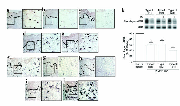
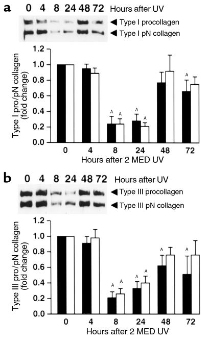
The aged appearance of skin following repeated exposure to solar ultraviolet (UV) irradiation stems largely from damage to cutaneous connective tissue, which is composed primarily of type I and type III collagens. We report here that a single exposure to UV irradiation causes significant loss of procollagen synthesis in human skin. Expression of type I and type III procollagens is substantially reduced within 24 hours after a single UV exposure, even at UV doses that cause only minimal skin reddening. Daily UV exposures over 4 days result in sustained reductions of both type I and type III procollagen protein levels for at least 24 hours after the final UV exposure. UV inhibition of type I procollagen synthesis is mediated in part by c-Jun, which is induced by UV irradiation and interferes with procollagen transcription. Pretreatment of human skin in vivo with all-trans retinoic acid inhibits UV induction of c-Jun and protects skin against loss of procollagen synthesis. We have reported previously that UV irradiation induces matrix-degrading metalloproteinases in human skin and that pretreatment of skin with all-trans retinoic acid inhibits this induction. UV irradiation, therefore, damages human skin connective tissue by simultaneously inhibiting procollagen synthesis and stimulating collagen breakdown. All-trans retinoic acid protects against both of these deleterious effects and may thereby retard premature skin aging.


Figures

Figure 1
Figure 1
UV irradiation inhibits type I and type III procollagen gene expression in human skin in vivo. Nonirradiated and adjacent UV-irradiated (2 MED) human skin samples were obtained at the times indicated after UV irradiation. Skin was analyzed for type I(α1) and type III(α1) procollagen mRNA expression by digoxigenin-riboprobe in situ hybridization. (ae) Type I(α1) procollagen and (fj) type III(α1) procollagen mRNA expression in human skin. a and f: 0, b and g: 8, c and h: 24, d and i: 48, and e and j: 72 hours after irradiation. Data are representative of six subjects. Scale bar = 10 μm. In aj, solid black lines demarcate border between epidermis and dermal connective tissue. Areas outlined in black are shown in 2.5-fold enlargements. (k) Northern analysis of type I(α1), type I(α2), and type III(α1) procollagen mRNA levels in human skin. Total mRNA (20 μg/lane) was analyzed from nonirradiated and UV-irradiated (2 MED) human skin, obtained 24 hours after UV irradiation (2 MED), and 36B4 mRNA was used as an internal control. Inset shows representative Northern blot. Procollagen mRNA levels were normalized to 36B4 mRNA levels. n = 6–7. AP < 0.01 vs. no UV exposure.
Figure 2
Figure 2
UV reduces type I and type III procollagen protein levels in human skin in vivo. Nonirradiated and adjacent UV-irradiated (2 MED) human skin samples were obtained at the times indicated after UV irradiation. Skin was analyzed for type I(α1) and type III(α1) procollagen protein levels using Western blot method. (a) Type I procollagen (filled bars) and type I pN collagen (open bars) protein levels in human skin. Inset shows representative Western blot. (b) Type III procollagen (filled bars) and type III pN collagen (open bars) protein levels in human skin. Inset shows representative Western blot. n = 7–9 subjects. AP < 0.05 vs. no UV exposure.
Figure 3
Figure 3
Multiple UV exposures cause sustained reduction of type I and type III procollagen proteins in human skin in vivo. Human skin was exposed to one, two, three, or four doses of UV (1 MED) at 24-hour intervals. Skin biopsies were obtained 24 hours after each exposure. Skin samples were analyzed for type I(α1) and type III(α1) procollagen and pN collagen protein levels using Western blot method. (a) Type I procollagen (open circle, solid line) and type I pN collagen (filled circle, broken line) protein in human skin. Inset shows representative Western blot. n = 7 subjects. (b) Type III procollagen (open circle, solid line) and type III pN collagen (filled circle, broken line) protein in human skin. Inset shows representative Western blot. n = 6 subjects. AP < 0.05 vs. no UV exposure.
Figure 4
Figure 4
UVB/UVA2 and solar-simulated UV dose dependence for reduction of type I procollagen in human skin in vivo. Human skin was irradiated with the indicated doses of UV from either a UVB/UVA2 source or a solar simulator. Skin was obtained 24 hours after irradiation and analyzed for type I procollagen protein levels using the Western blot method. (a) UV spectra for Kodacel-filtered UVB/UVA2 source (broken line), solar simulator (thick solid line), and sunlight (thin solid line). (b) UVB/UVA2 dose dependence for reduction of type I procollagen (filled bars) and type I pN collagen (open bars) in human skin. Inset shows representative Western blot. n = 5 subjects. (c) Solar-simulated UV dose dependence for reduction of type I procollagen (filled bars) and type I pN collagen (open bars) in human skin. Inset shows representative Western blot. n = 5 subjects. AP < 0.05 vs. no UV exposure.
Figure 5
Figure 5
UV-induced c-Jun inhibits type I(α2) procollagen gene expression in primary human skin fibroblasts. (a) Time course of UV induction of c-Jun protein in human skin fibroblasts. Nuclear extracts were prepared from fibroblasts at the indicated times after UV irradiation (30 mJ/cm2). The c-Jun protein levels were determined using the Western blot method. Inset shows a representative Western blot. n = 5 subjects. AP < 0.5 vs. no UV exposure. (b) c-Jun mediates UV inhibition of type I(α2) procollagen gene-promoter activity in human skin fibroblasts. Fibroblasts were cotransfected with type I(α2) procollagen promoter CAT reporter alone or with either wild-type (WT) c-Jun or dominant-negative mutant (dN) c-Jun expression vectors, as indicated. CAT activity was determined 12 hours after irradiation (30 mJ/cm2). n = 5 subjects. AP ≤ 0.05 vs. UV exposure; BP ≤ 0.05 vs. control.
Figure 6
Figure 6
All-trans retinoic acid (RA) protects against UV-induced reduction of type I and type III procollagen mRNA expression in human skin in vivo. Human skin was pretreated with vehicle and 0.1% RA for 24 hours, then irradiated with UV (2 MED). Skin was obtained 24 hours after UV exposure. Type I (ad) and type III (eh) procollagen mRNA expression was determined by digoxigenin riboprobe in situ hybridization. a and e, vehicle-treated skin; b and f, RA-treated skin; c and g, vehicle-pretreated, UV-irradiated skin; d and h, RA-pretreated, UV-irradiated skin. Insets show enlargement of cells in the dermis. Solid black lines demarcate border between epidermis and connective tissue. Areas outlined in black are shown in 2.5-fold enlargements. Data displayed are representative of six subjects. Scale bar = 10 μm.
Figure 7
Figure 7
All-trans retinoic acid (RA) protects against UV-induced loss of type I and type III procollagen proteins in human skin in vivo. Human skin was pretreated with vehicle (VEH) and 0.1% RA for 24 hours, then irradiated with UV (2 MED). Skin was obtained 24 hours after UV exposure. (a) Type I procollagen (filled bars) and type I pN collagen (open bars) protein in human skin. Inset shows representative Western blot. (b) Type III procollagen (filled bars) and type III pN collagen (open bars) protein in human skin. Inset shows representative Western blot. n = 9 subjects. AP < 0.05 vs. vehicle-treated, nonirradiated skin.

References

    1. Kligman AM. Early destructive effects of sunlight on human skin. JAMA. 1969;210:2377–2380. - PubMed
    1. Bernstein EF, et al. Long-term sun exposure alters the collagen of the papillary dermis: comparison of sun-protected and photoaged skin by Northern analysis, immunohistochemical staining, and confocal laser scanning microscopy. J Am Acad Dermatol. 1996;34:209–218. - PubMed
    1. Lavker, R.M. 1995. Cutaneous aging: chronologic versus photoaging. In Photoaging. B.A. Gilchrest, editor. Blackwell Science. Cambridge, Massachusetts, USA. 123–135.
    1. Uitto, J. 1993. Collagen. In Dermatology in general medicine. Volume 1. T.B. Fitzpatrick, A.Z. Eisen, K. Wolff, I.M. Freedberg, and K.F. Austen, editors. McGraw-Hill. New York, New York, USA. 299–314.
    1. Talwar HS, Griffiths CEM, Fisher GJ, Hamilton TA, Voorhees JJ. Reduced type I and type III procollagens in photodamaged adult human skin. J Invest Dermatol. 1995;105:285–291. - PubMed

Publication types