. 2000 Sep 4;192(5):637-46.
doi: 10.1084/jem.192.5.637.

B cells of HIV-1-infected patients bind virions through CD21-complement interactions and transmit infectious virus to activated T cells

Affiliations

B cells of HIV-1-infected patients bind virions through CD21-complement interactions and transmit infectious virus to activated T cells

S Moir et al. J Exp Med. .

Abstract

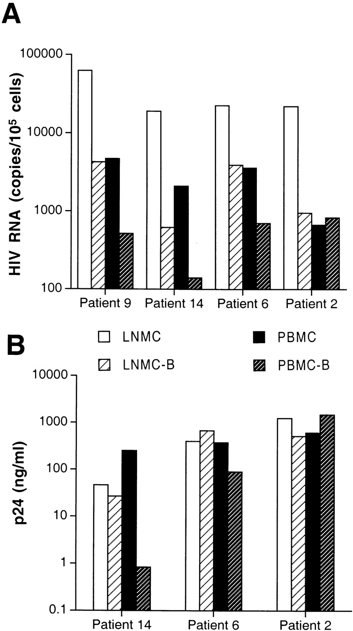
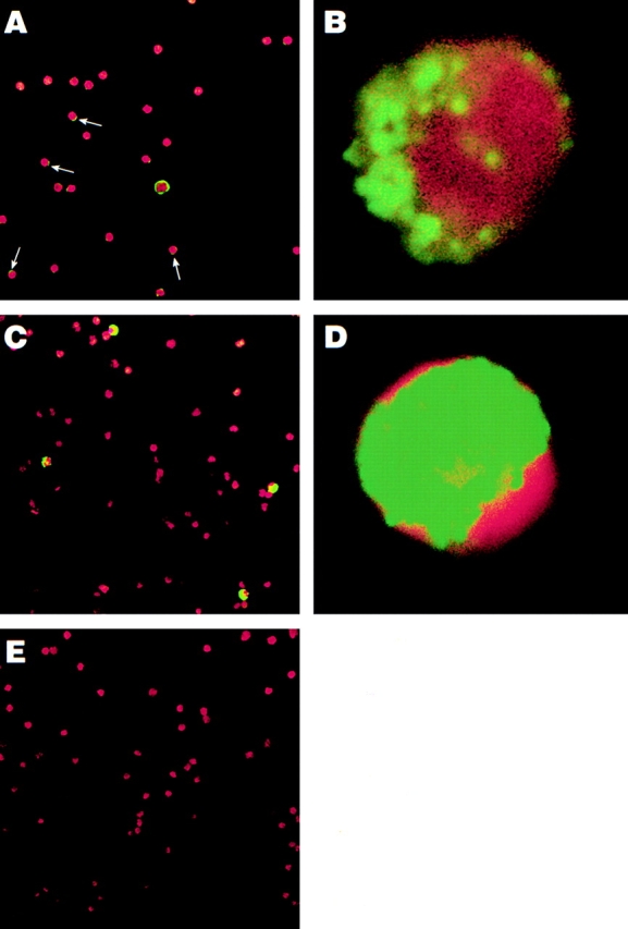
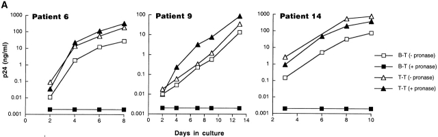
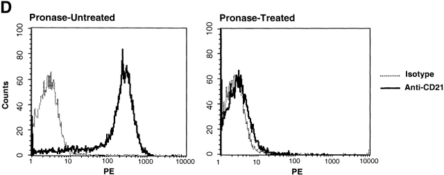
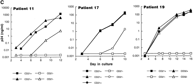
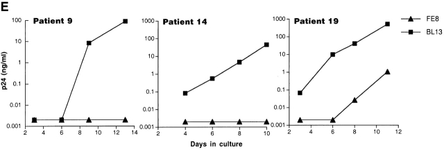
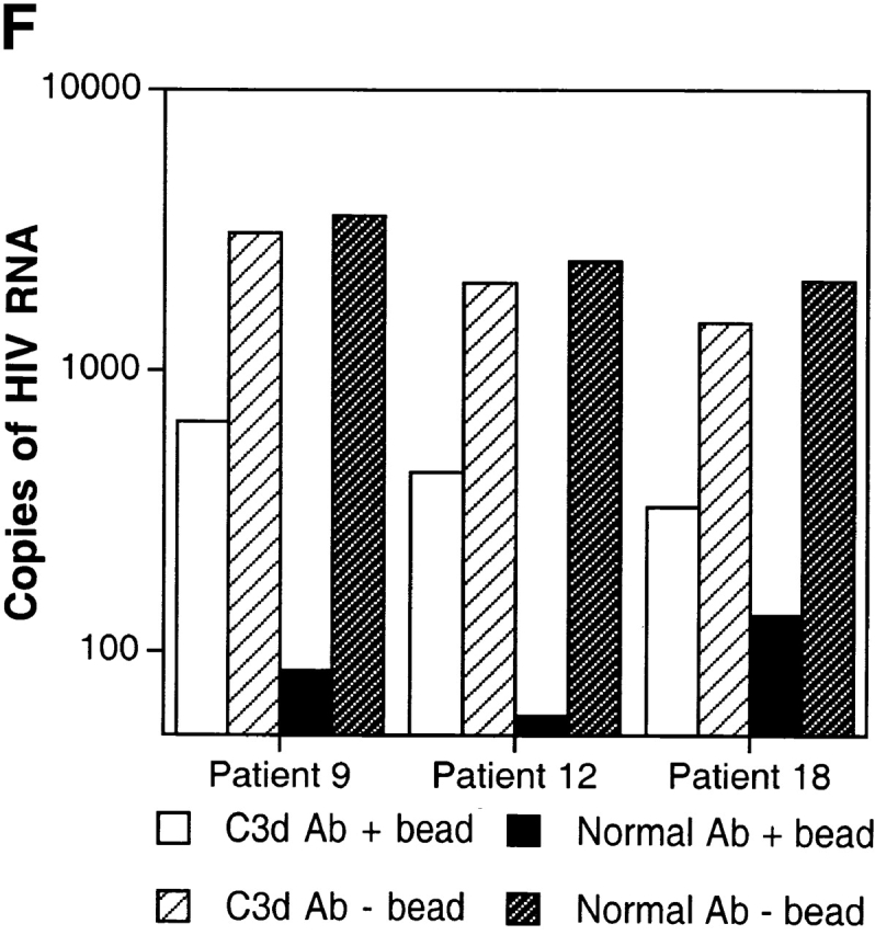
The impact of HIV-associated immunopathogenesis on B cells has been largely associated with indirect consequences of viral replication. This study demonstrates that HIV interacts directly with B cells in both lymphoid tissues and peripheral blood. B cells isolated from lymph node and peripheral blood mononuclear cells (PBMCs) of 4 and 23 chronically infected patients, respectively, demonstrated similar capacities to pass virus to activated HIV-negative PBMCs when compared with CD4(+) cells from the same patients. However, in contrast to T cells, virus associated with B cells was surface bound, as shown by its sensitivity to pronase and the staining pattern revealed by in situ amplification of HIV-1 RNA. Cell sorting and ligand displacing approaches established that CD21 was the HIV-binding receptor on B cells, and that this association was mediated through complement-opsonized virus. These B cells were also found to express significantly lower levels of CD21 compared with HIV-negative individuals, suggesting a direct perturbing effect of HIV on B cells. These findings suggest that B cells, although they themselves are not readily infected by HIV, are similar to follicular dendritic cells in their capacity to serve as extracellular reservoirs for HIV-1. Furthermore, B cells possess the added capability of circulating in peripheral blood and migrating through tissues where they can potentially interact with and pass virus to T cells.


Figures

Figure 1
Figure 1
Levels of cell-associated HIV-1 RNA and replication-competent virus in various cell types isolated from patients chronically infected with HIV-1. Highly enriched B cells identified as LNMC-B and PBMC-B were isolated from LNMCs and PBMCs, respectively, of (A) four patients and compared with corresponding unfractionated cells for levels of cellular HIV RNA, and (B) in three patients compared with corresponding CD8-depleted cells for levels of replication-competent virus. Coculture conditions included 106 HIV-infected cells and 106 indicator T cells, with the exception of LNMCs of patient 6, where cocultures were performed on 0.2 × 10⁶ input cells. Levels of HIV-1 p24 were measured in coculture supernatants on day 7.
Figure 2
Figure 2
Location of HIV RNA in and on cells isolated from an HIV-infected individual. In situ amplification for HIV-1 RNA (gag) was carried out on highly purified B cells from patient 9 (A and B), CD8/CD19-depleted PBMCs from patient 9 (C and D), and HIV-negative PBMCs (E). The FITC stains (HIV RNA, in green) were overlaid onto the corresponding DAPI stains (cellular DNA, in red) to create the final image. Arrows indicate where weak positive signals were observed on the surface of B cells. Original magnification: (A, C, and E) ×400; (B and D) ×1,000.
Figure 3
Figure 3
Characterization of the replication-competent virus associated with PBMC-derived B cells of HIV-infected patients. (A) B cells and T cells isolated from three HIV-infected individuals were pretreated with pronase followed by coculture with indicator T cells. (B, top) Flow cytometric comparison of CD21 expression on representative HIV-negative B cells and presorted B cells of patient 17, followed by (B, bottom) post-sorted CD20+/CD21+ and CD20+/CD21 fractions of patient 17. (C) B cells of three HIV-infected patients sorted into CD21+ and CD21 fractions (and CD32+ versus CD32 fractions in the case of patient 19) and cocultured with indicator T cells. Colors identify the sorted fraction and symbols identify the number of coculture wells plated for a given sorted fraction. (D) Flow cytometric comparison of CD21 expression on pronase-treated and -untreated B cells of patient 9. (E) B cells of three HIV-infected patients preincubated with mAbs FE8 and BL13 and cocultured with indicator T cells. (F) FE8-displaced HIV from 2 × 10⁶ B cells of three HIV-infected patients, treated with rabbit anti–human C3d (C3d Ab) or normal rabbit serum (Normal Ab), and captured with magnetized anti-rabbit antibodies. Fractions captured on magnet (+ bead) and fractions not captured (− bead) were tested for HIV-1 RNA by bDNA.
Figure 3
Figure 3
Characterization of the replication-competent virus associated with PBMC-derived B cells of HIV-infected patients. (A) B cells and T cells isolated from three HIV-infected individuals were pretreated with pronase followed by coculture with indicator T cells. (B, top) Flow cytometric comparison of CD21 expression on representative HIV-negative B cells and presorted B cells of patient 17, followed by (B, bottom) post-sorted CD20+/CD21+ and CD20+/CD21 fractions of patient 17. (C) B cells of three HIV-infected patients sorted into CD21+ and CD21 fractions (and CD32+ versus CD32 fractions in the case of patient 19) and cocultured with indicator T cells. Colors identify the sorted fraction and symbols identify the number of coculture wells plated for a given sorted fraction. (D) Flow cytometric comparison of CD21 expression on pronase-treated and -untreated B cells of patient 9. (E) B cells of three HIV-infected patients preincubated with mAbs FE8 and BL13 and cocultured with indicator T cells. (F) FE8-displaced HIV from 2 × 10⁶ B cells of three HIV-infected patients, treated with rabbit anti–human C3d (C3d Ab) or normal rabbit serum (Normal Ab), and captured with magnetized anti-rabbit antibodies. Fractions captured on magnet (+ bead) and fractions not captured (− bead) were tested for HIV-1 RNA by bDNA.

Similar articles

Cited by

References

    1. Burton G.F., Masuda A., Heath S.L., Smith B.A., Tew J.G., Szakal A.K. Follicular dendritic cells (FDC) in retroviral infectionhost/pathogen perspectives. Immunol. Rev. 1997;156:185–197. - PubMed
    1. Chun T.W., Fauci A.S. Latent reservoirs of HIVobstacles to the eradication of virus. Proc. Natl. Acad. Sci. USA. 1999;96:10958–10961. - PMC - PubMed
    1. Haase A.T. Population biology of HIV-1 infectionviral and CD4+ T cell demographics and dynamics in lymphatic tissues. Annu. Rev. Immunol. 1999;17:625–656. - PubMed
    1. Geijtenbeek T.B., Kwon D.S., Torensma R., van Vliet S.J., van Duijnhoven G.C., Middel J., Cornelissen I.L., Nottet H.S., KewalRamani V.N., Littman D.R. DC-SIGN, a dendritic cell-specific HIV-1-binding protein that enhances trans-infection of T cells. Cell. 2000;100:587–597. - PubMed
    1. Liao Z., Roos J.W., Hildreth J.E. Increased infectivity of HIV type 1 particles bound to cell surface and solid-phase ICAM-1 and VCAM-1 through acquired adhesion molecules LFA-1 and VLA-4. AIDS Res. Hum. Retroviruses. 2000;16:355–366. - PubMed

MeSH terms