

Blockade of T lymphocyte costimulation with cytotoxic T lymphocyte-associated antigen 4-immunoglobulin (CTLA4Ig) reverses the cellular pathology of psoriatic plaques, including the activation of keratinocytes, dendritic cells, and endothelial cells
- PMID: 10974034
- PMCID: PMC2193278
- DOI: 10.1084/jem.192.5.681
Blockade of T lymphocyte costimulation with cytotoxic T lymphocyte-associated antigen 4-immunoglobulin (CTLA4Ig) reverses the cellular pathology of psoriatic plaques, including the activation of keratinocytes, dendritic cells, and endothelial cells
Abstract
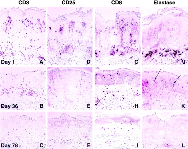
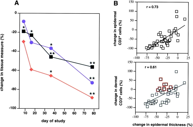
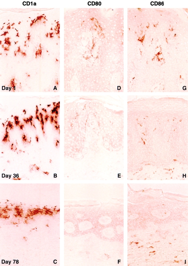
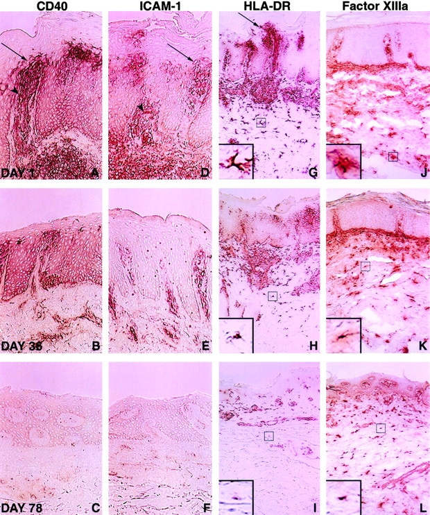
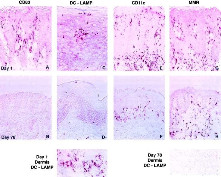
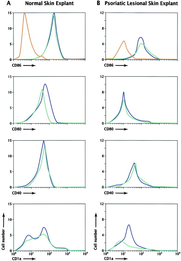
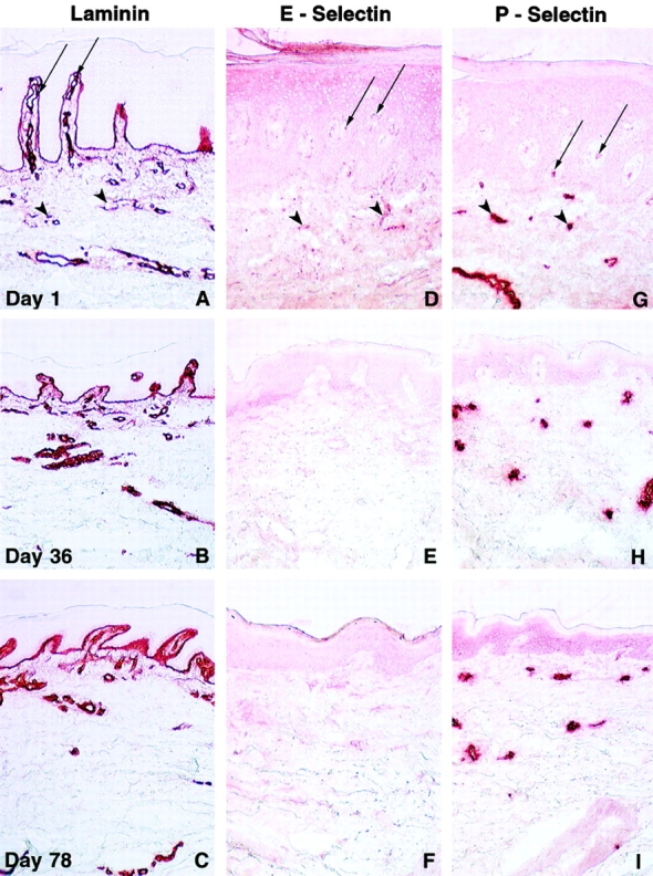
Efficient T cell activation is dependent on the intimate contact between antigen-presenting cells (APCs) and T cells. The engagement of the B7 family of molecules on APCs with CD28 and CD152 (cytotoxic T lymphocyte-associated antigen 4 [CTLA-4]) receptors on T cells delivers costimulatory signal(s) important in T cell activation. We investigated the dependence of pathologic cellular activation in psoriatic plaques on B7-mediated T cell costimulation. Patients with psoriasis vulgaris received four intravenous infusions of the soluble chimeric protein CTLA4Ig (BMS-188667) in a 26-wk, phase I, open label dose escalation study. Clinical improvement was associated with reduced cellular activation of lesional T cells, keratinocytes, dendritic cells (DCs), and vascular endothelium. Expression of CD40, CD54, and major histocompatibility complex (MHC) class II HLA-DR antigens by lesional keratinocytes was markedly reduced in serial biopsy specimens. Concurrent reductions in B7-1 (CD80), B7-2 (CD86), CD40, MHC class II, CD83, DC-lysosomal-associated membrane glycoprotein (DC-LAMP), and CD11c expression were detected on lesional DCs, which also decreased in number within lesional biopsies. Skin explant experiments suggested that these alterations in activated or mature DCs were not the result of direct toxicity of CTLA4Ig for DCs. Decreased lesional vascular ectasia and tortuosity were also observed and were accompanied by reduced presence of E-selectin, P-selectin, and CD54 on vascular endothelium. This study highlights the critical and proximal role of T cell activation through the B7-CD28/CD152 costimulatory pathway in maintaining the pathology of psoriasis, including the newly recognized accumulation of mature DCs in the epidermis.
Figures
References
-
- Paukkonen K., Naukkarinen A., Horsmanheimo M. The development of manifest psoriatic lesions is linked with the invasion of CD8+ T cells and CD11c+ macrophages into the epidermis. Arch. Dermatol. Res. 1992;284:375–379. - PubMed
-
- Demiden A., Taylor J.R., Grammer S.F., Streilein J.W. T-lymphocyte-activating properties of epidermal antigen-presenting cells from normal and psoriatic skinevidence that psoriatic epidermal antigen-presenting cells resemble cultured normal Langerhans cells. J. Invest. Dermatol. 1991;97:454–460. - PubMed
-
- Banchereau J., Steinman R.M. Dendritic cells and the control of immunity. Nature. 1998;392:245–252. - PubMed
-
- Cerio R., Griffiths C.E.M., Cooper K.D., Nickoloff B.J., Headington J.T. Characterization of factor XIIIa positive dermal dendritic cells in normal and inflamed skin. Br. J. Dermatol. 1989;121:421–431. - PubMed
MeSH terms
Substances
LinkOut - more resources
Full Text Sources
Other Literature Sources
Medical
Research Materials
Miscellaneous
