. 2000 Sep 4;192(5):695-704.
doi: 10.1084/jem.192.5.695.

Inhibition of E-selectin gene expression by transforming growth factor beta in endothelial cells involves coactivator integration of Smad and nuclear factor kappaB-mediated signals

Affiliations

Inhibition of E-selectin gene expression by transforming growth factor beta in endothelial cells involves coactivator integration of Smad and nuclear factor kappaB-mediated signals

M R DiChiara et al. J Exp Med. .

Abstract

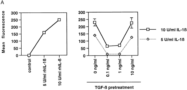
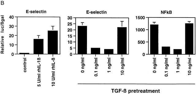
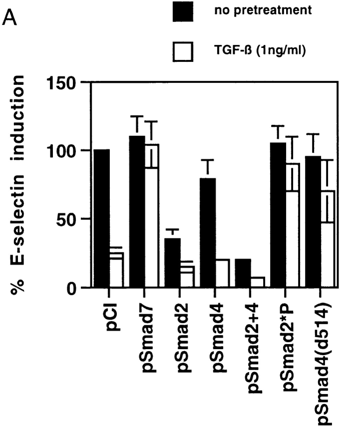
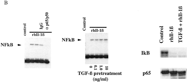
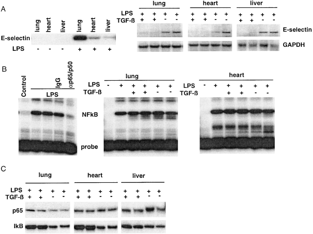
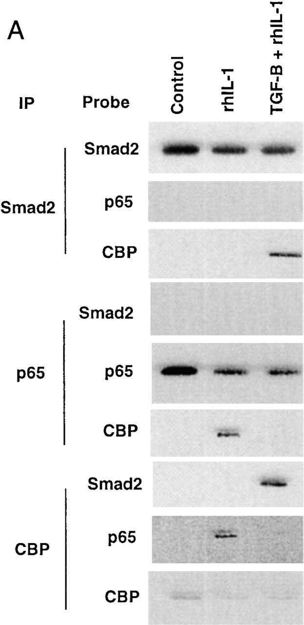
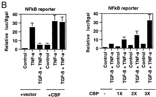
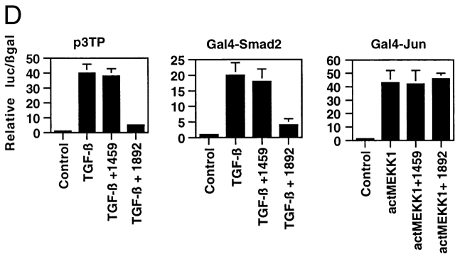
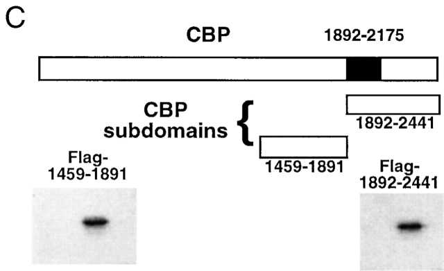
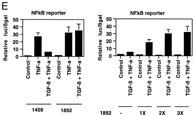
Transforming growth factor (TGF)-beta(1) is a pleiotropic cytokine/growth factor that is thought to play a critical role in the modulation of inflammatory events. We demonstrate that exogenous TGF-beta(1) can inhibit the expression of the proinflammatory adhesion molecule, E-selectin, in vascular endothelium exposed to inflammatory stimuli both in vitro and in vivo. This inhibitory effect occurs at the level of transcription of the E-selectin gene and is dependent on the action of Smad proteins, a class of intracellular signaling proteins involved in mediating the cellular effects of TGF-beta(1). Furthermore, we demonstrate that these Smad-mediated effects in endothelial cells result from a novel competitive interaction between Smad proteins activated by TGF-beta(1) and nuclear factor kappaB (NFkappaB) proteins activated by inflammatory stimuli (such as cytokines or bacterial lipopolysaccharide) that is mediated by the transcriptional coactivator cyclic AMP response element-binding protein (CREB)-binding protein (CBP). Augmentation of the limited amount of CBP present in endothelial cells (via overexpression) or selective disruption of Smad-CBP interactions (via a dominant negative strategy) effectively antagonizes the ability of TGF-beta(1) to block proinflammatory E-selectin expression. These data thus demonstrate a novel mechanism of interaction between TGF-beta(1)-regulated Smad proteins and NFkappaB proteins regulated by inflammatory stimuli in vascular endothelial cells. This type of signaling mechanism may play an important role in the immunomodulatory actions of this cytokine/growth factor in the cardiovascular system.


Figures

Figure 1
Figure 1
(A) Inducible expression of E-selectin protein on the surface of cultured endothelial cells in response to Il-1β can be inhibited by pretreatment with active TGF-β1. The level of cell surface E-selectin protein was determined by fluorescence immunoassay at baseline and 4 h after treatment with 5 or 10 U/ml of rhIL-1β (left). To assess the effect of TGF-β1 pretreatment, the cells were exposed to the indicated doses of active TGF-β1 for 6 h before cytokine treatment. The right panel demonstrates that pretreatment of HUVECs with intermediate doses of TGF-β1 markedly diminished cytokine-induced E-selectin expression. (B) Pretreatment of TGF-β can inhibit cytokine-induced transcription of the E-selectin promoter and a simplified NFκB-dependent promoter in endothelial cells. Luciferase reporter constructs containing either the E-selectin promoter (left and middle) or a promoter consisting of a series of tandem NFκB sites (right) were transiently transfected into HUVECs. The cells were subsequently stimulated with rhIL-1β for 4 h, and luciferase levels were measured and normalized to β-galactosidase activity (from a cotransfected CMV-β gal reporter vector). As shown in the left panel, both 5 and 10 U/ml of rhIL-1β effectively induce transcription from the E-selectin promoter. In the middle and right panels, the cells were pretreated with the indicated doses of active TGF-β 1 for 6 h before cytokine stimulation (5 U/ml). Intermediate doses of TGF-β1 were effective at inhibiting cytokine-induced, NFκB-mediated transcription.
Figure 1
Figure 1
(A) Inducible expression of E-selectin protein on the surface of cultured endothelial cells in response to Il-1β can be inhibited by pretreatment with active TGF-β1. The level of cell surface E-selectin protein was determined by fluorescence immunoassay at baseline and 4 h after treatment with 5 or 10 U/ml of rhIL-1β (left). To assess the effect of TGF-β1 pretreatment, the cells were exposed to the indicated doses of active TGF-β1 for 6 h before cytokine treatment. The right panel demonstrates that pretreatment of HUVECs with intermediate doses of TGF-β1 markedly diminished cytokine-induced E-selectin expression. (B) Pretreatment of TGF-β can inhibit cytokine-induced transcription of the E-selectin promoter and a simplified NFκB-dependent promoter in endothelial cells. Luciferase reporter constructs containing either the E-selectin promoter (left and middle) or a promoter consisting of a series of tandem NFκB sites (right) were transiently transfected into HUVECs. The cells were subsequently stimulated with rhIL-1β for 4 h, and luciferase levels were measured and normalized to β-galactosidase activity (from a cotransfected CMV-β gal reporter vector). As shown in the left panel, both 5 and 10 U/ml of rhIL-1β effectively induce transcription from the E-selectin promoter. In the middle and right panels, the cells were pretreated with the indicated doses of active TGF-β 1 for 6 h before cytokine stimulation (5 U/ml). Intermediate doses of TGF-β1 were effective at inhibiting cytokine-induced, NFκB-mediated transcription.
Figure 2
Figure 2
(A) TGF-β1–mediated inhibition of IL-1β–induced E-selectin expression in endothelial cells is Smad protein dependent. BAECs were transiently cotransfected with the E-selectin promoter construct and the indicated Smad protein expression constructs or an empty expression vector (pCi). The cells were subsequently stimulated for 4 h with TNF-α, with and without 6 h of pretreatment with TGF-β1 at 1 ng/ml. The level of induction of the E-selectin promoter has been normalized to that seen in the absence of TGF-β1 pretreatment (in the presence of the cotransfected empty expression vector) and expressed as 100%. In the presence of cotransfected empty expression vector (pCI), there is a marked inhibition of E-selectin promoter induction by TGF-β1 pretreatment, as described in the legend to Fig. 1. Coexpression of Smad7, an inhibitory Smad protein, or of Smad2*P or Smad4 (d514), which can act as dominant negative inhibitors, markedly attenuates the TGF-β1–mediated inhibition. In contrast, coexpression of wild-type Smad2, Smad4, or both Smad2 and -4 significantly enhances the level of inhibition seen and is able to induce varying levels of inhibition even in the absence of exogenous TGF-β1 pretreatment. All of the Smad expression constructs encode epitope-tagged species that were expressed at comparable levels as monitored by immunoblot (data not shown). (B) Cytokine-induced activation of NFκB appears preserved in the presence of TGF-β1 pretreatment. HUVEC monolayers were treated with 10 U/ml of rhIL-1β for 30 min and lysed, and a nuclear extract was prepared. Specific NFκB-mediated DNA binding activity was assessed by mobility shift assay and compared with that present in unstimulated HUVEC. As shown in the left panel, there is a specific shifted band present in the rhIL-1β–treated lanes but not in the control (untreated) lane that can be abolished by immunodepletion of the extract with a mixture of anti-p65/antip50 antisera but not nonspecific IgG. As shown in the middle panel, rhIL-1β–induced, NFκB-mediated DNA binding activity (as assessed by this gel shift) appears preserved even in the presence of TGF-β pretreatment. The panels at the right represent immunoblots for IκB and P65 protein levels. The former is markedly depleted by rhIL-1β treatment, even in the presence of TGF-β1 pretreatment. p65 levels appeared stable under these conditions. These findings suggest that IκB degradation and NFκB-mediated DNA binding are not affected by TGF-β1 pretreatment in HUVECs.
Figure 2
Figure 2
(A) TGF-β1–mediated inhibition of IL-1β–induced E-selectin expression in endothelial cells is Smad protein dependent. BAECs were transiently cotransfected with the E-selectin promoter construct and the indicated Smad protein expression constructs or an empty expression vector (pCi). The cells were subsequently stimulated for 4 h with TNF-α, with and without 6 h of pretreatment with TGF-β1 at 1 ng/ml. The level of induction of the E-selectin promoter has been normalized to that seen in the absence of TGF-β1 pretreatment (in the presence of the cotransfected empty expression vector) and expressed as 100%. In the presence of cotransfected empty expression vector (pCI), there is a marked inhibition of E-selectin promoter induction by TGF-β1 pretreatment, as described in the legend to Fig. 1. Coexpression of Smad7, an inhibitory Smad protein, or of Smad2*P or Smad4 (d514), which can act as dominant negative inhibitors, markedly attenuates the TGF-β1–mediated inhibition. In contrast, coexpression of wild-type Smad2, Smad4, or both Smad2 and -4 significantly enhances the level of inhibition seen and is able to induce varying levels of inhibition even in the absence of exogenous TGF-β1 pretreatment. All of the Smad expression constructs encode epitope-tagged species that were expressed at comparable levels as monitored by immunoblot (data not shown). (B) Cytokine-induced activation of NFκB appears preserved in the presence of TGF-β1 pretreatment. HUVEC monolayers were treated with 10 U/ml of rhIL-1β for 30 min and lysed, and a nuclear extract was prepared. Specific NFκB-mediated DNA binding activity was assessed by mobility shift assay and compared with that present in unstimulated HUVEC. As shown in the left panel, there is a specific shifted band present in the rhIL-1β–treated lanes but not in the control (untreated) lane that can be abolished by immunodepletion of the extract with a mixture of anti-p65/antip50 antisera but not nonspecific IgG. As shown in the middle panel, rhIL-1β–induced, NFκB-mediated DNA binding activity (as assessed by this gel shift) appears preserved even in the presence of TGF-β pretreatment. The panels at the right represent immunoblots for IκB and P65 protein levels. The former is markedly depleted by rhIL-1β treatment, even in the presence of TGF-β1 pretreatment. p65 levels appeared stable under these conditions. These findings suggest that IκB degradation and NFκB-mediated DNA binding are not affected by TGF-β1 pretreatment in HUVECs.
Figure 3
Figure 3
(A) Induction of E-selectin mRNA in rat tissues by LPS can be inhibited by systemic pretreatment with TGF-β1. As described in Materials and Methods, rats received 4 mg/kg of LPS intraperitoneally, and 6 h later the indicated tissues were harvested and processed for RNA and protein extracts as described. (A) As shown by Northern analysis (left panel), E-selectin mRNA is not detected in control tissues but is detectable in the lung, liver, and hearts 6 h after LPS administration. In the right panels, tissues from LPS-treated animals that had received either vehicle or TGF-β1 pretreatment were similarly analyzed. Each pair of lanes represents two independently treated animals. Pretreatment with TGF-β1 markedly attenuated E-selectin mRNA induction (upper blots) but did not affect glyceraldehyde 3-phosphate dehydrogenase (GAPDH) mRNA levels (lower blots). (B) NFκB-mediated DNA binding activity appears preserved after TGF-β1 pretreatment in vivo. The lungs and hearts of LPS-treated rats were processed to obtain nuclear extracts for DNA mobility shift analyses with the NFκB-specific oligo probes as described in the legend to Fig. 2. In the left panel, a specific shifted band is detected in lysates from LPS-treated rat lung that is not present in lysates from control lung and that is attenuated by immunodepletion with antip65/p50 antisera but not nonimmune IgG. The middle and right panels are lysates from lung and heart, respectively, from the animals that have received LPS with or without TGF-β1 pretreatment. Each pair of lanes represents two independently treated animals. TGF-β1 pretreatment in vivo does not appear to affect LPS-induced NFκB-mediated DNA binding in these tissues. (C) Western analysis for p65 and IκB levels in tissues from LPS and TGF-β1–pretreated rats. The levels of immunoreactive p65 and IκB in the rat tissues were assessed by immunoblot. Each pair of lanes represents two independently treated animals. Levels of p65 were not significantly altered in response to any of the treatment protocols. The levels of immunoreactive IκB appeared slightly depressed by LPS treatment in the absence of TGF-β1 pretreatment.
Figure 4
Figure 4
(A) Smad2 but not p65 is recruited into a CBP-containing complex in response to TGF-β1 pretreatment followed by rhIL-1β treatment of HUVECs. HUVEC monolayers were treated with 10 U/ml of rhIL-1β for 30 min with and without pretreatment with TGF-β1 at 1 ng/ml for 6 h. Protein lysates were then prepared and immunoprecipitated with anti-Smad2, anti-p65, or anti-CBP antisera as indicated. These immunoprecipitates were then analyzed by Western blot with the antisera indicated. (B) Overexpression of CBP reverses TGF-β1–mediated inhibition of TNF-α–induced NFκB-mediated transcription. BAEC monolayers were cotransfected with the simplified NFκB-dependent promoter and either empty expression vector or vector encoding wild-type CBP (left). They were then treated with TNF-α (100 U/ml) both with and without TGF-β1 pretreatment (1 ng/ml), as described, and luciferase activity was assayed 12 h later. Overexpression of CBP reversed the ability of TGF-β1 pretreatment to inhibit NFκB-mediated transcription. The right panel shows cells that were transfected with the NFκB-dependent promoter together with increasing ratios of the CBP expression plasmid and subsequently pretreated with TGF-β1 before TNF-α stimulation to demonstrate that this effect is dose dependent. (C) Expression of subdomains of CBP. The region of interaction between CBP and Smad2 was mapped to amino acids 1892–2175, and is shown schematically. Two subdomains of CBP corresponding to amino acids 1459–1891 or 1892–2441, respectively, were subcloned by PCR and expressed as epitope (Flag)-tagged species and detected by anti-Flag immunoblot, as shown. (D) Overexpression of a specific subdomain of CBP can act as selective inhibitor of Smad-dependent transcription when overexpressed. BAECs were transfected with either the TGF-β1 responsive reporter construct p3TP-lux (left), a Gal4-Smad2 fusion protein expression construct together with a Gal4 reporter (middle), or a Gal4-Jun fusion protein expression construct together with the Gal4 reporter (right), and were stimulated by either soluble TGF-β1 (5 U/ml for 18 h) or coexpression of a constitutively active form of mitogen-activated protein kinase kinase 1 (actMEKK1). The effect of overexpression of the subdomains of CBP described in the legend to Fig. 4 C was assessed by cotransfecting these expression constructs as listed. Overexpression of the subdomain corresponding to the Smad interaction domain (1892–2441) was able to inhibit the TGF-β/Smad-dependent promoters but not the Gal4-Jun–mediated promoter. The adjacent subdomain of CBP (1459–1891) did not act as an inhibitor of these promoters. (E) Overexpression of 1892 but not 1459 can reverse TGF-β1–mediated inhibition of NFκB-dependent transcription. BAECs were cotransfected with the simplified NFκB-dependent promoter and the indicated subdomains of CBP (left). The cells were subsequently stimulated with TNF-α with and without TGF-β1 pretreatment, as above. In the presence of the overexpressed 1892 subdomain of CBP, the TGF-β–mediated inhibition of the NFκB-dependent promoter is not observed. The right panel shows cells that were transfected with the NFκB reporter and increasing ratios of the 1892 subdomain expression vector, then pretreated with TGF-β1 before TNF-α stimulation, as above. Increasing doses of the 1892 expression vector result in a progressive decrease in the ability of TGF-β1 pretreatment to inhibit the cytokine induction of this promoter.
Figure 4
Figure 4
(A) Smad2 but not p65 is recruited into a CBP-containing complex in response to TGF-β1 pretreatment followed by rhIL-1β treatment of HUVECs. HUVEC monolayers were treated with 10 U/ml of rhIL-1β for 30 min with and without pretreatment with TGF-β1 at 1 ng/ml for 6 h. Protein lysates were then prepared and immunoprecipitated with anti-Smad2, anti-p65, or anti-CBP antisera as indicated. These immunoprecipitates were then analyzed by Western blot with the antisera indicated. (B) Overexpression of CBP reverses TGF-β1–mediated inhibition of TNF-α–induced NFκB-mediated transcription. BAEC monolayers were cotransfected with the simplified NFκB-dependent promoter and either empty expression vector or vector encoding wild-type CBP (left). They were then treated with TNF-α (100 U/ml) both with and without TGF-β1 pretreatment (1 ng/ml), as described, and luciferase activity was assayed 12 h later. Overexpression of CBP reversed the ability of TGF-β1 pretreatment to inhibit NFκB-mediated transcription. The right panel shows cells that were transfected with the NFκB-dependent promoter together with increasing ratios of the CBP expression plasmid and subsequently pretreated with TGF-β1 before TNF-α stimulation to demonstrate that this effect is dose dependent. (C) Expression of subdomains of CBP. The region of interaction between CBP and Smad2 was mapped to amino acids 1892–2175, and is shown schematically. Two subdomains of CBP corresponding to amino acids 1459–1891 or 1892–2441, respectively, were subcloned by PCR and expressed as epitope (Flag)-tagged species and detected by anti-Flag immunoblot, as shown. (D) Overexpression of a specific subdomain of CBP can act as selective inhibitor of Smad-dependent transcription when overexpressed. BAECs were transfected with either the TGF-β1 responsive reporter construct p3TP-lux (left), a Gal4-Smad2 fusion protein expression construct together with a Gal4 reporter (middle), or a Gal4-Jun fusion protein expression construct together with the Gal4 reporter (right), and were stimulated by either soluble TGF-β1 (5 U/ml for 18 h) or coexpression of a constitutively active form of mitogen-activated protein kinase kinase 1 (actMEKK1). The effect of overexpression of the subdomains of CBP described in the legend to Fig. 4 C was assessed by cotransfecting these expression constructs as listed. Overexpression of the subdomain corresponding to the Smad interaction domain (1892–2441) was able to inhibit the TGF-β/Smad-dependent promoters but not the Gal4-Jun–mediated promoter. The adjacent subdomain of CBP (1459–1891) did not act as an inhibitor of these promoters. (E) Overexpression of 1892 but not 1459 can reverse TGF-β1–mediated inhibition of NFκB-dependent transcription. BAECs were cotransfected with the simplified NFκB-dependent promoter and the indicated subdomains of CBP (left). The cells were subsequently stimulated with TNF-α with and without TGF-β1 pretreatment, as above. In the presence of the overexpressed 1892 subdomain of CBP, the TGF-β–mediated inhibition of the NFκB-dependent promoter is not observed. The right panel shows cells that were transfected with the NFκB reporter and increasing ratios of the 1892 subdomain expression vector, then pretreated with TGF-β1 before TNF-α stimulation, as above. Increasing doses of the 1892 expression vector result in a progressive decrease in the ability of TGF-β1 pretreatment to inhibit the cytokine induction of this promoter.

References

    1. Bevilacqua M.P., Stengelin S., Gimbrone M.A.J., Jr., Seed B. Endothelial leukocyte adhesion molecule 1an inducible receptor for neutrophils related to complement regulatory proteins and lectins. Science. 1989;243:1160–1165. - PubMed
    1. Collins T., Read M.A., Neish A.S., Whitley M.Z., Thanos D., Maniatis T. Transcriptional regulation of endothelial cell adhesion moleculesNF-kappa B and cytokine-inducible enhancers. FASEB J. 1995;9:899–909. - PubMed
    1. Read M.A., Neish A.S., Luscinskas F.W., Palombella V.J., Maniatis T., Collins T. The proteasome pathway is required for cytokine-induced endothelial-leukocyte adhesion molecule expression. Immunity. 1995;2:493–506. - PubMed
    1. Read M.A., Whitley M.Z., Gupta S., Pierce J.W., Best J., Davis R.J., Collins T. Tumor necrosis factor alpha-induced E-selectin expression is activated by the nuclear factor-kappaB and c-JUN N-terminal kinase/p38 mitogen-activated protein kinase pathways. J. Biol. Chem. 1997;272:2753–2761. - PubMed
    1. Whitley M.Z., Thanos D., Read M.A., Maniatis T., Collins T. A striking similarity in the organization of the E-selectin and beta interferon gene promoters. Mol. Cell. Biol. 1994;14:6464–6475. - PMC - PubMed

Publication types