. 2000 Sep 4;192(5):729-40.
doi: 10.1084/jem.192.5.729.

Akt-dependent cytokine production in mast cells

Affiliations

Akt-dependent cytokine production in mast cells

J Kitaura et al. J Exp Med. .

Abstract

Cross-linking of FcepsilonRI induces the activation of three protein tyrosine kinases, Lyn, Syk, and Bruton's tyrosine kinase (Btk), leading to the secretion of a panel of proinflammatory mediators from mast cells. This study showed phosphorylation at Ser-473 and enzymatic activation of Akt/protein kinase B, the crucial survival kinase, upon FcepsilonRI stimulation in mouse mast cells. Phosphorylation of Akt is regulated positively by Btk and Syk and negatively by Lyn. Akt in turn can regulate positively the transcriptional activity of interleukin (IL)-2 and tumor necrosis factor (TNF)-alpha promoters. Transcription from the nuclear factor kappaB (NF-kappaB), nuclear factor of activated T cells (NF-AT), and activator protein 1 (AP-1) sites within these promoters is under the control of Akt activity. Accordingly, the signaling pathway involving IkappaB-alpha, a cytoplasmic protein that binds NF-kappaB and inhibits its nuclear translocation, appears to be regulated by Akt in mast cells. Catalytic activity of glycogen synthase kinase (GSK)-3beta, a serine/threonine kinase that phosphorylates NF-AT and promotes its nuclear export, seems to be inhibited by Akt. Importantly, Akt regulates the production and secretion of IL-2 and TNF-alpha in FcepsilonRI-stimulated mast cells. Altogether, these results revealed a novel function of Akt in transcriptional activation of cytokine genes via NF-kappaB, NF-AT, and AP-1 that contributes to the production of cytokines.


Figures

Figure 1
Figure 1
Activation of Akt in mast cells by growth factor or FcεRI stimulation. (A) BMMCs sensitized overnight with anti-DNP IgE were stimulated with 100 ng/ml DNP-HSA for the indicated amounts of time. Cells were lysed, and lysates were analyzed by SDS-PAGE followed by immunoblotting with antiphospho-Akt antibody that specifically recognizes the phosphorylated Ser-473 residue and its flanking sequence. The same blot was reprobed with anti-Akt antibody (left). Shown is the result representative of at least five independent experiments. Cell lysates were immunoprecipitated with anti-Akt, and immune complexes were subjected to kinase assays using histone H2B as an exogenous substrate (right). The kinase result is representative of three similar experiments. Stim., stimulation. (B) IgE-sensitized BMMCs were pretreated with the indicated concentrations of genistein, wortmannin, or LY294002 for 15 min before antigen (100 ng/ml DNP-HSA) stimulation (Stim.) for 10 min. Cell lysates were analyzed for Ser-473 phosphorylation as above.
Figure 2
Figure 2
PTK dependence on Akt activation induced by FcεRI cross-linking. (A) BMMCs derived from wt, lyn /−, and btk /lyn /− mice were sensitized by IgE and stimulated with antigen for the indicated amounts of time. Cells were analyzed for Akt Ser-473 phosphorylation and Akt amounts. Stim., stimulation. (B) wt and btk /− BMMCs and (C) Syk-deficient (syk−) and Syk-reconstituted (syk+) Syk-deficient RBL-2H3 cells were similarly analyzed. Similar results shown in A–C were reproduced in two more independent experiments. Stim., stimulation.
Figure 2
Figure 2
PTK dependence on Akt activation induced by FcεRI cross-linking. (A) BMMCs derived from wt, lyn /−, and btk /lyn /− mice were sensitized by IgE and stimulated with antigen for the indicated amounts of time. Cells were analyzed for Akt Ser-473 phosphorylation and Akt amounts. Stim., stimulation. (B) wt and btk /− BMMCs and (C) Syk-deficient (syk−) and Syk-reconstituted (syk+) Syk-deficient RBL-2H3 cells were similarly analyzed. Similar results shown in A–C were reproduced in two more independent experiments. Stim., stimulation.
Figure 3
Figure 3
Akt regulation of IL-2 and TNF-α promoter activities. wt BMMCs were transfected with IL-2/luc or TNF-α/luc reporter plasmids together with an empty vector or a vector coding for wt, K179M, or AAA Akt. 24 h later, cells were sensitized with IgE overnight. Cells were then stimulated with antigen for the last 8 h before luciferase assays. The IL-2/luc results are representative of three transfection experiments, and the TNF-α/luc results are representative of two experiments. Stim., stimulation.
Figure 4
Figure 4
Akt regulation of the NF-κB pathway. (A) wt BMMCs were transfected with an NF-κB/luc reporter plasmid together with an empty vector or a vector encoding wt, E40K, or K179M Akt. 24 h later, cells were sensitized with IgE overnight. Cells were then stimulated with antigen for the last 8 h before luciferase assays. Representative results out of three transfection experiments are shown. (B) wt BMMCs were cotransfected with NF-κB/luc and either an empty vector, DN IκB-α (IκBαM), or DN IKKα (K44M). Results are representative of two experiments. Stim., stimulation. (C) wt and lyn /− BMMCs were sensitized by IgE and stimulated with antigen for the indicated amounts of time. Cell lysates were analyzed by immunoblotting with antiphospho–IκB-α (phosphorylated Ser-32) antibody followed by reprobing with anti–IκB-α. Results are representative of two similar experiments. Stim., stimulation.
Figure 4
Figure 4
Akt regulation of the NF-κB pathway. (A) wt BMMCs were transfected with an NF-κB/luc reporter plasmid together with an empty vector or a vector encoding wt, E40K, or K179M Akt. 24 h later, cells were sensitized with IgE overnight. Cells were then stimulated with antigen for the last 8 h before luciferase assays. Representative results out of three transfection experiments are shown. (B) wt BMMCs were cotransfected with NF-κB/luc and either an empty vector, DN IκB-α (IκBαM), or DN IKKα (K44M). Results are representative of two experiments. Stim., stimulation. (C) wt and lyn /− BMMCs were sensitized by IgE and stimulated with antigen for the indicated amounts of time. Cell lysates were analyzed by immunoblotting with antiphospho–IκB-α (phosphorylated Ser-32) antibody followed by reprobing with anti–IκB-α. Results are representative of two similar experiments. Stim., stimulation.
Figure 5
Figure 5
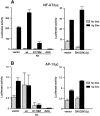
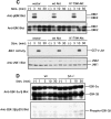
Akt regulation of NF-AT transcription factor. wt BMMCs were transfected with (A) NF-AT/luc or (B) AP-1/luc reporter plasmids together with an empty vector or a vector encoding wt Akt, K179M Akt, AAA Akt, or DN GSK-3β. 24 h later, cells were sensitized with IgE overnight. Cells were then stimulated with antigen for the last 8 h before luciferase assays. Shown are the representative results of two transfection experiments. Stim., stimulation. (C) wt BMMCs stably transfected with an empty vector, wt Akt or K179M Akt, were IgE sensitized and stimulated with antigen. Cell lysates were either directly analyzed by SDS-PAGE followed by immunoblotting with anti-phospho-ERK (for ERK assays) or immunoprecipitated with anti-JNK1 antibody before being subjected to kinase assays using GST-c-Jun as an exogenous substrate (for JNK1 assays). Equal expression of ERK1, ERK2, JNK1, and JNK2 was confirmed by probing immunoblots of total cell lysates with appropriate antibodies. Stim., stimulation. (D) wt and lyn /− BMMCs were stimulated with IgE and antigen. Cell lysates were analyzed by immunoblotting with phosphospecific anti–GSK-3β (pSer9) antibody. The same blot was then reprobed with anti–GSK-3α/β antibody. Stim., stimulation.
Figure 5
Figure 5
Akt regulation of NF-AT transcription factor. wt BMMCs were transfected with (A) NF-AT/luc or (B) AP-1/luc reporter plasmids together with an empty vector or a vector encoding wt Akt, K179M Akt, AAA Akt, or DN GSK-3β. 24 h later, cells were sensitized with IgE overnight. Cells were then stimulated with antigen for the last 8 h before luciferase assays. Shown are the representative results of two transfection experiments. Stim., stimulation. (C) wt BMMCs stably transfected with an empty vector, wt Akt or K179M Akt, were IgE sensitized and stimulated with antigen. Cell lysates were either directly analyzed by SDS-PAGE followed by immunoblotting with anti-phospho-ERK (for ERK assays) or immunoprecipitated with anti-JNK1 antibody before being subjected to kinase assays using GST-c-Jun as an exogenous substrate (for JNK1 assays). Equal expression of ERK1, ERK2, JNK1, and JNK2 was confirmed by probing immunoblots of total cell lysates with appropriate antibodies. Stim., stimulation. (D) wt and lyn /− BMMCs were stimulated with IgE and antigen. Cell lysates were analyzed by immunoblotting with phosphospecific anti–GSK-3β (pSer9) antibody. The same blot was then reprobed with anti–GSK-3α/β antibody. Stim., stimulation.
Figure 6
Figure 6
Akt regulates cytokine secretion in FcεRI-stimulated mast cells. Retroviruses coding for HA-tagged wt or K179M Akt were used to overexpress Akt proteins in stably transfected wt BMMCs. (A) Expression of HA-tagged Akt and endogenous Akt proteins was confirmed by immunoblot analysis of lysates or immune complexes precipitated with anti-HA or anti-Akt. Stable transfectants were sensitized with anti-DNP IgE and stimulated with DNP-HSA for the indicated periods of time. Cell lysates were also analyzed for Akt Ser-473 phosphorylation. Stim., stimulation; vec, vector. (B) Transfectants were sensitized with anti-DNP IgE and stimulated with the indicated concentrations of DNP-HSA for 20 h. IL-2 and TNF-α secreted into culture media were measured by ELISA. Values of secreted cytokines varied <10% from the averages among triplicate samples, and therefore SDs were not shown. Similar results were observed in another set of transfectants.
Figure 6
Figure 6
Akt regulates cytokine secretion in FcεRI-stimulated mast cells. Retroviruses coding for HA-tagged wt or K179M Akt were used to overexpress Akt proteins in stably transfected wt BMMCs. (A) Expression of HA-tagged Akt and endogenous Akt proteins was confirmed by immunoblot analysis of lysates or immune complexes precipitated with anti-HA or anti-Akt. Stable transfectants were sensitized with anti-DNP IgE and stimulated with DNP-HSA for the indicated periods of time. Cell lysates were also analyzed for Akt Ser-473 phosphorylation. Stim., stimulation; vec, vector. (B) Transfectants were sensitized with anti-DNP IgE and stimulated with the indicated concentrations of DNP-HSA for 20 h. IL-2 and TNF-α secreted into culture media were measured by ELISA. Values of secreted cytokines varied <10% from the averages among triplicate samples, and therefore SDs were not shown. Similar results were observed in another set of transfectants.

Similar articles

Cited by

References

    1. Jones P.F., Jakubowicz T., Pitossi F.J., Maurer F., Hemmings B.A. Molecular cloning and identification of a serine/threonine protein kinase of the second-messenger subfamily. Proc. Natl. Acad. Sci. USA. 1991;88:4171–4175. - PMC - PubMed
    1. Coffer P.J., Woodgett J.R. Molecular cloning and characterization of a novel putative protein-serine kinase related to the cAMP-dependent and protein kinase C families. Eur. J. Biochem. 1991;201:475–481. - PubMed
    1. Bellacosa A., Testa J.R., Staal S.P., Tsichlis P.N. A retroviral oncogene, akt, encoding a serine-threonine kinase containing an SH2-like region. Science. 1991;254:274–277. - PubMed
    1. Franke T.F., Kaplan D.R., Cantley L.C. PI3Kdownstream AKTion blocks apoptosis. Cell. 1997;88:435–437. - PubMed
    1. Marte B.M., Downward J. PKB/Aktconnecting phosphoinositide 3-kinase to cell survival and beyond. Trends Biochem. Sci. 1997;22:355–358. - PubMed

Publication types

MeSH terms