. 2000 Sep 4;192(5):755-60.
doi: 10.1084/jem.192.5.755.

Perforin-mediated cytotoxicity is critical for surveillance of spontaneous lymphoma

Affiliations

Perforin-mediated cytotoxicity is critical for surveillance of spontaneous lymphoma

M J Smyth et al. J Exp Med. .

Abstract

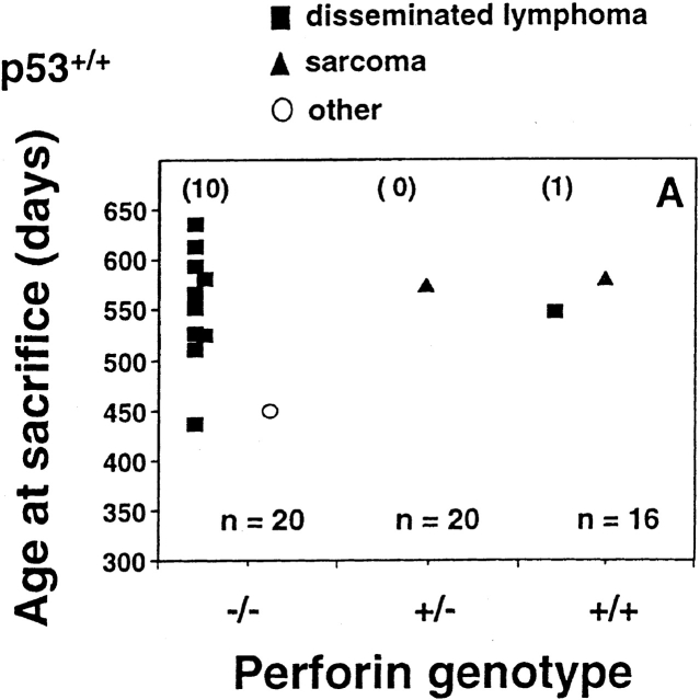
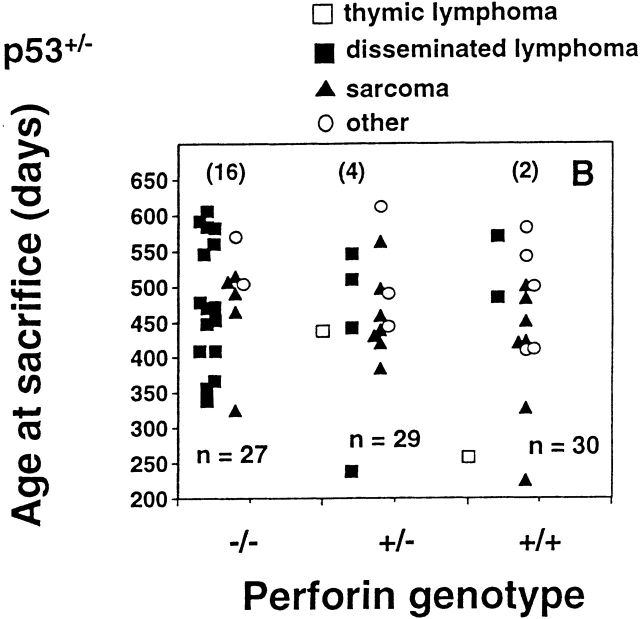
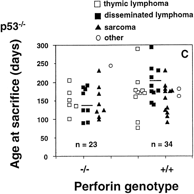
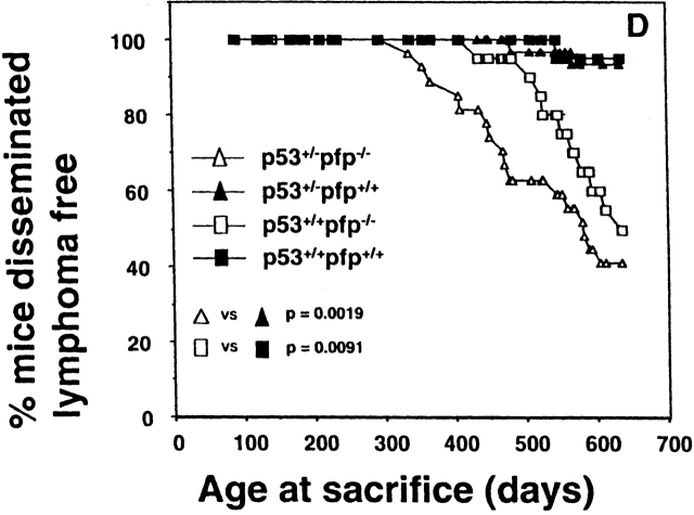
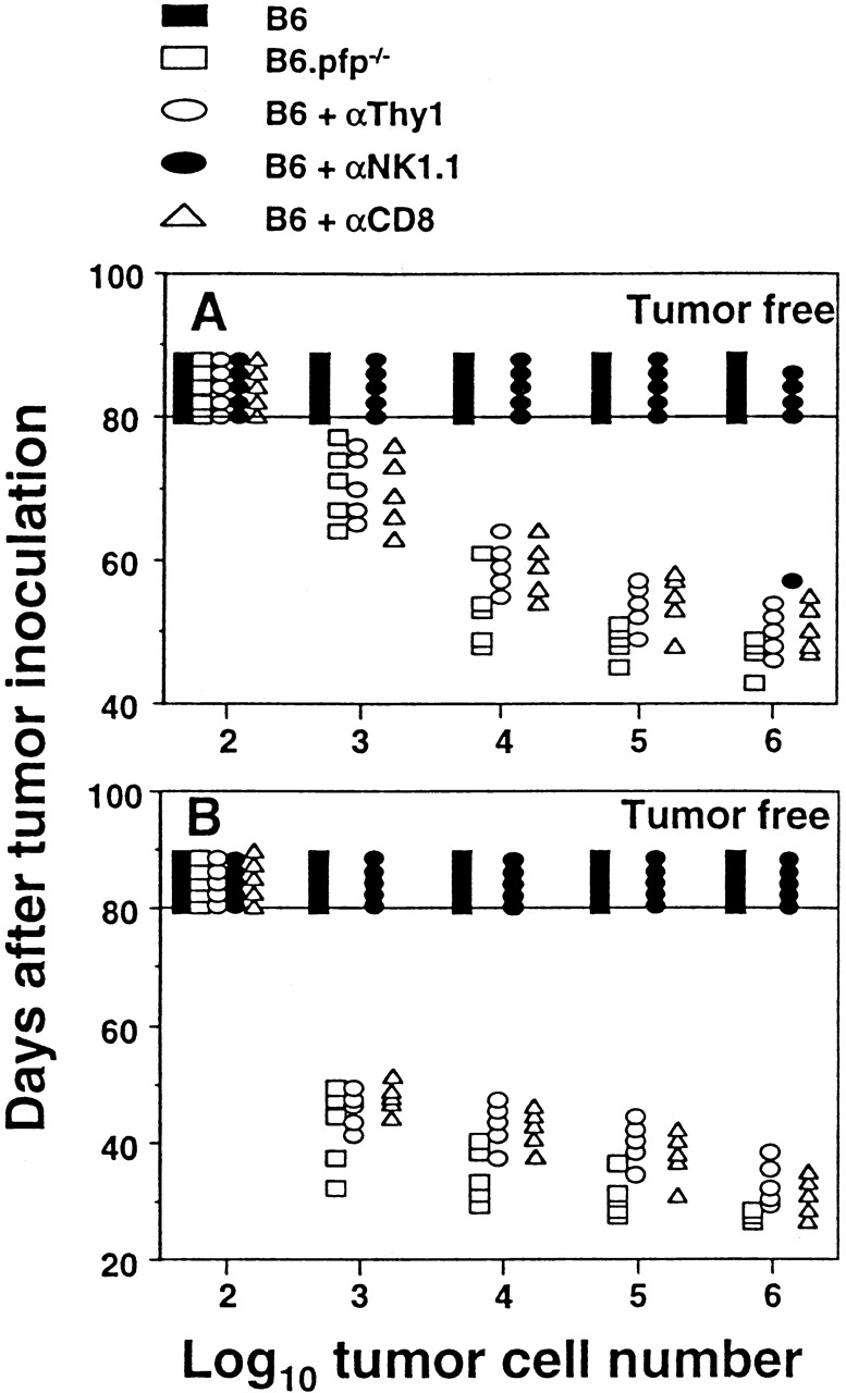
Immune surveillance by cytotoxic lymphocytes against cancer has been postulated for decades, but direct evidence for the role of cytotoxic lymphocytes in protecting against spontaneous malignancy has been lacking. As the rejection of many experimental cancers by cytotoxic T lymphocytes and natural killer cells is dependent on the pore-forming protein perforin (pfp), we examined pfp-deficient mice for increased cancer susceptibility. Here we show that pfp-deficient mice have a high incidence of malignancy in distinct lymphoid cell lineages (T, B, NKT), indicating a specific requirement for pfp in protection against lymphomagenesis. The susceptibility to lymphoma was accentuated by simultaneous lack of expression of the p53 gene, mutations in which also commonly predispose to human malignancies, including lymphoma. In contrast, the incidence and age of onset of sarcoma was unaffected in p53-deficient mice. Pfp-deficient mice were at least 1,000-fold more susceptible to these lymphomas when transplanted, compared with immunocompetent mice in which tumor rejection was controlled by CD8(+) T lymphocytes. This study is the first that implicates direct cytotoxicity by lymphocytes in regulating lymphomagenesis.


Figures

Figure 1
Figure 1
Perforin protects mice from spontaneous lymphomas. The appearance of tumors was recorded in mice derived from a pfp+/−p53+/− intercross. Groups of mice (n = 16–34) were evaluated on a weekly basis and when moribund, tumor type (□, thymic lymphoma; ▪, disseminated lymphoma; ▴, sarcoma; ○, other tumors) recorded against the age at time of death/autopsy (in days). The perforin genotype (pfp−/−, pfp+/−, or pfp+/+) is also displayed on the x-axis. (A) p53+/+, (B) p53+/−, or (C) p53−/−. The MA of p53−/− mice developing disseminated lymphoma is depicted by the crossbar in C. All p53−/− mice studied developed tumors and the numbers of p53+/− and p53+/+ mice developing lymphomas are shown in parentheses. In D, the percentage of pfp−/− and pfp+/+ mice free of disseminated lymphoma (from A and B) are plotted against their age at time of death in days, and P values (Fisher's exact test) comparing the proportions of disseminated lymphoma between pfp−/− and pfp+/+ mice are indicated.
Figure 1
Figure 1
Perforin protects mice from spontaneous lymphomas. The appearance of tumors was recorded in mice derived from a pfp+/−p53+/− intercross. Groups of mice (n = 16–34) were evaluated on a weekly basis and when moribund, tumor type (□, thymic lymphoma; ▪, disseminated lymphoma; ▴, sarcoma; ○, other tumors) recorded against the age at time of death/autopsy (in days). The perforin genotype (pfp−/−, pfp+/−, or pfp+/+) is also displayed on the x-axis. (A) p53+/+, (B) p53+/−, or (C) p53−/−. The MA of p53−/− mice developing disseminated lymphoma is depicted by the crossbar in C. All p53−/− mice studied developed tumors and the numbers of p53+/− and p53+/+ mice developing lymphomas are shown in parentheses. In D, the percentage of pfp−/− and pfp+/+ mice free of disseminated lymphoma (from A and B) are plotted against their age at time of death in days, and P values (Fisher's exact test) comparing the proportions of disseminated lymphoma between pfp−/− and pfp+/+ mice are indicated.
Figure 2
Figure 2
Spontaneous lymphomas arising in pfp−/−p53+/− mice are malignant, immunogenic, and rejected by CD8+ T cells. Elimination of lymphomas transplanted directly intraperitoneally (102–106 cells in 0.2 ml PBS, as indicated) into untreated B6 or B6.pfp−/− mice or B6 mice (5/group) depleted of Thy-1+, NK1.1+, or CD8+ cells using mAb 24. Each symbol depicts an individual mouse. A, PN53H-1; B, PN53H-7.

References

    1. Burnet F.M. The concept of immunological surveillance. Prog. Exp. Tumor Res. 1970;13:1–27. - PubMed
    1. Thomas L. On immunosurveillance in human cancer. Yale J. Biol. Med. 1982;55:329–333. - PMC - PubMed
    1. Melief C.J., Kast W.M. Cytotoxic T lymphocyte therapy of cancer and tumor escape mechanisms. Semin. Cancer Biol. 1991;2:347–354. - PubMed
    1. Whiteside T.L., Vujanovic N.L., Herberman R.B. Natural killer cells and tumor therapy. Curr. Top. Microbiol. Immunol. 1998;230:221–244. - PubMed
    1. Smyth M.J., Ortaldo J.R., Shinkai Y., Yagita H., Nakata M., Okumura K., Young H.A. Interleukin 2 induction of pore-forming protein gene expression in human peripheral blood CD8+ T cells. J. Exp. Med. 1990;171:1269–1281. - PMC - PubMed

Publication types