. 2000 Sep 4;192(5):761-8.
doi: 10.1084/jem.192.5.761.

Lymphocyte CC chemokine receptor 9 and epithelial thymus-expressed chemokine (TECK) expression distinguish the small intestinal immune compartment: Epithelial expression of tissue-specific chemokines as an organizing principle in regional immunity

Affiliations

Lymphocyte CC chemokine receptor 9 and epithelial thymus-expressed chemokine (TECK) expression distinguish the small intestinal immune compartment: Epithelial expression of tissue-specific chemokines as an organizing principle in regional immunity

E J Kunkel et al. J Exp Med. .

Abstract

The immune system has evolved specialized cellular and molecular mechanisms for targeting and regulating immune responses at epithelial surfaces. Here we show that small intestinal intraepithelial lymphocytes and lamina propria lymphocytes migrate to thymus-expressed chemokine (TECK). This attraction is mediated by CC chemokine receptor (CCR)9, a chemoattractant receptor expressed at high levels by essentially all CD4(+) and CD8(+) T lymphocytes in the small intestine. Only a small subset of lymphocytes in the colon are CCR9(+), and lymphocytes from other tissues including tonsils, lung, inflamed liver, normal or inflamed skin, inflamed synovium and synovial fluid, breast milk, and seminal fluid are universally CCR9(-). TECK expression is also restricted to the small intestine: immunohistochemistry reveals that intense anti-TECK reactivity characterizes crypt epithelium in the jejunum and ileum, but not in other epithelia of the digestive tract (including stomach and colon), skin, lung, or salivary gland. These results imply a restricted role for lymphocyte CCR9 and its ligand TECK in the small intestine, and provide the first evidence for distinctive mechanisms of lymphocyte recruitment that may permit functional specialization of immune responses in different segments of the gastrointestinal tract. Selective expression of chemokines by differentiated epithelium may represent an important mechanism for targeting and specialization of immune responses.


Figures
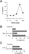
Figure 1
Figure 1
Human LPL chemotaxis to human TECK is mediated by CCR9. (A) Whole populations of LPLs isolated from the human small intestine chemotax efficiently to hTECK. TECK-induced chemotaxis is mediated exclusively by the chemokine receptor CCR9 since (B) the anti-CCR9 Ab 3C3 (40 μg/ml) blocks chemotaxis to hTECK whereas (C) the same Ab does not block chemotaxis to hI-TAC, which signals through CXCR3 expressed on virtually all LPLs. Data in A are the mean ± SD of duplicate wells (n = 3) and data in B and C are the mean ± SD of triplicate wells (n = 2). 250 nM TECK and 100 nM I-TAC were used in B and C.
Figure 2
Figure 2
CCR9 expression is highly specific for CD4+ and CD8+ lymphocytes infiltrating the small intestine. FACS® analysis reveals that CCR9 is expressed on virtually all CD4+ and CD8+ lymphocytes in the (A) IEL and (B) LPL compartments of the jejunum. Interestingly, CCR9 was not detected on lymphocytes isolated from (C) normal lung, (D) hepatitis C virus (HCV)-infected liver, (E) breast milk, (F) normal skin, (G) inflamed skin (delayed-type hypersensitivity [DTH]), (H) rheumatoid arthritis (RA) synovium, and (I) rheumatoid arthritis synovial fluid, and (J) a smaller percentage of colon lymphocytes expresses CCR9 compared with jejunal lymphocytes (2,000 cells in each panel). (A–I) Isotype-matched control Ab staining shown as dotted lines. Percentage of CCR9+ cells based on a marker encompassing 5% of isotype control–stained cells in both histogram and dot blots. Data are representative of three jejunum, four colon, four liver, two breast milk, four normal skin, four inflamed skin, four synovium, and four synovial fluid samples.
Figure 2
Figure 2
CCR9 expression is highly specific for CD4+ and CD8+ lymphocytes infiltrating the small intestine. FACS® analysis reveals that CCR9 is expressed on virtually all CD4+ and CD8+ lymphocytes in the (A) IEL and (B) LPL compartments of the jejunum. Interestingly, CCR9 was not detected on lymphocytes isolated from (C) normal lung, (D) hepatitis C virus (HCV)-infected liver, (E) breast milk, (F) normal skin, (G) inflamed skin (delayed-type hypersensitivity [DTH]), (H) rheumatoid arthritis (RA) synovium, and (I) rheumatoid arthritis synovial fluid, and (J) a smaller percentage of colon lymphocytes expresses CCR9 compared with jejunal lymphocytes (2,000 cells in each panel). (A–I) Isotype-matched control Ab staining shown as dotted lines. Percentage of CCR9+ cells based on a marker encompassing 5% of isotype control–stained cells in both histogram and dot blots. Data are representative of three jejunum, four colon, four liver, two breast milk, four normal skin, four inflamed skin, four synovium, and four synovial fluid samples.
Figure 3
Figure 3
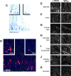
TECK is highly expressed by small intestinal epithelium but not epithelia in other tissues. (A and B) Dot blot and northern blot analysis of TECK mRNA expression in various human mucosal and epithelial tissues demonstrates highly preferential expression of TECK message in the thymus and various segments of the small intestine, but not other epithelial tissues such as the skin, kidney, trachea, lung, placenta, bladder, adrenal gland, thyroid gland, salivary gland, prostate gland, and mammary gland. GAPDH, glyceraldehyde 3-phosphate dehydrogenase. (C) Further analysis by in situ hybridization localizes TECK mRNA expression specifically to the intestinal epithelium in the crypts of Lieberkühn and (D and E) immunohistochemistry localizes TECK protein to the epithelium of the crypts of Lieberkühn and villi in the small intestine. No TECK protein reactivity is detected in the (F) colon or in (G) epithelia from a variety of other human tissues including (G) normal skin, (H) inflamed psoriatic skin, (I) lung bronchioles, (J) salivary gland, or (K) stomach. Ubiquitin staining of the dot blot array revealed that mRNA loading of each tissue was relatively consistent. In situ data are representative of three jejunum samples, and immunohistochemistry data are representative of five jejunum, five colon, four normal skin, three inflamed skin (two psoriatic and one hives), three lung, three salivary gland, and four stomach samples. (D–K) Arrowheads or asterisks highlight epithelia.
Figure 3
Figure 3
TECK is highly expressed by small intestinal epithelium but not epithelia in other tissues. (A and B) Dot blot and northern blot analysis of TECK mRNA expression in various human mucosal and epithelial tissues demonstrates highly preferential expression of TECK message in the thymus and various segments of the small intestine, but not other epithelial tissues such as the skin, kidney, trachea, lung, placenta, bladder, adrenal gland, thyroid gland, salivary gland, prostate gland, and mammary gland. GAPDH, glyceraldehyde 3-phosphate dehydrogenase. (C) Further analysis by in situ hybridization localizes TECK mRNA expression specifically to the intestinal epithelium in the crypts of Lieberkühn and (D and E) immunohistochemistry localizes TECK protein to the epithelium of the crypts of Lieberkühn and villi in the small intestine. No TECK protein reactivity is detected in the (F) colon or in (G) epithelia from a variety of other human tissues including (G) normal skin, (H) inflamed psoriatic skin, (I) lung bronchioles, (J) salivary gland, or (K) stomach. Ubiquitin staining of the dot blot array revealed that mRNA loading of each tissue was relatively consistent. In situ data are representative of three jejunum samples, and immunohistochemistry data are representative of five jejunum, five colon, four normal skin, three inflamed skin (two psoriatic and one hives), three lung, three salivary gland, and four stomach samples. (D–K) Arrowheads or asterisks highlight epithelia.

Similar articles

Cited by

References

    1. Zlotnik A., Yoshie O. Chemokinesa new classification system and their role in immunity. Immunity. 2000;12:121–127. - PubMed
    1. Campbell J.J., Butcher E.C. Chemokines in tissue-specific and microenvironment-specific lymphocyte homing. Curr. Opin. Immunol. 2000;12:336–341. - PubMed
    1. Campbell J.J., Haraldsen G., Pan J., Rottman J., Qin S., Ponath P.D., Andrew D.P., Warnke R., Ruffing N., Kassam N. The chemokine receptor CCR4 in vascular recognition by cutaneous but not intestinal memory T cells. Nature. 1999;400:776–780. - PubMed
    1. Morales J., Homey B., Vicari A.P., Hudak S., Oldham E., Hedrick J.A., Orozco R., Copeland N.G., Jenkins N.A., McEvoy L.M., Zlotnik A. CTACK, a skin-associated chemokine that preferentially attracts skin-homing memory T cells. Proc. Natl. Acad. Sci. USA. 1999;96:14470–14475. - PMC - PubMed
    1. Zabel B.A., Agace W.W., Campbell J.J., Heath H.M., Parent D., Roberts A.I., Ebert E.C., Kassam N., Qin S., Zovko M. Human G protein–coupled receptor GPR-9-6/CC chemokine receptor 9 is selectively expressed on intestinal homing T lymphocytes, mucosal lymphocytes, and thymocytes and is required for thymus-expressed chemokine–mediated chemotaxis. J. Exp. Med. 1999;190:1241–1255. - PMC - PubMed

Publication types