Review
. 2000 Sep;64(3):607-23.
doi: 10.1128/MMBR.64.3.607-623.2000.

Lytic cycle of Toxoplasma gondii

Affiliations
Review

Lytic cycle of Toxoplasma gondii

M W Black et al. Microbiol Mol Biol Rev. 2000 Sep.

Abstract

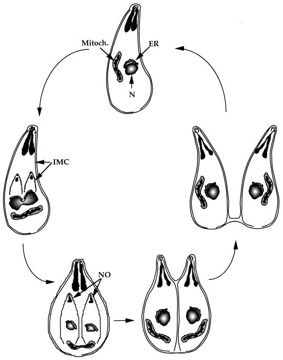
Toxoplasma gondii is an obligate intracellular pathogen within the phylum Apicomplexa. This protozoan parasite is one of the most widespread, with a broad host range including many birds and mammals and a geographic range that is nearly worldwide. While infection of healthy adults is usually relatively mild, serious disease can result in utero or when the host is immunocompromised. This sophisticated eukaryote has many specialized features that make it well suited to its intracellular lifestyle. In this review, we describe the current knowledge of how the asexual tachyzoite stage of Toxoplasma attaches to, invades, replicates in, and exits the host cell. Since this process is closely analogous to the way in which viruses reproduce, we refer to it as the Toxoplasma "lytic cycle."


Figures

FIG. 1
FIG. 1
Diagram of the Toxoplasma life cycle. The sexual cycle (top section) is initiated when a member of the feline family ingests either oocysts or tissues that are infected with bradyzoite cysts. This cycle is limited to the feline intestine and results in the shedding of oocysts in the cat's feces (middle section). Following oocyst maturation (activated after being excreted from the cat), the oocysts become highly infectious and survive in the environment for months and possibly years. Any warm-blooded animal that ingests these infectious oocysts becomes a host for the asexual cycle (bottom section). The sporozoites that are released from the oocyst will infect the intestinal epithelium and differentiate into the tachyzoite stage. After an acute infection, characterized by the dissemination of tachyzoites throughout the body, tissue cysts arise as a result of differentiation to the bradyzoite stage. Upon ingestion of these tissue cysts in raw or undercooked meat from a chronically infected host, the bradyzoites will infect the intestinal epithelium of the next susceptible host and differentiate back to the tachyzoite stage to complete the asexual cycle. If the ingesting animal is a cat, the bradyzoites can differentiate into the sexual stages, thereby completing the full life cycle. The sexual and asexual components of the life cycle are potentially independent; in particular, the asexual phase can theoretically cycle between intermediate hosts ad infinitum. The degree to which the sexual and asexual portions of the overall life cycle feed into one another in nature is not known.
FIG. 2
FIG. 2
Toxoplasma organelles. The major organelles of the asexually reproducing tachyzoite stage are shown. Mitoch., mitochondrion.
FIG. 3
FIG. 3
The Toxoplasma cytoskeleton. This diagram illustrates the dominant features of the parasite's cytoskeletal arrangement. The anterior end of the parasite is enlarged in the box to the left so as to illustrate the preconoidal rings (CR), the conoid (C), the two apical microtubules (M), and the polar ring (PR) from which 22 subpellicular microtubules (SPM) emanate. The IMC is located just beneath the plasma membrane from the anterior to the posterior poles and is interrupted only by the micropore (MP) located in the middle of the parasite body. This pore is believed to be the primary portal through which endocytosis takes place.
FIG. 4
FIG. 4
Trajectory of Toxoplasma gliding motility. As parasites glide across a surface, they leave behind a trail of surface proteins. These trails were detected using immunofluorescence directed against the major surface protein SAG1. The circular trajectory is a result of the crescent shape of the parasite and its forward corkscrew motility, which is believed to be dictated, at least in part, by the subpellicular microtubules (106).
FIG. 5
FIG. 5
Toxoplasma invasion. (A) Illustration of the critical events associated with host cell invasion. Secretion from the rhoptries is represented by the emptied vesicles and release of membranous material into the forming PV. As the parasite squeezes into the cell (as indicated by the constriction on the parasite body), a moving junction (MJ) forms between the parasite surface and host plasmalemma. (B) Still frames from a video of Toxoplasma invading a human fibroblast. The timing of the event is indicated in the lower right corner of each frame and was virtually complete 10 s after attachment. As in the cartoon, the constriction is apparent as the parasite crosses through the host cytoskeleton (indicated by arrowheads).
FIG. 6
FIG. 6
Current models for the gliding motility exhibited by T. gondii. (A) Graphic illustration of the linear motion model (77). In this model, a transmembrane attachment protein(s) binds to an extracellular ligand and associates with myosin via its cytoplasmic tail. Myosin binds to the actin microfilaments located just beneath the plasmalemma and ratchets the transmembrane protein down the length of the parasite. The microfilaments are aligned to the orientation of the subpellicular microtubules via trans-IMC linkages, which results in the characteristic forward corkscrew locomotion. (B) The second model suggests that the IMC plays a more active role in motility (104). This model localizes both actin and myosin beneath the IMC in direct contact with the subpellicular microtubules. Instead of myosin, the IMPs associated with the IMC bind to the cytoplasmic tail of the attachment protein(s). These IMPs are connected to a secondary network of filaments that encapsulates the individual flattened vesicles. Myosin binds to either the same IMPs or distinct particles that are on the inner face of the IMC and move along microfilaments associated with the subpellicular microtubules. As these myosin-associated IMPs move along the actin tracks, they pull the secondary network like a conveyor belt. Since the outer surface IMPs are also connected to this filamentous network, they transduce this force to the attachment protein and move it down the length of the vesicle.
FIG. 7
FIG. 7
Diagram of endodyogeny. As a parasite begins to divide, two IMCs begin to develop in the middle of the cell from what appears to be a rudimentary conoid and microtubule-organizing center (polar ring). As the IMC extends from these structures, the nucleus (N) and mitochondrion (Mitoch.) divide into these membranous outlines. Nascent apical organelles (NO) develop within the anterior poles as the daughter cells grow. Eventually, the entire cytoplasm is divided between the daughters and the IMC of the mother dissociates. A cleavage furrow divides the cells from the anterior pole. This division continues down the length of the cells until it reaches the posterior end, where it can leave a residual body connecting the two daughters.
FIG. 8
FIG. 8
Ionophore-induced egress. Four video frames of human fibroblasts infected with Toxoplasma are shown starting about 30 h postinfection. The first frame (A) shows a typical rosette of 16 parasites that are positioned with their anterior poles pointing away from the center. The clock in the upper left corner of each frame was activated at the time of replacing the medium with Hanks balanced salt solution containing 1 μM A23187 (a calcium ionophore) to induce egress. The frames were obtained at ∼10-s intervals (starting at ∼20 s postinduction in frame A) to illustrate the speed of the response to A23187. By ∼31 s (B), the parasites are beginning to move within the PV, and at ∼40 s (C) they are leaving the PV (see arrow) and beginning to cross through the host cytoplasm. The last frame (D), taken at ∼49 s postinduction, shows two parasites apparently passing through the cytoskeleton of the host as suggested by the severe constriction on the bodies of the parasites (arrows).
FIG. 9
FIG. 9
The Toxoplasma lytic cycle. This diagram depicts the five events that are discussed in the text which are responsible for the acute phase of toxoplasmosis. The first event is attachment of Toxoplasma to its host cell (step 1). This step may involve two forms of contact: an initial transient interaction that signals microneme secretion followed by a firm adherence that utilizes the transmembrane micronemal proteins (e.g., MIC2). As the parasite glides across the surface, it is reoriented to make contact with the host surface at its apical end and to initiate invasion (step 2). During the invasion event, the PV is formed and modified by secretion from both the rhoptries and, later, the dense granules. After the parasite enters its host, it stops moving and the vacuole closes at its posterior end by pinching off via a fission pore. The newly formed PV immediately recruits host mitochondria and portions of the host ER (step 3). This close association between host organelles and the PV is likely to play a role in acquiring essential nutrients (possibly lipids) for intracellular growth. The parasites undergo several rounds of replication (step 4) until they receive a signal from either the parasite itself or the host cell, resulting in egress (step 5). Host cell egress is typically a destructive process that lyses the host cell and releases motile parasites. These parasites quickly invade neighboring cells to complete the cycle.

References

    1. Achbarou A, Mercereau P, Sadak A, Gortier B, Leriche M A, Camus D, Dubremetz J F. Differential targeting of dense granule proteins in the parasitophorous vacuole of Toxoplasma gondii. Parasitology. 1991;3:321–329. - PubMed
    1. Achbarou A, Mercereau-Puijalon O, Autheman J M, Fortier B, Camus D, Dubremetz J F. Characterization of microneme proteins of Toxoplasma gondii. Mol Biochem Parasitol. 1991;47:223–233. - PubMed
    1. Aikawa M, Komata Y, Asai T, Midorikawa O. Transmission and scanning electron microscopy of host cell entry by Toxoplasma gondii. Am J Pathol. 1977;87:285–295. - PMC - PubMed
    1. Aikawa M, Miller L H, Johnson J, Rabbege J. Erythrocyte entry by malarial parasites: a moving junction between erythrocyte and parasite. J Cell Biol. 1978;77:72–82. - PMC - PubMed
    1. Ajioka J W, Boothroyd J C, Brunk B P, Hehl A, Hillier L, Manger I D, Marra M, Overton G C, Roos D S, Wan K L, Waterston R, Sibley L D. Gene discovery by EST sequencing in Toxoplasma gondii reveals sequences restricted to the Apicomplexa. Genome Res. 1998;8:18–28. - PubMed

Publication types

LinkOut - more resources