. 2000 Aug 22;254(1-2):209-17.
doi: 10.1016/s0378-1119(00)00264-x.

Synthesis of a bacteriophage MB78 late protein by novel ribosomal frameshifting

Affiliations

Synthesis of a bacteriophage MB78 late protein by novel ribosomal frameshifting

V Kolla et al. Gene. .

Abstract

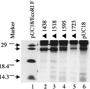
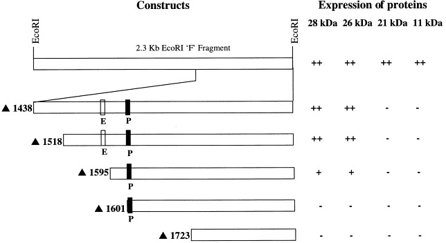
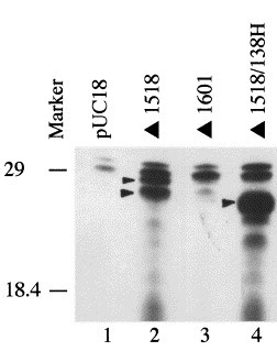
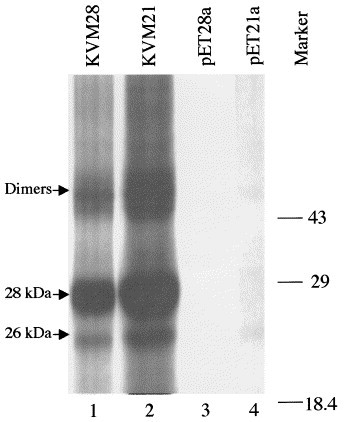
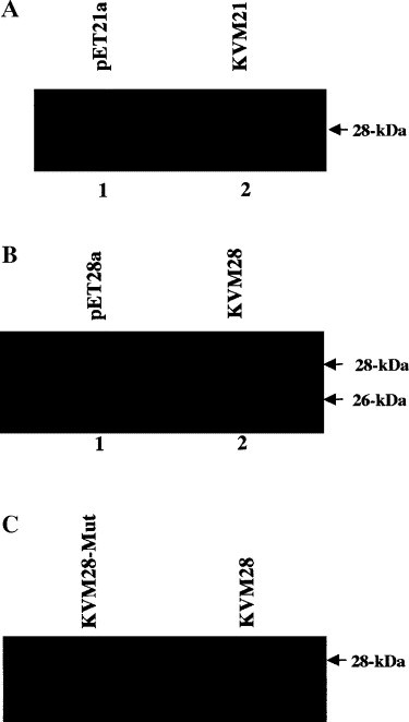
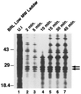
MB78 is a virulent phage of Salmonella typhimurium that possesses a number of interesting features, making it a suitable organism to study the regulation of gene expression. A detailed physical map of this phage genome has been constructed and is being extensively studied at the molecular level. Here, we demonstrate the expression of two late proteins of bacteriophage MB78 derived from the same gene as a result of possible ribosomal frameshifting. In vitro transcription-translation yields a major protein that migrates as 28kDa, whereas in vivo expression using pET expression vectors yields two equally expressed proteins of molecular sizes 28 and 26kDa. A putative slippery sequence TTTAAAG and a pseudoknot structure, two essential cis elements required for the classical ribosomal frameshifting, are identified in the reading frame. Mutations created at the slippery sequence resulted in a single 28kDa protein and completely abolished the expression of 26kDa protein. Thus, we have produced the first evidence that ribosomal frameshifting occurs in bacteriophage MB78 of Salmonella typhimurium.


Figures

Fig. 1
Fig. 1
In vivo expression of the minicells. Minicells produced from DS410, transformed with EcoRI in pUC18, deletion mutants and pUC18 were labeled with 35S-methionine, as described in Section 2. Samples were denatured and analyzed on 12.5% SDS-polyacrylamide gel followed by fluorography. Lane 1, EcoRI F in pUC18; lanes 2–5 represent expression of different plasmid DNAs in minicells; lane 2, F ▴1438; lane 3, F ▴1518; lane 4, F ▴1595; lane 5, F ▴1723; lane 6, pUC18. Molecular weight standards in kDa (BRL: low molecular weight marker) are marked on the left-hand side. Positions of 28 and 26 kDa proteins have been marked with dots (.) between lanes 2 and 3. F ▴ followed by a number indicates the number of bases deleted from the full-length EcoRI ‘F’ fragment.
Fig. 2
Fig. 2
Schematic representation of the different deleted constructs of the EcoRI ‘F’ fragment and the extent of expression, shown arbitrarily with restriction sites in pUC18 vector. The putative promoter, enhancer sequences and restriction sites are marked arbitrarily. The number of bases deleted in each construct is also shown on the left-hand side.
Fig. 3
Fig. 3
Sequence analysis. (A) Probable secondary structure of mRNA derived from 3′ 138 nucleotides. (B) Probable slippery sequence and downstream pseudoknot structure. The slippery sequence essential for the frameshift is marked in a box, and the bold nucleotides represent the stop codon where mutations were created. Nucleotides involved in the pseudoknot structure are connected.
Fig. 4
Fig. 4
In vivo minicell expression of truncated 28 and 26 kDa proteins. Minicells were labeled as described in Fig. 1. Lane 1, pUC18 in DS410; lane 2, F ▴1518 in DS410; lane 3, F ▴1601 in DS410; lane 4, F ▴1518/138H in DS410. Molecular weight standards (kDa) are from GIBCO-BRL.
Fig. 5
Fig. 5
In vitro transcription and translation. The coding region for 26 and 28 kDa proteins was amplified by PCR and cloned in bacterial expression vectors, pET21a and pET28a. Recombinant DNAs were translated in vitro in the presence of [35S]-methionine in rabbit reticulocyte lysate, as described in Section 2. One to three microliters of these samples, as indicated, were applied on to SDS-polyacrylamide gels after denaturing in a sample buffer by boiling. A major translated protein product (28 kDa) and a minor protein are marked with arrows. The position of possible dimers is also marked with an arrow. Lanes 3 and 4 represent the translation of empty pET28a and pET21a vectors, respectively.
Fig. 6
Fig. 6
Expression of 28 and 26 kDa proteins in BL-21 DE3. (A) pET21a vector. (B) pET28a vector. (C) Mutated construct in pET28a. Recombinant constructs and empty vectors were transformed into BL-21 DE3 bacteria, as indicated. Cells were induced in the presence of IPTG, an inducing substance in certain bacteria, at a final concentration of 1 mM and allowed to grow for additional 2–3 h. Subsequently, cells were collected and washed with 1× PBS and sonicated to lyse in the presence of protease inhibitors: (aprotinin, apopain, and PMSF). The clear supernatant was applied to Ni+2columns and purified as per the manufacturer's instructions (Clontech). Finally, the purified proteins, bound to resin, were denatured by boiling in a suitable volume of 2× sample buffer and subjected to SDS-PAGE followed by Western blot. The presence of 26 and or 28 kDa proteins was detected using the ECL kit (Amersham). The molecular weight of the proteins is marked with arrows.
Fig. 7
Fig. 7
Kinetic study of 28 and 26 kDa proteins. Exponentially growing LT2 cells in minimal medium (M9) at 37°C were infected with phage MB78 at an m.o.i. 10. The infected cells (400 μl) were pulse-labeled for 3 min at different times after infection. The cells were collected by centrifugation, washed with 500 μl of medium and finally suspended in 25 μl of M9 medium. Samples collected at different times as indicated were lysed in an equal volume of 2× sample buffer and applied on to a 12.5% SDS-polyacrylamide gel. Lane 1, uninfected (UI) LT2 cells; lanes 2–7, cells infected with phage MB78 after 2, 5, 10, 15, 30 and 45 min, respectively. BRL low-molecular-weight markers are represented in the left lane. The 28 and 26 kDa proteins are marked with two arrows.

Similar articles

Cited by

References

    1. Atkinson J., Dodge M., Gallant J. Secondary structures and starvation-induced frameshifting. Mol. Microbiol. 1997;26:747–753. - PubMed
    1. Belcourt M.F., Farabaugh P.J. Ribosomal frameshifting in the yeast retrotransposon Ty: tRNAs induce slippage on a 7 nucleotide minimal site. Cell. 1990;62:339–352. - PMC - PubMed
    1. Blinkowa A.L., Walker J.R. Programmed ribosomal frameshifting generates the Escherichia coli DNA polymerase III gamma subunit from within the tau subunit reading frame. Nucleic Acids Res. 1990;18:1725–1729. - PMC - PubMed
    1. Bonner W.M. Fluorography for the detection of radioactivity in gels. Meth. Enzymol. 1984;104:460–465. - PubMed
    1. Brault V., Miller W.A. Translational frameshifting mediated by a viral sequence in plant cells. Proc. Natl. Acad. Sci. USA. 1992;89:2262–2266. - PMC - PubMed

Publication types

MeSH terms

LinkOut - more resources