. 2000 Sep;157(3):709-15.
doi: 10.1016/S0002-9440(10)64583-X.

Coexpression of BMI-1 and EZH2 polycomb group genes in Reed-Sternberg cells of Hodgkin's disease

Affiliations

Coexpression of BMI-1 and EZH2 polycomb group genes in Reed-Sternberg cells of Hodgkin's disease

F M Raaphorst et al. Am J Pathol. 2000 Sep.

Abstract

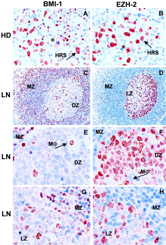
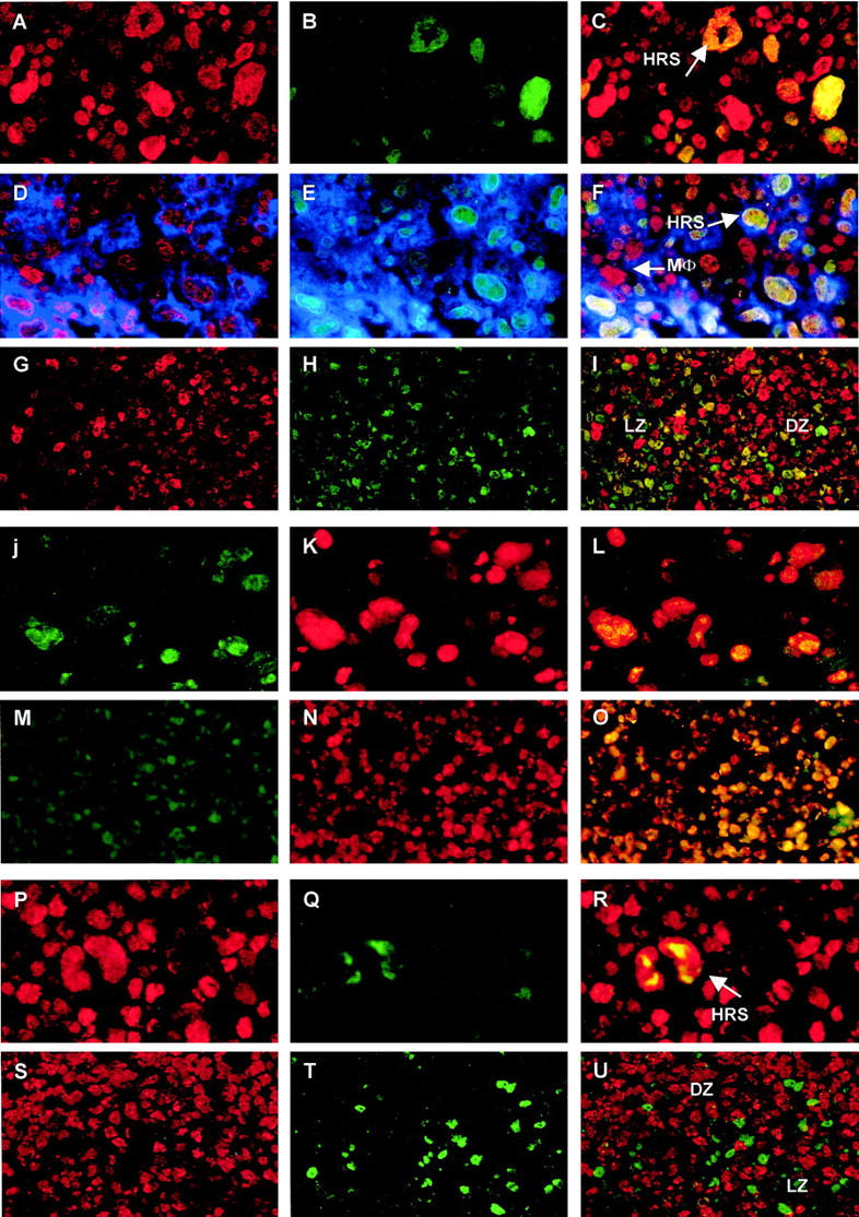
The human BMI-1 and EZH2 polycomb group (PcG) proteins are constituents of two distinct complexes of PcG proteins with gene regulatory activity. PcG proteins ensure correct embryonic development by suppressing homeobox genes, and they also contribute to regulation of lymphopoiesis. The two PcG complexes are thought to regulate different target genes and probably have different tissue distributions. Altered expression of PcG genes is linked to transformation in cell lines and induction of tumors in mutant mice, but the role of PcG genes in human cancers is relatively unexplored. Using antisera specific for human PcG proteins, we used immunohistochemistry and immunofluorescence to detect BMI-1 and EZH2 PcG proteins in Reed-Sternberg cells of Hodgkin's disease (HRS). The expression patterns were compared to those in follicular lymphocytes of the lymph node, the normal counterparts of HRS cells. In the germinal center, expression of BMI-1 is restricted to resting Mib-1/Ki-67(-) centrocytes, whereas EZH2 expression is associated with dividing Mib-1/Ki-67(+) centroblasts. By contrast, HRS cells coexpress BMI-1, EZH2, and Mib-1/Ki-67. Because HRS cells are thought to originate from germinal center lymphocytes, these observations suggests that Hodgkin's disease is associated with coexpression of BMI-1 and EZH2 in HRS cells.


Figures

Figure 1.
Figure 1.
Immunohistochemical analysis of BMI-1 and EZH2 expression in HRS cells and germinal center follicular lymphocytes in the lymph node. A and B: Staining of Reed-Sternberg (HRS) cells for BMI-1 (A) and EZH2 (B). HRS cells express both BMI-1 and EZH2, whereas infiltrating lymphocytes stain for BMI-1 but not for EZH2. Note that dividing cells express BMI-1 in the cytoplasm, whereas EZH2 remains associated with condensed chromosomes (indicated by * in A and B). C, E, and G: Expression of BMI-1 in germinal centers of the lymph node. D, F, and H: Expression of EZH2 in germinal centers of the lymph node. C and D: Overview of lymph node germinal center. E and F: Detail of germinal center showing dark zone (DZ) and mantle zone (MZ). G and H: Detail of germinal center showing light zone (LZ) and MZ. Cb, centroblast; cc, centrocyte; LN, lymph node; MΦ, macrophage. Note that the mutually exclusive expression of BMI-1 and EZH2 is particularly notable in the MZ, DZ, and MΦ staining profiles (C−H), whereas HRS cells stain for both PcG proteins (A and B). Original magnifications, ×400 (A and B), ×200 (C and D), and ×630 (E−H).
Figure 2.
Figure 2.
A−I: Expression of BMI-1 and EZH2 in HRS cells (A−F) and LN (G−I). BMI-1 (A and D; red fluorescence) and EZH2 (B and E; green fluorescence) are detected in HRS cells, whereas infiltrating lymphocytes are BMI-1+ (A and B) and generally do not express EZH2 (B and E). Coexpression of BMI-1 and EZH2 produces a yellow nuclear signal (C and F). These nuclei belong to HRS cells since these express CD30 (blue signal in D−F). Occassional EZH2-expressing cells with a compact nucleus also express CD30 (E), but are BMI-1; these are probably activated lymphocytes. Note that EZH2+ lymphocytes in the infiltrate may be remaining centroblasts from lymph node follicles. CD30/BMI-1+ cells with a large nucleus are probably macrophages (MΦ in F). In follicular lymphocytes (G−I), BMI-1 is mainly detected in the LZ (G), whereas EZH2 is primarily detected in the DZ (H). Coexpression of BMI-1 and EZH2 is sometimes observed at the interface of LZ and DZ, but occurs at a very low frequency (I). Expression of EZH2 and Mib-1/Ki-67 in HRS cells (J−L) and LN (M−O): Expression of EZH2 occurs in both HRS cells (J; green fluorescence) and LN centroblasts situated in the DZ (M), and coincides with detection of Mib-1/Ki-67 (K and N). Combination of the red and green fluorescent signal produces yellow nuclei (L and O), supportive of Mib-1/Ki-67 and EZH2 coexpression in HRS cells and centroblasts. Expression of BMI-1 and Mib-1/Ki-67 in HRS cells and LN: BMI-1 expression occurs in HRS cells and the surrounding infiltrate (P). HRS cells also express Mib-1/Ki-67 (Q, green fluorescence), producing a yellow signal in the HRS cell when red and green fluorescence are combined (R). By contrast, BMI-1 (S, red fluorescence) and Mib-1/Ki-67 expression (T, green fluorescence) are separated in follicular lymphocytes (U) and infiltrating lymphocytes surrounding an HRS cell (R). For abbreviations, see Figure 1 ▶ legend. Original magnifications, ×400.

References

    1. Cossman J, Messineo C, Bagg A: Reed-Sternberg cell: survival in a hostile sea. Lab Invest 1998, 78:229-235 - PubMed
    1. Cossman J, Annunziata CM, Barash S, Staudt L, Dillon P, He WW, Ricciardi-Castagnoli P, Rosen CA, Carter KC: Reed-Sternberg cell genome expression supports a B cell lineage. Blood 1999, 94:411-416 - PubMed
    1. Foss HD, Reusch R, Demel G, Lenz G, Anagnostopoulos I, Hummel M, Stein H: Frequent expression of the B cell-specific activator protein in Reed-Sternberg cells of classical Hodgkin’s disease provides further evidence for its B cell origin. Blood 1999, 94:3108-3113 - PubMed
    1. Carbone A, Gloghini A, Gaidano G, Franceschi S, Capello D, Drexler HG, Falini B, Dalla-Favera R: Expression status of BCL-6 and syndecan-1 identifies distict histogenic subtypes of Hodgkin’s disease. Blood 1998, 92:2220-2228 - PubMed
    1. Tamaru J, Hummel M, Zemlin M, Kalvelage B, Stein H: Hodgkin’s disease with a B cell phenotype often shows a VDJ rearrangement and somatic mutation in the VH genes. Blood 1994, 84:708-715 - PubMed

MeSH terms