. 2000 Sep;157(3):877-83.
doi: 10.1016/s0002-9440(10)64601-9.

Beta-catenin, an inducer of uncontrolled cell proliferation and migration in malignancies, is localized in the cytoplasm of vascular endothelium during neovascularization after myocardial infarction

Affiliations

Beta-catenin, an inducer of uncontrolled cell proliferation and migration in malignancies, is localized in the cytoplasm of vascular endothelium during neovascularization after myocardial infarction

W M Blankesteijn et al. Am J Pathol. 2000 Sep.

Abstract

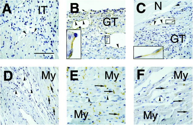
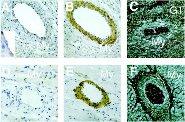
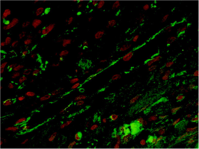
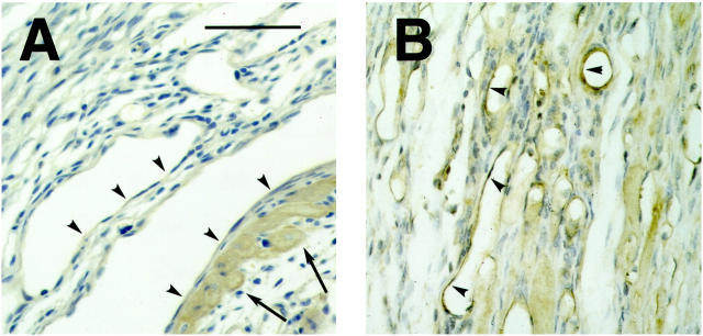
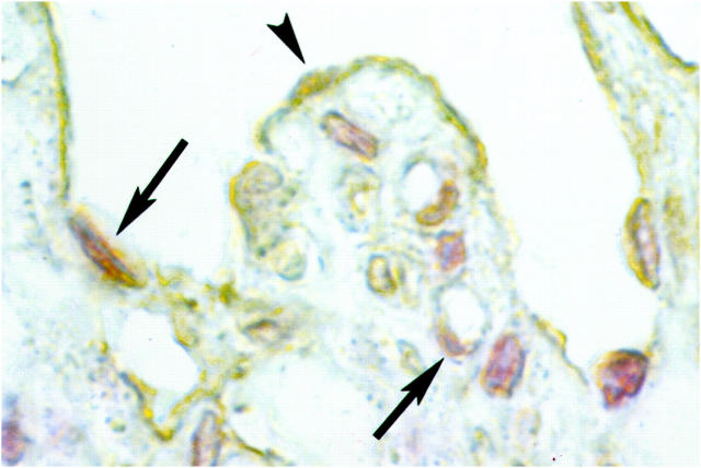
Beta-catenin is a protein involved in cell-cell adhesion and proliferation. In neoplastic diseases, defects in the regulation of the cellular beta-catenin content and cytoplasmic accumulation of the protein contribute to the uncontrolled cell proliferation and migration. Whether beta-catenin plays a role in the controlled proliferative and migratory responses to injury, eg, of vascular endothelial cells during neovascularization after myocardial infarction (MI), is not known. In the present study, we examined the localization of beta-catenin in the infarcted rat heart at different time points after MI. Cytoplasmic beta-catenin was observed in the endothelial cells of the newly formed and pre-existing blood vessels in the infarct area in the first week after MI, but not in the uninjured parts of the heart and not at later time points. Adenomatous polyposis coli (APC) protein was also detected; interaction of APC with beta-catenin has been reported to be critical in epithelial tube formation in vitro. Moreover, the expression of dishevelled-1, an upstream regulatory molecule of the cellular beta-catenin content, was observed in vascular endothelial cells in the infarct area. These findings suggest a role for the beta-catenin-APC complex in the proliferation and migration of vascular endothelial cells during neovascularization of the infarct area.


Figures

Figure 1.
Figure 1.
Time course of cytoplasmic β-catenin localization in small blood vessels in the area of infarction in sections of infarcted rat hearts. Arrowheads indicate the vascular endothelial cell layer whereas arrows indicate the intercalated disks of cardiomyocytes. A: Two days after MI neovascularization has not yet started. The small vessels in the infarct area do not show β-catenin staining, probably because these vessels are remnants of the vasculature present before infarction. The border zone of the infarct consists of inflammatory tissue (IT). Scale bar, 100 μm. B: Four days after MI many newly formed small vessels were present in the border zone of the infarct area. In the cytoplasm of the endothelial cells β-catenin staining was observed (see high-power magnification in inset). In contrast, the granulation tissue (GT), which is formed in the border zone of the infarct, did not show detectable β-catenin staining. C: Seven days after MI, the cytoplasm of the vascular endothelial cells of small vessels in the infarct area was still positively staining for β-catenin (inset shows high-power magnification). The granulation tissue again was negative. D: Twenty-one days after MI, no β-catenin staining could be detected in most of the vascular endothelial cells of the small vessels. The intercalated disks of the cardiomyocytes (My) stained positively for β-catenin. E: A small vessel in a noninfarcted area of the heart, 4 days after MI, is shown. No β-catenin staining of the cytoplasm of the vascular endothelial cells could be observed. The intercalated disks of the myocytes were positively staining for β-catenin. F: Small vessel in a noninfarcted part of the heart, 7 days after MI. Again no positive staining of vascular endothelial cells was observed.
Figure 2.
Figure 2.
A: β-catenin staining of endothelial cells of a larger, pre-existing artery in the area of infarction, 4 days after MI. Around the artery a thin layer of surviving cardiomyocytes (My) was present, confirming that the artery was present before infarction. Scale bar, 100 μm. B: Immunohistochemistry for α-smooth muscle actin (ASMA) shows staining of the vascular smooth muscle cells in the media of the same artery, but not of the endothelial cell layer or the cardiomyocytes. C: Dark-field image of an in situ hybridization with a dishevelled-1 (dvl-1) probe of a section of rat heart, 4 days after MI. The photographic grains are represented as white spots. Note that grains are localized over the vascular endothelium (arrowhead), whereas the amount of grains over the cardiomyocytes (My) is lower. A high density of grains was observed over the granulation tissue (GT) in the border zone of the infarct. D and E: Muscular arteries in the noninfarcted parts of the myocardium did not show endothelial β-catenin staining, but the vascular smooth muscle cells stained positively for (ASMA). F: No dvl-1 expression was observed in muscular arteries in the noninfarcted areas of the heart.
Figure 3.
Figure 3.
Confocal microscopy of a small vessel in the infarct area of a rat heart, 7 days after MI. β-Catenin staining is depicted in green and the nuclear counterstain (propidium iodide) is depicted in red. Note that the β-catenin staining is present in the cytoplasm of the vascular endothelial cells, whereas β-catenin staining in the nuclei could not be detected.
Figure 4.
Figure 4.
Localization of the APC protein in vascular endothelium during infarct healing in the rat. APC staining was observed in the endothelial cells of newly formed vessels around the infarct area at 7 days after MI (A). In contrast to the β-catenin staining, APC staining was still observed in these cells 21 days after MI (B).
Figure 5.
Figure 5.
High-power magnification of immunohistochemistry for BrdU and β-catenin in a small vessel and capillaries in the infarct area of a rat heart, 4 days after MI. BrdU-positive nuclei of proliferating cells are depicted in red, and β-catenin is shown in brown. Some of the vascular endothelial cells that contain β-catenin in their cytoplasm have a BrdU-positive nucleus (arrows), but β-catenin-positive cells without nuclear BrdU incorporation were also observed (arrowhead).

References

    1. Takeichi M: Cadherins: a molecular family important in selective cell-cell adhesion. Annu Rev Biochem 1990, 59:237-252 - PubMed
    1. Geiger B, Ayalon O: Cadherins. Annu Rev Cell Biol 1992, 8:307-332 - PubMed
    1. Gumbiner BM: Cell adhesion: the molecular basis of tissue architecture and morphogenesis. Cell 1996, 84:345-357 - PubMed
    1. Shapiro L: The multi-talented β-catenin makes its first appearance. Structure 1997, 5:1265-1268 - PubMed
    1. Dale TC: Signal transduction by the Wnt family of ligands. Biochem J 1998, 329:209-223 - PMC - PubMed

Publication types

MeSH terms