. 2000 Oct;74(19):8938-45.
doi: 10.1128/jvi.74.19.8938-8945.2000.

Association of human immunodeficiency virus type 1 Vif with RNA and its role in reverse transcription

Affiliations

Association of human immunodeficiency virus type 1 Vif with RNA and its role in reverse transcription

M Dettenhofer et al. J Virol. 2000 Oct.

Abstract

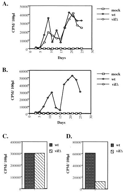
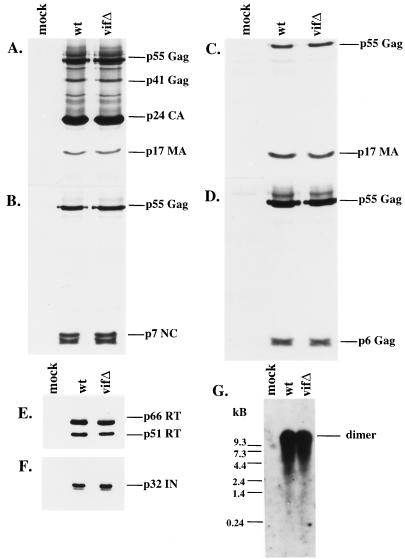
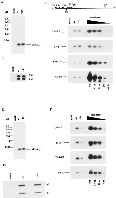
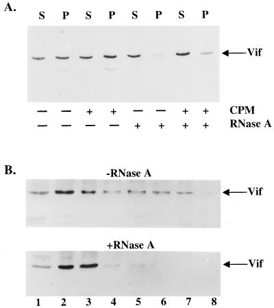
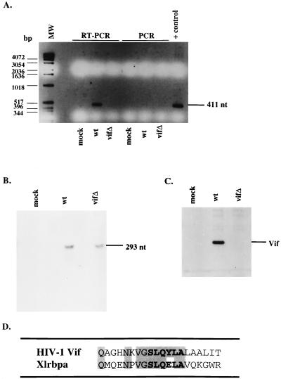
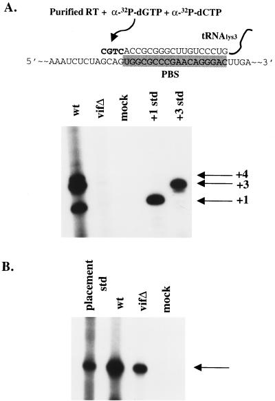
The vif gene of human immunodeficiency virus type 1 (HIV-1) is essential for viral replication, although the functional target of Vif remains elusive. HIV-1 vif mutant virions derived from nonpermissive H9 cells displayed no significant differences in the amount, ratio, or integrity of their protein composition relative to an isogenic wild-type virion. The amounts of the virion-associated viral genomic RNA and tRNA(3)(Lys) were additionally present at normal levels in vif mutant virions. We demonstrate that Vif associates with RNA in vitro as well as with viral genomic RNA in virus-infected cells. A functionally conserved lentivirus Vif motif was found in the double-stranded RNA binding domain of Xenopus laevis, Xlrbpa. The natural intravirion reverse transcriptase products were markedly reduced in vif mutant virions. Moreover, purified vif mutant genomic RNA-primer tRNA complexes displayed severe defects in the initiation of reverse transcription with recombinant reverse transcriptase. These data point to a novel role for Vif in the regulation of efficient reverse transcription through modulation of the virion nucleic acid components.


Figures

FIG. 1
FIG. 1
Viral growth curves and exogenous and endogenous RT reactions. Cell culture supernatants from COS-7 cells transfected with wild-type (wt) or vif mutant (vifΔ) viral constructs were used to initiate infections. After infection of Jurkat (A) and H9 T-cell (B) lines, virion production was monitored by RT activity in cell culture supernatants. Exogenous (C) and endogenous (D) RT reactions utilized H9-derived virions (wild type and vif mutant), which were normalized by viral protein content as displayed in Fig. 2. CPM, counts per minute.
FIG. 2
FIG. 2
Protein and genomic RNA composition of wild-type (wt) and vif mutant (vifΔ) virions from H9 cells. Protein profiles of virions derived from chronically infected H9 cells expressing wild-type and mutant constructs were evaluated by SDS-PAGE, and immunoblots were stained with HIV-1-positive patient serum (A), antiserum against p7NC (B), antiserum against p17MA (C), antiserum against p6gag (D), a monoclonal antibody against RT (E), or antiserum against IN (F). (G) Virion-associated genomic RNA was hybridized with a probe directed against the 5′ LTR and gag coding regions and visualized by autoradiography.
FIG. 3
FIG. 3
Intravirion nucleic acid profiles from Jurkat and H9 cells expressing wild-type (wt) or vif mutant (vifΔ) constructs. Nucleic acids were extracted from Jurkat cell-derived virions. (A and B) tRNA3Lys was analyzed by Northern blot hybridization (A), and (B) viral genomic RNA content was analyzed by slot blot analysis (B) with two separate dilutions (3 and 1 μl) of each sample. (C) Natural virion-associated RT products were detected by PCR with specific primers, followed by hybridization with a probe directed against the 5′LTR and gag coding region. The primers are described in Materials and Methods. Control standards were amplified with the indicated primers and the HXB2 construct as the template in serial 10-fold dilutions. Similar analysis was performed for (D) tRNA3Lys (D), virion-associated genomic RNA (E), and natural intravirion RT products (F) from virions derived from H9 cells expressing wild-type or vif mutant constructs.
FIG. 4
FIG. 4
RNA association with HIV-1 Vif in vitro and in vivo. (A) HIV-1 Vif was in vitro translated with rabbit reticulocyte lysates, followed by incubation with canine pancreatic microsomes (CPM), RNase A, or both in combination. Vif-containing lysates were subjected to centrifugation at 160,000 × g through a sucrose cushion in order to separate samples as supernatants (S) and pellets (P). (B) H9 cells infected with HXB2NEO (wild type) were lysed in the presence of 0.5% Triton X-100, and postnuclear supernatants were incubated in the presence (bottom panel) or absence (top panel) of RNase A. Lysates were layered on top of a 15 to 50% sucrose gradient and subjected to centrifugation, and fractions were collected. Fraction numbers are labeled, with fraction 1 being the top of the gradient.
FIG. 5
FIG. 5
Association of Vif with viral genomic RNA. (A) H9 cell lysates from mock-infected cells or cells expressing the wild type (wt) or vif mutant (vifΔ) were subjected to immunoprecipitation with antiserum against Vif. Immunoprecipitated pellets were either directly PCR amplified with primers directed against the HIV-1 genome, or cDNA synthesis was performed first with avian myeloblastosis virus RT, followed by PCR. Direct PCR amplification were performed on the HXB2 construct as a positive control. MW, molecular weight markers; nt, nucleotides. (B) RNA of H9 cell lysates from mock-infected cells or cells expressing the wild type or vif mutant were subjected to RNase protection with a probe to the HIV-1 genome. (C) Western blot analysis of cell lysates was performed with antiserum against Vif. (D) Amino acid sequence alignment of HIV-1 Vif and Xlrbpa. Residues in boldface display the highly conserved Vif sequence among lentiviruses. Shaded residues display sequence identity between HIV-1 Vif and Xlrbpa.
FIG. 6
FIG. 6
Primer placement and initiation of reverse transcription. (A) In vitro initiation of reverse transcription was assessed with [32P]dCTP and [32P]dGTP from H9-derived virion-associated nucleic acids. Positive controls for +1- and +3-base extensions are indicated. The diagram depicts viral genomic RNA with bound tRNA3Lys on the PBS (shaded box). Addition of recombinant RT and dCTP and dGTP results in +1-, +3-, and +4-base extensions. Std, standard. (B) In vivo tRNA3Lys primer placement for H9-derived wild-type (wt) and vif mutant (vifΔ) virions. An arrow indicates the primer placement product for a 6-base extension.

References

    1. Bouyac M, Rey F, Nascimbeni M, Courcoul M, Sire J, Blanc D, Clavel F, Vigne R, Spire B. Phenotypically Vif− human immunodeficiency virus type 1 is produced by chronically infected restrictive cells. J Virol. 1997;71:2473–2477. - PMC - PubMed
    1. Burd C G, Dreyfuss G. Conserved structures and diversity of functions of RNA-binding proteins. Science. 1994;265:615–621. - PubMed
    1. Cen S, Huang Y, Khorchid A, Darlix J-L, Wainberg M A, Kleiman L. The role of Pr55gag in annealing of tRNA3Lys to human immunodeficiency virus type 1 genomic RNA. J Virol. 1999;73:4485–4488. - PMC - PubMed
    1. Chatterji U, Grant C K, Elder J H. Feline immunodeficiency virus Vif localizes to the nucleus. J Virol. 2000;74:2533–2540. - PMC - PubMed
    1. Cobrinik D, Soskey L, Leis J. A retroviral RNA secondary structure required for efficient initiation of reverse transcription. J Virol. 1988;62:3622–3630. - PMC - PubMed

Substances

LinkOut - more resources