. 2000 Oct;74(19):9175-83.
doi: 10.1128/jvi.74.19.9175-9183.2000.

A glutaredoxin, encoded by the G4L gene of vaccinia virus, is essential for virion morphogenesis

Affiliations

A glutaredoxin, encoded by the G4L gene of vaccinia virus, is essential for virion morphogenesis

C L White et al. J Virol. 2000 Oct.

Abstract

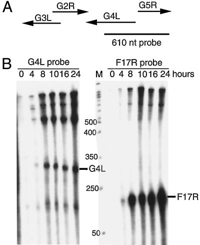
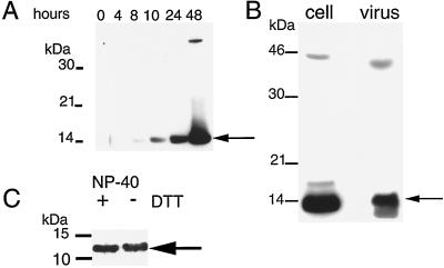
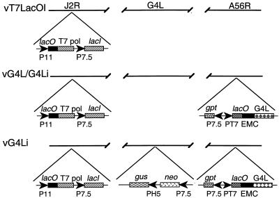
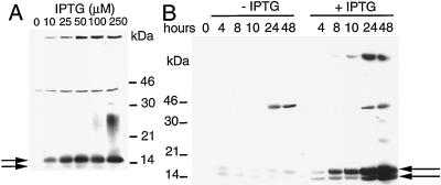
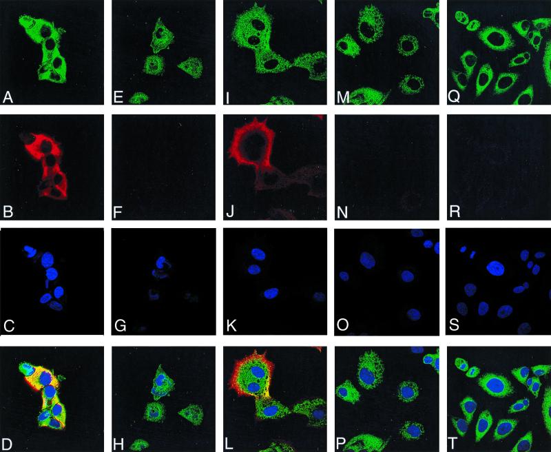
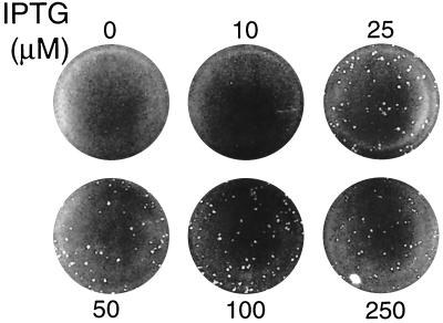
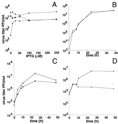
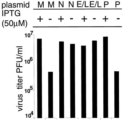
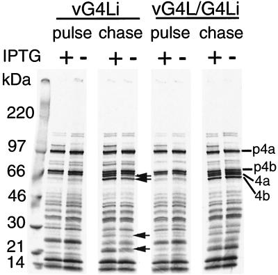
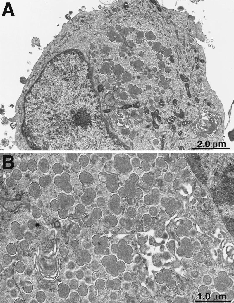
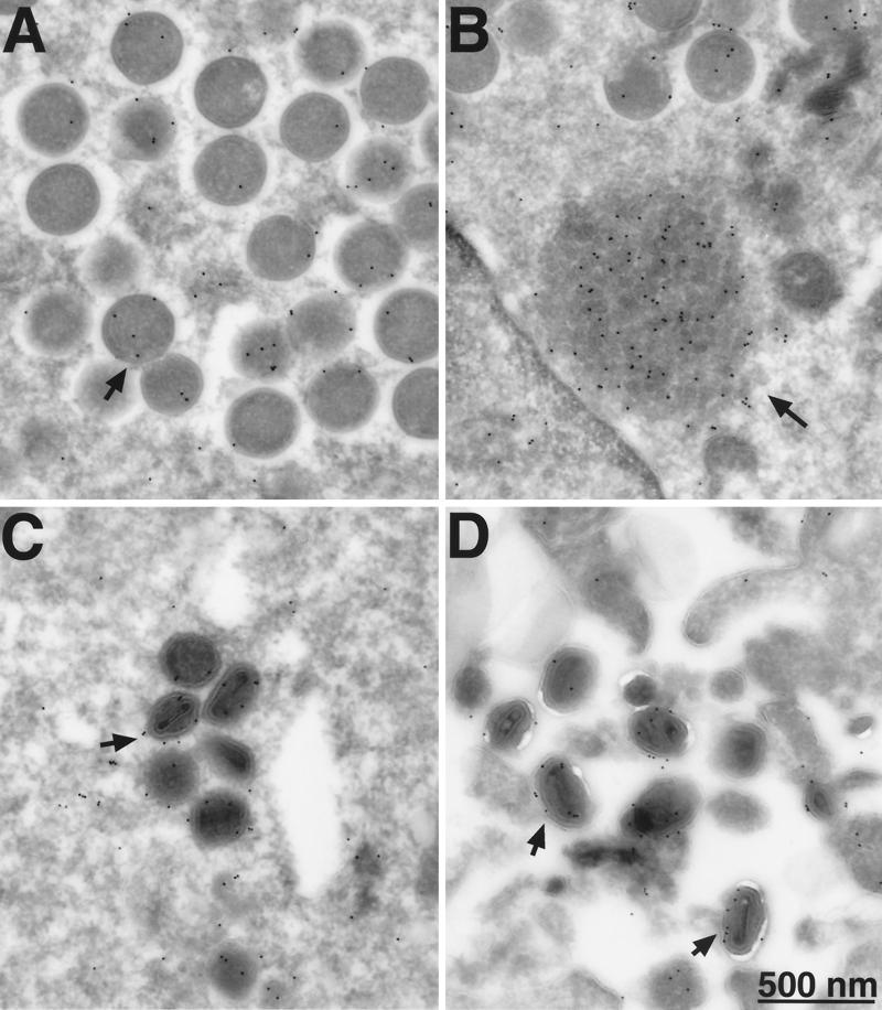
Vaccinia virus encodes two glutaredoxins, O2L and G4L, both of which exhibit thioltransferase and dehydroascorbate reductase activities in vitro. Although O2L was previously found to be dispensable for virus replication, we now show that G4L is necessary for virion morphogenesis. RNase protection and Western blotting assays indicated that G4L was expressed at late times after infection and was incorporated into mature virus particles. Attempts to isolate a mutant virus with a deleted G4L gene were unsuccessful, suggesting that the protein was required for virus replication. This interpretation was confirmed by the construction and characterization of a conditional lethal recombinant virus with an inducible copy of the G4L gene replacing the original one. Expression of G4L was proportional to the concentration of inducer, and the amount of glutaredoxin could be varied from barely detectable to greater than normal amounts of protein. Immunogold labeling revealed that the induced G4L protein was associated with immature and mature virions and adjacent cytoplasmic depots. In the absence of inducer, the production of infectious virus was severely inhibited, though viral late protein synthesis appeared unaffected except for decreased maturation-dependent proteolytic processing of certain core components. Electron microscopy of cells infected under nonpermissive conditions revealed an accumulation of crescent membranes on the periphery of electron-dense globular masses but few mature particles. We concluded that the two glutaredoxin homologs encoded by vaccinia virus have different functions and that G4L has a role in virion morphogenesis, perhaps by acting as a redox protein.


Figures

FIG. 1
FIG. 1
Transcriptional analysis. (A) Schematic diagrams of the G4L and adjacent ORFs with arrows indicating direction of transcription. The size and position of the uniformly 32P-labeled complementary G4L riboprobe relative to the ORFs are shown. nt, nucleotide. (B) RNase protection assays of G4L and F17R transcripts. Total RNA, extracted from 0 to 24 h after vaccinia virus infection of BS-C-1 cells, was hybridized with the 32P-labeled riboprobes specific for G4L or F17R sequences. Following RNase digestion, the protected probe fragments were analyzed by PAGE and autoradiography. The marker track (M) is a 50-nucleotide end-labeled DNA ladder (sizes in nucleotides on the left). The predicted protected fragment for each gene is indicated on the right.
FIG. 2
FIG. 2
Synthesis of the G4L protein and association with purified virions. (A) SDS-PAGE and Western blotting analysis of G4L synthesis. BS-C-1 cells were infected with vaccinia virus at a multiplicity of 5. At the indicated times after infection, cells were lysed with SDS and analyzed by Western blotting using antiserum directed to the C-terminal 13 amino acids of the G4L ORF. Proteins were detected by chemiluminescence. The G4L protein (arrow) and marker proteins are indicated on the right and left, respectively. (B) Association of the G4L protein with purified virions. Sucrose gradient-purified vaccinia virus (107 PFU) and lysates of cells infected for 24 h were analyzed by Western blotting as described for panel A. (C) Detergent extraction of G4L protein from purified vaccinia virus. Approximately 107 PFU of sucrose gradient-purified WR virus was incubated with buffer containing NP-40 with or without DTT. Insoluble material was removed by centrifugation, and the supernatants were analyzed as described for panel A. Only the lower part of the gel is shown. The G4L protein bands are indicated by arrows in all panels.
FIG. 3
FIG. 3
Schematic diagram of recombinant vaccinia viruses. The genomes of recombinant vaccinia viruses vT7lacOI, vG4L/G4Li, and vG4Li are shown with the J2R (thymidine kinase), G4L, and A56R (HA) loci depicted. Insertions into these loci are indicated below. Abbreviations: P11, vaccinia virus late promoter; P7.5, vaccinia virus early-late promoter; PH5, vaccinia virus early-late promoter; PT7, bacteriophage T7 promoter; T7 pol, T7 RNA polymerase; lacO, E. coli lac operator; lacI, E. coli lac repressor gene; EMC, encephalomyocarditis virus cap-independent translation enhancer element; neo, neomycin resistance gene; gus, color selection marker; gpt, mycophenolic acid resistance gene.
FIG. 4
FIG. 4
Inducer-dependent expression of G4L. The figure shows the effect of IPTG concentration. BS-C-1 cells were infected with 5 PFU of vG4Li per cell and incubated with 0 to 250 μM IPTG and harvested at 24 h (A) or incubated with 0 or 50 μM IPTG and harvested at the times indicated (B). The cells were lysed with SDS and analyzed by Western blotting as described in the legend to Fig. 2. The positions of marker proteins are indicated at the sides. The upper and lower arrows point to epitope-tagged and nontagged G4L proteins, respectively.
FIG. 5
FIG. 5
Detection of G4L by immunofluorescence. HeLa cells were uninfected (Q to T) or infected with vG4Li in the presence of inducer for 10 h (A to D) or 16 h (I to L) or in the absence of inducer for 10 h (E to H) or 16 h (M to P). After 10 or 16 h, the cells were fixed; permeabilized; stained with G4L peptide antiserum and an antirabbit rhodamine conjugate (B, F, J, N, and R), an anti-PDI MAb and an anti-mouse Oregon green conjugate (A, E, I, M, and Q), or Hoechst stain (blue) (C, G, K, O, and S); and viewed by confocal microscopy. Merged images of cells stained with anti-PDI, anti-G4L, and Hoechst stain are also shown (D, H, L, P, and T).
FIG. 6
FIG. 6
IPTG dependence of plaque formation. Cell monolayers were inoculated with vG4Li and overlaid with medium containing 0 to 250 μM IPTG. After incubation at 37°C for 48 h, the plates were stained with crystal violet.
FIG. 7
FIG. 7
Effect of IPTG on virus yields. (A) Effect of IPTG concentration on 24-h yields. BS-C-1 cells were infected with WR (⧫), vG4L/G4Li (■), or vG4Li (▴) at a multiplicity of 5 and incubated with medium containing 0 to 250 μM IPTG for 24 h. Virus yields were determined by plaque assay in the presence of 50 μM IPTG for all viruses. (B to D) Time course of virus production in the presence (filled symbols) or absence (unfilled symbols) of 50 μM IPTG. The virus was WR (B), vG4L/G4Li (C), or vG4Li (D). Virus titers were determined by plaque assay as described for panel A.
FIG. 8
FIG. 8
trans complementation of vG4Li. BS-C-1 cells were infected with vG4Li at a multiplicity of 5 in the presence (+) or absence (−) of 50 μM IPTG and then mock transfected (M), transfected with pUC-19 (P), or transfected with plasmids containing the G4L gene under its natural promoter (N) or a synthetic early-late viral promoter (E/L). After 24 h, the cells were harvested and the virus titers were determined by plaque assay in the presence of 50 μM IPTG.
FIG. 9
FIG. 9
Synthesis and processing of viral late proteins. BS-C-1 cells were infected at a multiplicity of 5 in the presence (+) or absence (−) of 50 μM IPTG. At 8.75 h after infection, the cells were labeled with [35S]methionine for 1 h. One set of cells (pulse) were harvested, and another set were incubated with excess unlabeled methionine for an additional 14 h (chase). The proteins were analyzed by SDS-PAGE and autoradiography. Arrows indicate bands that increase during the chase in the presence of IPTG. The positions of the major core precursor proteins (p4a and p4b) and their mature processed forms (4a and 4b) are indicated on the right.
FIG. 10
FIG. 10
Interruption of virus morphogenesis. BS-C-1 cells were infected with vG4Li in the absence of IPTG. After 36 h, cells were fixed and embedded in Epon. Ultrathin sections were prepared for transmission electron microscopy. Crescent membranes adjacent to electron-dense globules are shown at low (A) and high (B) magnifications.
FIG. 11
FIG. 11
Electron microscopy of immunogold-labeled G4L protein. BS-C-1 cells were infected with vG4Li in the presence of 50 μM IPTG. Frozen sections were stained with a MAb to the HA epitope tag and protein A-conjugated gold grains and examined by electron microscopy. Fields show predominantly immature virions (A), depot containing G4L (B), intracellular mature virions (C), and extracellular virions (D). Arrows point to representative gold grains.

Similar articles

Cited by

References

    1. Afonso C L, Tulman E R, Lu Z, Oma E, Kutish G F, Rock D L. The genome of Melanoplus sanguinipes entomopoxvirus. J Virol. 1999;73:533–552. - PMC - PubMed
    1. Afonso C L, Tulman E R, Lu Z, Zsak L, Kutish G F, Rock D L. The genome of fowlpox virus. J Virol. 2000;74:3815–3831. - PMC - PubMed
    1. Ahn B Y, Moss B. Glutaredoxin homolog encoded by vaccinia virus is a virion-associated enzyme with thioltransferase and dehydroascorbate reductase activities. Proc Natl Acad Sci USA. 1992;89:7060–7064. - PMC - PubMed
    1. Baldick C J, Jr, Moss B. Characterization and temporal regulation of mRNAs encoded by vaccinia virus intermediate-stage genes. J Virol. 1993;67:3515–3527. - PMC - PubMed
    1. Bordier C. Phase separation of integral membrane proteins in Triton X-114 solution. J Biol Chem. 1981;256:1604–1607. - PubMed

LinkOut - more resources