. 2000 Oct;74(19):9184-96.
doi: 10.1128/jvi.74.19.9184-9196.2000.

Endocytosis and nuclear trafficking of adeno-associated virus type 2 are controlled by rac1 and phosphatidylinositol-3 kinase activation

Affiliations

Endocytosis and nuclear trafficking of adeno-associated virus type 2 are controlled by rac1 and phosphatidylinositol-3 kinase activation

S Sanlioglu et al. J Virol. 2000 Oct.

Abstract

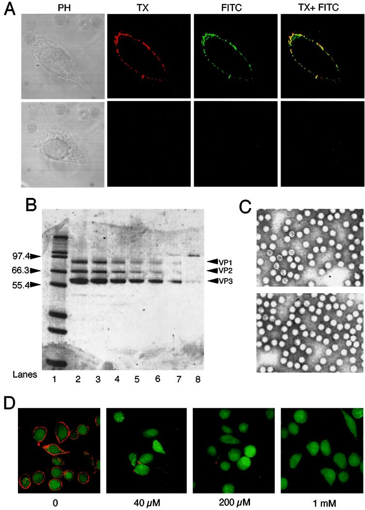
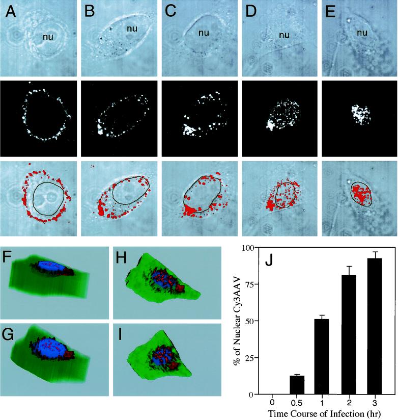
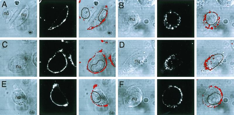
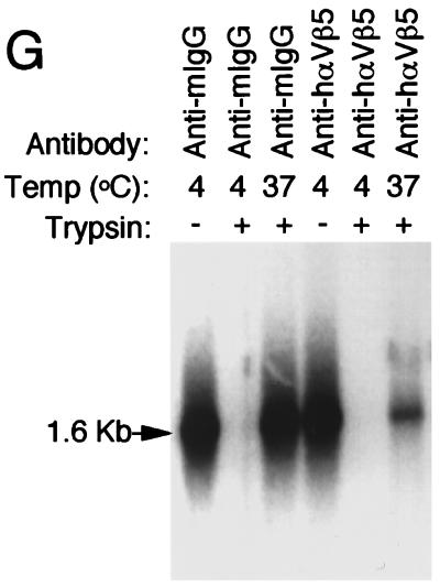
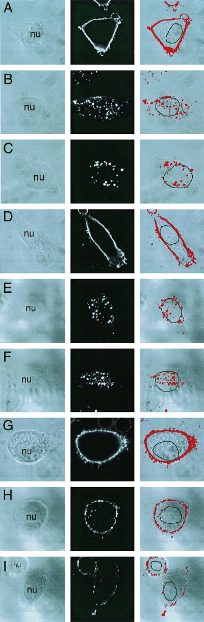
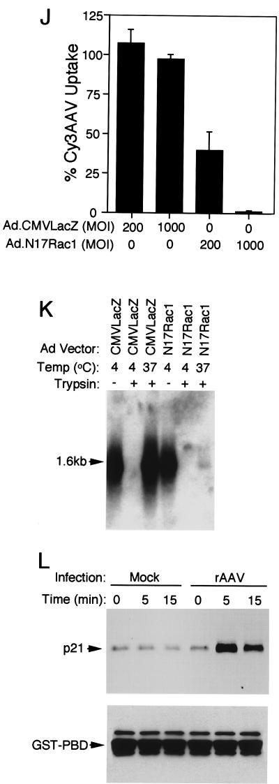
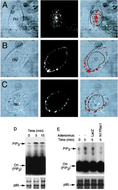
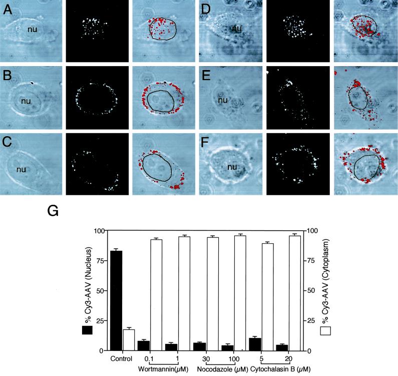
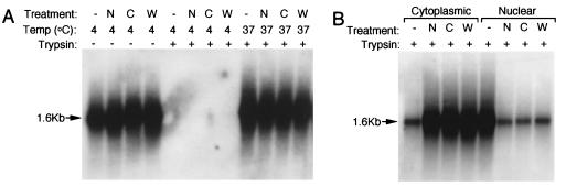
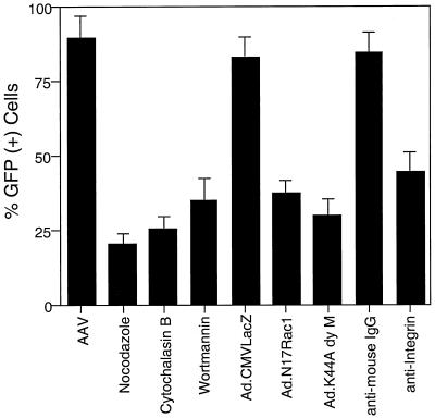
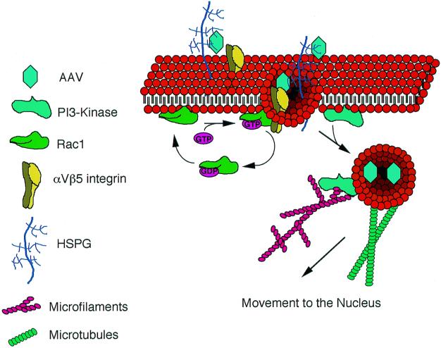
Adeno-associated virus (AAV) is a single-stranded DNA parvovirus that causes no currently known pathology in humans. Despite the fact that this virus is of increasing interest to molecular medicine as a vector for gene delivery, relatively little is known about the cellular mechanisms controlling infection. In this study, we have examined endocytic and intracellular trafficking of AAV-2 using fluorescent (Cy3)-conjugated viral particles and molecular techniques. Our results demonstrate that internalization of heparan sulfate proteoglycan-bound AAV-2 requires alphaVbeta5 integrin and activation of the small GTP-binding protein Rac1. Following endocytosis, activation of a phosphatidylinositol-3 (PI3) kinase pathway was necessary to initiate intracellular movement of AAV-2 to the nucleus via both microfilaments and microtubules. Inhibition of Rac1 using a dominant N17Rac1 mutant led to a decrease in AAV-2-mediated PI3 kinase activation, indicating that Rac1 may act proximal to PI3 kinase during AAV-2 infection. In summary, our results indicate that alphaVbeta5 integrin-mediated endocytosis of AAV-2 occurs through a Rac1 and PI3 kinase activation cascade, which directs viral movement along the cytoskeletal network to the nucleus.


Figures

FIG. 1
FIG. 1
Cy3 conjugation to AAV capsids. HeLa cells were infected with rAAV labeled with Cy3 as explained in Materials and Methods. (A) Fluorescent colocalization of Cy3AAV (left panel in red) with AAV capsid proteins stained with FITC-labeled anti-cap B1 antibody (center panel in green). The two channels are superimposed in the right panel, demonstrating colocalization of Cy3AAV and capsid proteins (in yellow). SDS-PAGE analysis, following staining with Sypro-Orange, was used to evaluate the purity of the rAAV preparation used for Cy3 labeling (B). Lane 1 contains molecular size markers, and the sizes (in kilodaltons) are given to the left of the gel. Various amounts of rAAV from 20 to 1 μl were loaded onto lanes 2 to 7 in a 12% polyacrylamide–Tris–glycine gel (Novex). The locations of capsid proteins VP-1, -2, and -3 are marked to the right of the gel. Lane 8 contains 50 ng of β-galactosidase protein as a reference. (C) Ultrastructure of unlabeled (upper panel) and Cy3-conjugated AAV (lower panel) by transmission electron microscopy. The binding of Cy3-labeled AAV to its receptor on HeLa cells was tested for competition by increasing amounts of heparin (D). Numbers below each panel indicate the doses of heparin used in these assays.
FIG. 2
FIG. 2
Time course of AAV translocation to the nucleus. HeLa cells were incubated with Cy3AAV at 4°C for 1 h to promote virus binding. Following binding, cells were either immediately washed and fixed (A) or washed and then shifted to 37°C for 0.5 (B), 1 (C), 2 (D), or 3 (E) h prior to fixation. Cells were then analyzed by confocal fluorescent microscopy. Each image is a stack of three consecutive 0.5-μm layers, with each panel representing confocal phase contrast (top), gray scale Cy3 (middle), and superimposed images (bottom). The nucleus (nu) of each cell is marked for clarity. 3-D reconstructed images were generated from 25 (F and G) or 5 (H and I) consecutive 0.5-μm confocal layers obtained after both phase-contrast and red channel scans from a cell infected for 2 h at 37°C. Cellular architecture was generated from the individual phase-contrast confocal images, with the nucleus pseudocolored in blue and the cytoplasm in green. The same cell is represented in each panel with a different angle of rotation. The percentage of nucleus-associated Cy3AAV particles was determined using morphometric image analysis as explained in Materials and Methods (n = 7 cells analyzed for each time point) and is graphically represented in panel J. Values in panel J are the mean ± standard error of the mean (SEM) percentage of particles associated with the nucleus for each time point.
FIG. 3
FIG. 3
αVβ5 integrin is required for the endocytosis of AAV-2 virus. Cy3AAV was incubated alone (A and B) or in the presence of control anti-mouse IgG antibody (C and D) or anti-human αVβ5 integrin-blocking antibody (E and F) at 4°C for 1 h. Cells were then either washed and fixed immediately (A, C, and E) or washed and incubated for an additional 45 min at 37°C (B, D, and F). Representative confocal photomicrographs are shown for each condition. Each of the panels shows the confocal phase-contrast image (left), gray scale stack of Cy3AAV images (middle), and superimposed images (right). Each confocal image is a stack of six consecutive 0.5-μm layers that intersect the nucleus. The nucleus (nu) of each cell is marked for clarity. Southern blotting of Hirt DNA isolated from rAAV-infected cells was performed as an alternative approach to evaluate rAAV endocytosis in cells treated with anti-mouse IgG or anti-human αVβ5 integrin (G). HeLa cells were infected with unlabeled AV.GFP3ori virus (MOI of 1,000 DNA particles/cell) for 1 h at 4°C, washed, and shifted to 37°C for 2 h. Cells were harvested by either direct scraping to determine viral binding (− trypsin) or by trypsinization to remove extracellular virus (+ trypsin). Hirt DNA was prepared from cells treated with the various conditions, and Southern blots were hybridized with P32-labeled EGFP cDNA probe. Single-stranded rAAV genomes are marked by a 1.6-kb hybridizing band.
FIG. 3
FIG. 3
αVβ5 integrin is required for the endocytosis of AAV-2 virus. Cy3AAV was incubated alone (A and B) or in the presence of control anti-mouse IgG antibody (C and D) or anti-human αVβ5 integrin-blocking antibody (E and F) at 4°C for 1 h. Cells were then either washed and fixed immediately (A, C, and E) or washed and incubated for an additional 45 min at 37°C (B, D, and F). Representative confocal photomicrographs are shown for each condition. Each of the panels shows the confocal phase-contrast image (left), gray scale stack of Cy3AAV images (middle), and superimposed images (right). Each confocal image is a stack of six consecutive 0.5-μm layers that intersect the nucleus. The nucleus (nu) of each cell is marked for clarity. Southern blotting of Hirt DNA isolated from rAAV-infected cells was performed as an alternative approach to evaluate rAAV endocytosis in cells treated with anti-mouse IgG or anti-human αVβ5 integrin (G). HeLa cells were infected with unlabeled AV.GFP3ori virus (MOI of 1,000 DNA particles/cell) for 1 h at 4°C, washed, and shifted to 37°C for 2 h. Cells were harvested by either direct scraping to determine viral binding (− trypsin) or by trypsinization to remove extracellular virus (+ trypsin). Hirt DNA was prepared from cells treated with the various conditions, and Southern blots were hybridized with P32-labeled EGFP cDNA probe. Single-stranded rAAV genomes are marked by a 1.6-kb hybridizing band.
FIG. 4
FIG. 4
AAV endocytosis requires Rac1 activation. A dominant inhibitor of Rac1 (N17Rac1) was used to evaluate the involvement of the small GTPase Rac1 in AAV endocytosis. HeLa cells were either uninfected (A to C) or infected with Ad.CMVLacZ (D to F) or Ad.N17Rac1 (G to I) virus at an MOI of 1,000 (D, F, G, and I) or 200 (E and H) particles/cell 48 h prior to incubation with Cy3AAV at 4°C for 1 h. Following incubation with Cy3AAV, cells were either washed and immediately fixed to examine viral binding (A, D, and G) or shifted to at 37°C for 45 min to examine endocytosis (B, C, E, F, H, and I). Representative confocal photomicrographs are given for each condition, representing six 0.5-μm stacked layers that intersect the nucleus. Each of the panels shows the confocal phase contrast image (left), gray scale stack of Cy3AAV images (middle), and superimposed images (right). The nucleus (nu) of each cell is marked for clarity. Panel J represents the mean percentage ± SEM (n = 7) of Cy3AAV particles internalized within a 45-min time period for each condition, as determined by computer-aided image analysis. Southern blotting of Hirt DNA isolated from rAAV-infected cells was performed as an alternative approach to evaluate rAAV endocytosis in Ad.N17Rac1- and Ad.CMVLacZ-infected (1,000 DNA particles/cell) HeLa cells (K). At 48 h following adenoviral infection, HeLa cells were infected with unlabeled AV.GFP3ori virus (MOI of 1,000 DNA particles/cell) for 1 h at 4°C, washed, and either harvested directly or shifted to 37°C for 2 h prior to harvesting. Cells were harvested by either direct scraping to determine viral binding (− trypsin) or by trypsinization to remove extracellular virus (+ trypsin). Hirt DNA was prepared from the various conditions, and Southern blots were hybridized with a 32P-labeled EGFP cDNA probe. Single-stranded rAAV genomes are marked by a 1.6-kb hybridizing band. Rac1 activation assays were performed as described in Materials and Methods. A Western blot detecting GST-PBD-precipitated GTP-bound Rac1 is given in panel L. Cells were infected with unlabeled tgAAVCF virus (MOI of 5,000 DNA particles/cell) or mock infected with vehicle alone for the exposure times indicated above each lane, then 500 μg of HeLa cell lysate from each condition was precipitated with GST-PBD and evaluated by Western blot against anti-Rac1 antibodies. The p21 band marked by an arrow is Rac1. The filters were also probed with anti-GST antibodies as a loading control. The position of GST-PBD protein is marked by an arrow below the Rac1 Western blot.
FIG. 4
FIG. 4
AAV endocytosis requires Rac1 activation. A dominant inhibitor of Rac1 (N17Rac1) was used to evaluate the involvement of the small GTPase Rac1 in AAV endocytosis. HeLa cells were either uninfected (A to C) or infected with Ad.CMVLacZ (D to F) or Ad.N17Rac1 (G to I) virus at an MOI of 1,000 (D, F, G, and I) or 200 (E and H) particles/cell 48 h prior to incubation with Cy3AAV at 4°C for 1 h. Following incubation with Cy3AAV, cells were either washed and immediately fixed to examine viral binding (A, D, and G) or shifted to at 37°C for 45 min to examine endocytosis (B, C, E, F, H, and I). Representative confocal photomicrographs are given for each condition, representing six 0.5-μm stacked layers that intersect the nucleus. Each of the panels shows the confocal phase contrast image (left), gray scale stack of Cy3AAV images (middle), and superimposed images (right). The nucleus (nu) of each cell is marked for clarity. Panel J represents the mean percentage ± SEM (n = 7) of Cy3AAV particles internalized within a 45-min time period for each condition, as determined by computer-aided image analysis. Southern blotting of Hirt DNA isolated from rAAV-infected cells was performed as an alternative approach to evaluate rAAV endocytosis in Ad.N17Rac1- and Ad.CMVLacZ-infected (1,000 DNA particles/cell) HeLa cells (K). At 48 h following adenoviral infection, HeLa cells were infected with unlabeled AV.GFP3ori virus (MOI of 1,000 DNA particles/cell) for 1 h at 4°C, washed, and either harvested directly or shifted to 37°C for 2 h prior to harvesting. Cells were harvested by either direct scraping to determine viral binding (− trypsin) or by trypsinization to remove extracellular virus (+ trypsin). Hirt DNA was prepared from the various conditions, and Southern blots were hybridized with a 32P-labeled EGFP cDNA probe. Single-stranded rAAV genomes are marked by a 1.6-kb hybridizing band. Rac1 activation assays were performed as described in Materials and Methods. A Western blot detecting GST-PBD-precipitated GTP-bound Rac1 is given in panel L. Cells were infected with unlabeled tgAAVCF virus (MOI of 5,000 DNA particles/cell) or mock infected with vehicle alone for the exposure times indicated above each lane, then 500 μg of HeLa cell lysate from each condition was precipitated with GST-PBD and evaluated by Western blot against anti-Rac1 antibodies. The p21 band marked by an arrow is Rac1. The filters were also probed with anti-GST antibodies as a loading control. The position of GST-PBD protein is marked by an arrow below the Rac1 Western blot.
FIG. 5
FIG. 5
Trafficking of Cy3AAV to the nucleus is sensitive to wortmannin treatment. HeLa cells were either untreated (A) or treated with 0.1 μM (B) or 1 μM (C) wortmannin for 1 h at 37°C prior to infection with Cy3AAV. Following wortmannin treatment, Cy3AAV was bound to cells for 1 h at 4°C, then the cells were washed of excess virus and incubated at 37°C for an additional 2 h in the continued presence of wortmannin. Representative confocal photomicrographs are given for each condition, each representing six 0.5-μm stacked layers that intersect the nucleus. Each panel illustrates the confocal phase contrast (left), gray scale stack of Cy3AAV images (middle), and superimposed images (right). The nucleus (nu) of each cell is marked for clarity. The effect of rAAV infection on PI3K activation was evaluated as described in Materials and Methods (D and E). Cells were infected with unlabeled tgAAVCF virus at an MOI of 5,000 DNA particles/cell for 0, 5, and 15 min at 37°C. The PI3K complex was immunoprecipitated from cell lysates and assayed in the presence of [γ-32P]ATP and l-α-phosphatidylinositol followed by TLC and autoradiography (D, top). Faster migrating bands above the origin of migration (ori) indicate the product (PIP3). Western blot analysis was also performed using anti-PI3K p85 antibody, which indicates that equal amounts of PI3K complex were used for each reaction (lower panel of D). To evaluate whether functional Rac1 was necessary for PI3K activation, cells were infected with either Ad.N17Rac1 or Ad.CMVLacZ virus at an MOI of 1,000 particles/cell for 48 h prior to assaying PI3K activity (E). Conditions for infection and times of harvest are marked above each lane of the TLC autoradiogram. The bottom panel is a Western blot using anti-PI3K p85 antibody.
FIG. 6
FIG. 6
Nocodazole and cytochalasin B treatment reduces nuclear targeting of Cy3AAV. HeLa cells were either untreated (A and D) or treated with 30 μM nocodazole (B), 100 μM nocodazole (C), 5 μM cytochalasin B (E), or 20 μM cytochalasin B (F) for 1 h at 37°C prior to infection with Cy3AAV. Following nocodazole and cytochalasin B treatment, Cy3AAV was bound to cells for 1 h at 4°C, and the cells were then washed of excess virus and incubated at 37°C for an additional 2 h in the continued presence of inhibitor. Representative confocal photomicrographs are given for each condition, each representing six 0.5-μm stacked layers that intersect the nucleus. Each of the panels shows the confocal phase contrast (left), gray scale stack of Cy3AAV images (middle), and superimposed images (right). The nucleus (nu) of each cell is marked for clarity. Quantitative analysis of AAV nuclear trafficking is shown in panel G. The percentages of cytoplasmic and nuclear associated Cy3AAV particles were quantified by morphometric image analysis, as explained in Materials and Methods, following treatment of cells with various chemical agents. Solid bars indicate nucleus-associated Cy3AAV, while open bars indicate cytoplasmic Cy3AAV particles. Values are the mean ± SEM of 10 cells quantified for each condition.
FIG. 7
FIG. 7
Wortmannin, nocodazole, and cytochalasin B inhibit intracellular transport of AAV-2 to the nucleus but not endocytosis. HeLa cells were infected with unlabeled AV.GFP3ori virus (MOI of 1,000 DNA particles/cell) following treatment with wortmannin (W), nocodazole (N), or cytochalasin B (C). Protocols for chemical treatment were as described in Materials and Methods. Cells were infected for 1 h at 4°C, washed, and shifted to 37°C for 2 h. To determine the extent of viral endocytosis, cells were harvested after both 4 and 37°C incubation by either direct scraping to determine viral binding (− trypsin) or by trypsinization to remove extracellular virus (+ trypsin) (A). Alternatively, following 37°C incubations, cells were harvested by trypsinization and washed, and cytoplasmic and nuclear fractions were purified (B). Hirt DNA was prepared from cells or subcellular fractions following the various treatment conditions (as indicated above each lane), and Southern blots were hybridized with 32P-labeled EGFP cDNA probe. Single-stranded rAAV genomes are marked by a 1.6-kb hybridizing band.
FIG. 8
FIG. 8
Inhibitors of AAV-2 endocytosis and nuclear trafficking also reduce rAAV-mediated gene expression. HeLa cells were treated with various agents (indicated below the graph) which modulate AAV-2 endocytosis or nuclear trafficking, and the extent of AV.GFP3ori-mediated GFP expression was analyzed at 35 h postinfection by FACS analysis. For conditions involving infection with recombinant adenoviruses Ad.N17Rac1, Ad.K44Adynamin, and Ad.CMVLacZ, cells were infected at MOIs of 1,000 particles/cell 48 h prior to AV.GFP3ori infection. Treatments with nocodazole, cytochalasin B, wortmannin, anti-IgG, and anti-integrin were performed as described for Cy3 analyses. All conditions included infection with AV.GFP3ori virus at an MOI of 1,000 DNA particles/cell. FACS analysis data represent the mean ± SEM of four independent data points.
FIG. 9
FIG. 9
Schematic representation of AAV-2 endocytic and nuclear trafficking mechanisms. Receptor-mediated endocytosis of AAV-2 in HeLa cells is facilitated through binding to its receptor HSPG and interactions with αVβ5 integrin. Activation of Rac1, potentially by αVβ5 integrin, is required for efficient endocytosis of AAV virus. Following endocytosis, we propose that Rac1 activation leads to stimulation of PI3K pathways, which facilitate the functional rearrangements of the cytoskeleton (microfilaments and microtubules) required for the efficient nuclear targeting of AAV.

References

    1. Alexander I E, Russell D W, Miller A D. DNA-damaging agents greatly increase the transduction of nondividing cells by adeno-associated virus vectors. J Virol. 1994;68:8282–8287. - PMC - PubMed
    1. Ali R R, Reichel M B, Thrasher A J, Levinsky R J, Kinnon C, Kanuga N, Hunt D M, Bhattacharya S S. Gene transfer into the mouse retina mediated by an adeno-associated viral vector. Hum Mol Genet. 1996;5:591–594. - PubMed
    1. Andrews N C, Faller D V. A rapid micropreparation technique for extraction of DNA-binding proteins from limiting numbers of mammalian cells. Nucleic Acids Res. 1991;19:2499. - PMC - PubMed
    1. Bagrodia S, Taylor S J, Jordon K A, Van Aelst L, Cerione R A. A novel regulator of p21-activated kinases. J Biol Chem. 1998;273:23633–23636. - PubMed
    1. Bartlett J S, Samulski R J, McCown T J. Selective and rapid uptake of adeno-associated virus type 2 in brain. Hum Gene Ther. 1998;9:1181–1186. - PubMed

Publication types

MeSH terms

Substances