. 2000 Oct;74(19):9234-9.
doi: 10.1128/jvi.74.19.9234-9239.2000.

Human coronavirus 229E infects polarized airway epithelia from the apical surface

Affiliations

Human coronavirus 229E infects polarized airway epithelia from the apical surface

G Wang et al. J Virol. 2000 Oct.

Abstract

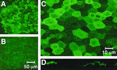
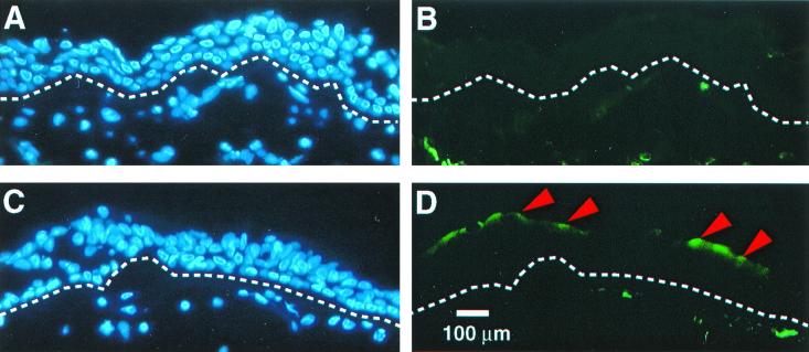
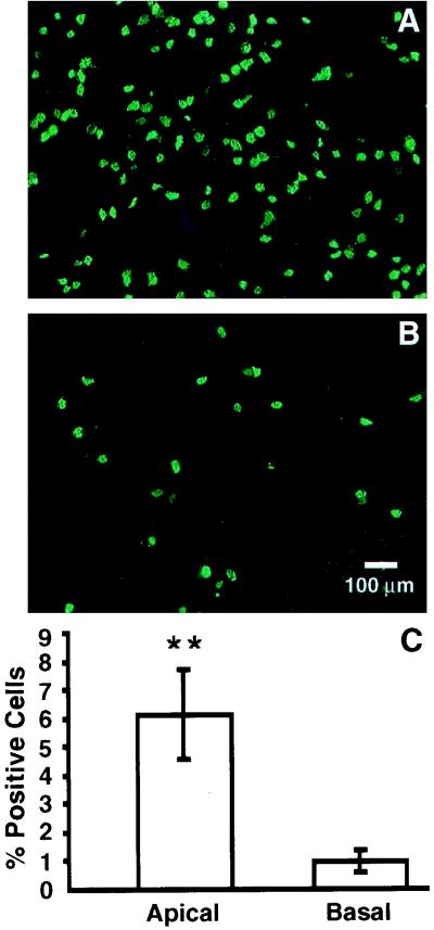
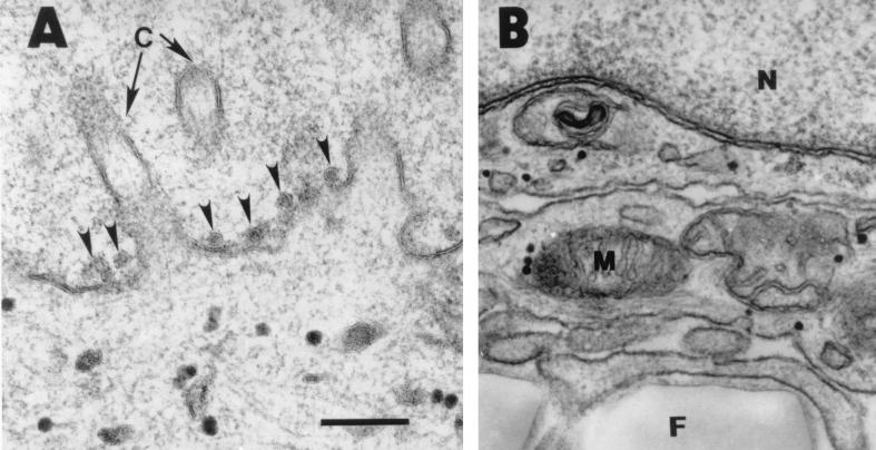
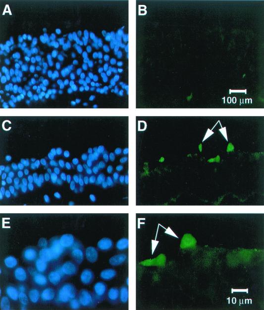
Gene transfer to differentiated airway epithelia with existing viral vectors is very inefficient when they are applied to the apical surface. This largely reflects the polarized distribution of receptors on the basolateral surface. To identify new receptor-ligand interactions that might be used to redirect vectors to the apical surface, we investigated the process of infection of airway epithelial cells by human coronavirus 229E (HCoV-229E), a common cause of respiratory tract infections. Using immunohistochemistry, we found the receptor for HCoV-229E (CD13 or aminopeptidase N) localized mainly to the apical surface of airway epithelia. When HCoV-229E was applied to the apical or basolateral surface of well-differentiated primary cultures of human airway epithelia, infection primarily occurred from the apical side. Similar results were noted when the virus was applied to cultured human tracheal explants. Newly synthesized virions were released mainly to the apical side. Thus, HCoV-229E preferentially infects human airway epithelia from the apical surface. The spike glycoprotein that mediates HCoV-229E binding and fusion to CD13 is a candidate for pseudotyping retroviral envelopes or modifying other viral vectors.


Figures

FIG. 1
FIG. 1
The HCoV-229E receptor aminopeptidase N is abundantly expressed on the apical surface of differentiated human airway epithelial cells. Nonpermeabilized, well-differentiated human airway epithelial cells were fixed and immunostained with a mouse anti-human CD13 antibody by directly applying the antibody to either the apical or basal surface. Samples were examined by confocal microscopy. (A) CD13 expression on apical surface; (B) CD13 expression on basal surface; (C) higher magnification view of apical surface demonstrating detailed stained cell boundaries; (D) x-z image construction showing characteristic apical cell surface staining pattern. When the primary antibody was omitted or mouse serum was substituted, no signal was detected (data not shown). Data shown are representative of two independent experiments done using cells derived from different donor lungs.
FIG. 2
FIG. 2
The HCoV-229E receptor preferentially localizes to the apical surface of human trachea. Tracheal tissues were immunostained with the anti-human CD13 monoclonal antibody as described in Materials and Methods. For panels A and B, normal mouse serum (control) was substituted for the primary antibody. Nuclear staining of the epithelia with DAPI (blue); dotted lines indicate location of basement membrane. In all cells stained with anti-CD13 antibody (panels C and D), CD13 expression was seen along the apical surface of some of the tracheal cells (arrowheads). Results are representative of two independent experiments performed using cells from different donor tissues.
FIG. 3
FIG. 3
HCoV-229E infects airway epithelial cells preferentially from the apical surface. Well-differentiated airway epithelial cells were inoculated with HCoV-229E at an MOI of 0.1 from the apical (A) or basal (B) surface for 16 h, and immunofluorescent staining for HCoV-229E proteins was performed to document the polarity of infection, as indicated by the percentage of HCoV-229E protein-expressing cells (C). The efficiency of infection was significantly greater from the apical surface (P < 0.05 by Student's t test).
FIG. 4
FIG. 4
Transmission electron microscopy of airway epithelia infected with HCoV-229E. (A) HCoV virions were frequently detected on the apical surface of epithelia (arrowheads) (A), but individual virions were not seen in vesicles or released at the basolateral surface (B). The lower portion of panel B shows the permeable membrane on which the epithelia were growing. A total of 100 cells from three epithelial preparations were examined. N, nucleus; M, mitochondria; F, permeable filter. Bar = 200 nm.
FIG. 5
FIG. 5
HCoV-229E infects human tracheal explants from the apical surface. Human tracheal explants were cultured overnight. HCoV-229E virus was applied to the mucosal surface, and 24 h later frozen sections of tissue were immunostained for HCoV-229E protein expression. The sections were counterstained with DAPI to identify cell nuclei (blue). Control specimens were processed immediately after application of the virus. No viral proteins were detected in controls (A and B), whereas samples infected with HCoV-229E for 24 h (C to F) showed scattered virus antigen-positive cells (indicated by arrows) at the apical surface (green).

References

    1. Arbour N, Ekande S, Cote G, Lachance C, Chagnon F, Tardieu M, Cashman N R, Talbot P J. Persistent infection of human oligodendrocytic and neuroglial cell lines by human coronavirus 229E. J Virol. 1999;73:3326–3337. - PMC - PubMed
    1. Blau D M, Compans R W. Entry and release of measles virus are polarized in epithelial cells. Virology. 1995;210:91–99. - PubMed
    1. Bozzola J J, Russell L D. Electron microscopy. 2nd ed. Sudbury, Mass: Jones and Bartlett Publishers; 1998.
    1. Bradburne A F, Bynoe M L, Tyrrell D A. Effects of a “new” human respiratory virus in volunteers. Br Med J. 1967;3:767–769. - PMC - PubMed
    1. Burns J C, Friedmann T, Driever W, Burrascano M, Yee J-K. Vesicular stomatitis virus G glycoprotein pseudotyped retroviral vectors: concentration to very high titer and efficient gene transfer into mammalian and nonmammalian cells. Proc Natl Acad Sci USA. 1993;90:8033–8037. - PMC - PubMed

Publication types

LinkOut - more resources