. 2000 Sep;11(9):3031-44.
doi: 10.1091/mbc.11.9.3031.

The unique catalytic subunit of sperm cAMP-dependent protein kinase is the product of an alternative Calpha mRNA expressed specifically in spermatogenic cells

Affiliations
Free PMC article

The unique catalytic subunit of sperm cAMP-dependent protein kinase is the product of an alternative Calpha mRNA expressed specifically in spermatogenic cells

J T Agustin et al. Mol Biol Cell. 2000 Sep.
Free PMC article

Abstract

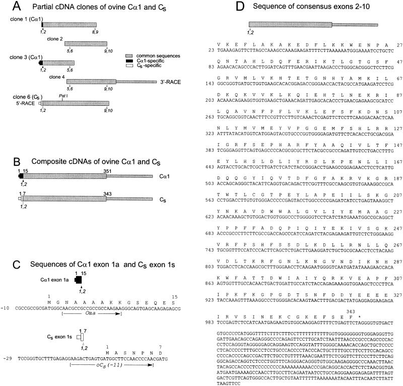
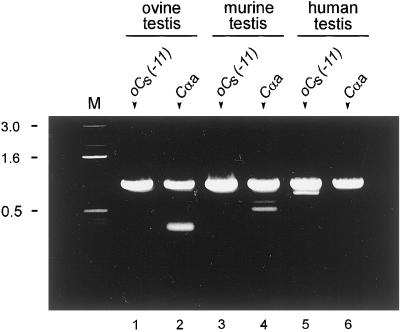
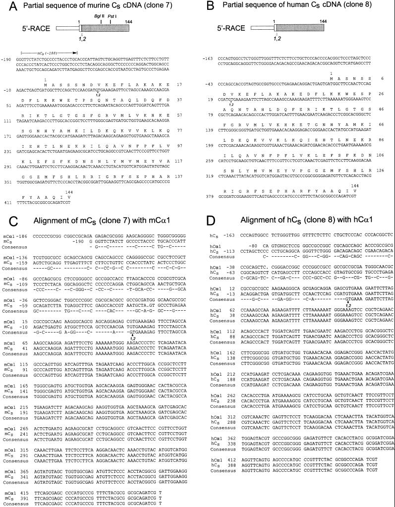
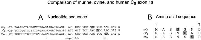
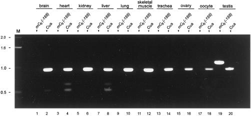
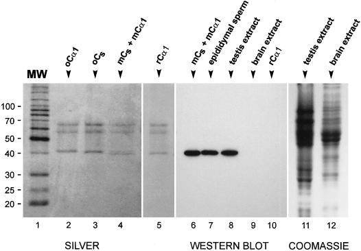
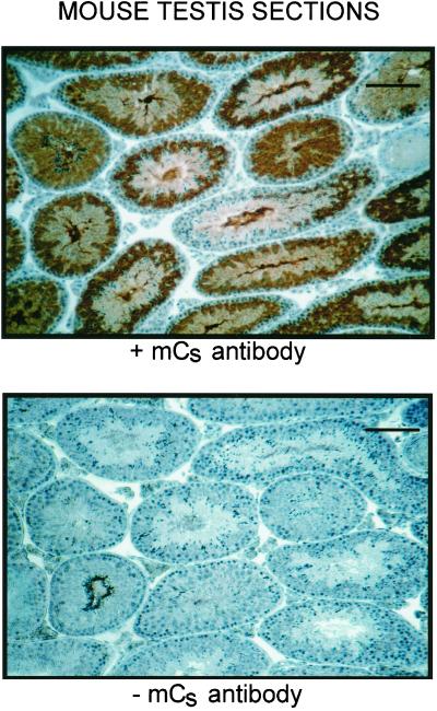
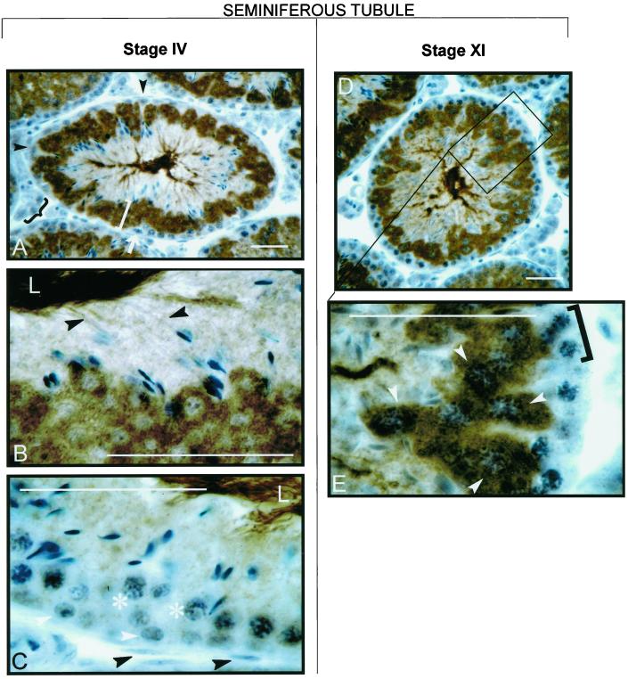
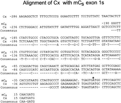
cAMP-dependent protein kinase has a central role in the control of mammalian sperm capacitation and motility. Previous protein biochemical studies indicated that the only cAMP-dependent protein kinase catalytic subunit (C) in ovine sperm is an unusual isoform, termed C(s), whose amino terminus differs from those of published C isoforms of other species. Isolation and sequencing of cDNA clones encoding ovine C(s) and Calpha1 (the predominant somatic isoform) now reveal that C(s) is the product of an alternative transcript of the Calpha gene. C(s) cDNA clones from murine and human testes also were isolated and sequenced, indicating that C(s) is of ancient origin and widespread in mammals. In the mouse, C(s) transcripts were detected only in testis and not in any other tissue examined, including ciliated tissues and ovaries. Finally, immunohistochemistry of the testis shows that C(s) first appears in pachytene spermatocytes. This is the first demonstration of a cell type-specific expression for any C isoform. The conservation of C(s) throughout mammalian evolution suggests that the unique structure of C(s) is important in the subunit's localization or function within the sperm.


Figures

Figure 1
Figure 1
Cloning and sequences of ovine Cα1 and Cs cDNAs. The initiating methionine is designated as amino acid residue 1, and the first base of the initiation codon ATG is designated as nucleotide 1. (A) Bars represent the five cDNA clones used to obtain the composite cDNAs and nucleotide sequences of ovine Cα1 and Cs. The ORFs are depicted as the wider portions of the bars. Sequences common to both Cα1 and Cs are gray, sequences specific for Cα1 are black, and sequences specific for Cs are white. For orientation, selected presumptive exon junctions based on the murine Cα genomic sequence (Chrivia et al., 1988) are marked below the bars (arrowheads). (B) Bars represent the composite cDNAs of ovine testis Cα1 and Cs. The numbers on top of the bars indicate the positions of amino acid residues encoded at the start (1) and ends (351 and 343) of the ORFs and the ends (15 and 7) of exon 1 of ovine Cα1 and Cs, respectively. Shading is as in A. (C) Partial nucleotide and predicted amino acid sequences of ovine Cα1 exon 1a and ovine Cs exon 1s. The positions of the forward primers a and oCs(−11) also are shown. (D) The nucleotide sequence of exons 2–10, which are identical for ovine Cα1 and Cs cDNAs, and their predicted amino acid sequence. The amino acid and nucleotide positions indicated (right and left margins, respectively) are those for Cs. Sequence data for ovine Cα1 and ovine Cs have been deposited in GenBank/EMBL/DDBJ under accession numbers AF238979 and AF238980, respectively.
Figure 2
Figure 2
Detection of Cα1 and Cs mRNA in various species. RT-PCR of total RNA from ovine and murine testes and PCR of cDNA from human testis. The forward primers used to generate the PCR products are shown at the top of the lanes: oCs(−11) to amplify Cs and a to amplify Cα1. The reverse primer in all cases was eR. The PCR products were subjected to electrophoresis in an 0.8% agarose gel and stained with ethidium bromide. Cα1 and Cs products were obtained from all three species. Controls in which the RT was omitted yielded no bands. Lane M, DNA molecular mass markers (in kilobases).
Figure 3
Figure 3
Partial nucleotide and amino acid sequences of murine Cs and human Cs cDNA. (A and B) Murine Cs cDNA (clone 7) and human Cs cDNA (clone 8) were obtained by 5′-RACE with the use of murine and human testis cDNAs as template. The shading and numbering are as in Figure 1. The BglII and PstI sites that are present in clone 7 but not in clone 8 are indicated. The murine and human Cs sequences are available from GenBank/EMBL/DDBJ under the accession numbers AF239743 and AF239744, respectively. (C and D) The cDNA sequences of clones 7 (mCs) and 8 (hCs) are compared with the corresponding regions of the murine Cα1 (mCα1) (Uhler et al., 1986a) and human Cα1 (hCα1) (Maldonado and Hanks, 1988) sequences. Dashes indicate nonconsensus nucleotides.
Figure 4
Figure 4
Comparison of exon 1s–encoded regions of ovine, murine, and human Cs. Partial cDNA nucleotide (A) and predicted amino acid (B) sequences of the murine (mCs), ovine (oCs), and human (hCs) versions of Cs exon 1s are aligned. The nonconsensus bases of the ORFs are highlighted, as are the amino acid residues that will result from these substitutions. The position of the primer oCs(−11) also is shown.
Figure 5
Figure 5
Detection of Cα1 and Cs mRNA in murine tissues. RT-PCR of total RNA from various murine tissues. The forward primers used to generate the PCR products are shown at the top of the lanes: mCs(−188) to amplify Cs and a to amplify Cα1. The reverse primer in all cases was eR. These primer sets are predicted to yield PCR products of 949 bases for murine Cα1 and 1119 bases for murine Cs. The PCR products were subjected to electrophoresis in an 0.8% agarose gel and stained with ethidium bromide. Transcripts encoding the Cα1 isoform are present in all the tissues analyzed, whereas Cs transcripts are detected only in the testis. Lane M, DNA molecular mass markers (in kilobases).
Figure 6
Figure 6
Specificity of the anti-mCs antibody. (Left) Silver-stained SDS-polyacrylamide gels of purified ovine Cα1 (oCα1), purified ovine Cs (oCs), a mixture of murine Cs and Cα1 (mCs + mCα1) isolated from murine testis, and mouse recombinant Cα1 (rCα1). Molecular mass markers (MW) are in kilodaltons. As reported previously (San Agustin et al., 1998), ovine Cs migrates slightly faster than ovine Cα1. The partially purified murine Cα1 and Cs, which are resolved as two bands at ∼40 kDa, appear to migrate slightly faster than their ovine homologues. The bands in the 60- to 70-kDa range are human keratin contaminants (San Agustin et al., 1998). (Center) Western blot probed with an affinity-purified antibody generated against an acetylated peptide corresponding to the unique amino terminus of murine Cs. The antibody reacts with a single protein in the mixture of murine Cs and Cα1 (mCs + mCα1), in murine epididymal sperm (1 × 10⁶ sperm), and in murine testis extract (50 μg of total protein) but does not react with any band in murine brain extract (30 μg of total protein) or with recombinant Cα1 (37 ng). (Right) SDS-polyacrylamide gel of murine testis extract (50 μg) and murine brain extract (30 μg) stained with Coomassie blue as loading control for lanes 8 and 9 of the Western blot.
Figure 7
Figure 7
Immunohistochemical staining of murine testis sections with the use of the anti-mouse Cs antibody. Cells stained brown are positive for Cs. Only germ cells at later stages of spermatogenesis stain with the antibody (top); because the cell associations seen in cross-sections of the seminiferous tubules vary depending on their stage in the spermatogenic cycle, the different tubules display different staining patterns. No staining is detected in the absence of the primary antibody (bottom). Bars, 100 μm.
Figure 8
Figure 8
Higher magnification of testis sections stained with anti-mouse Cs antibody. Bars, 20 μm. Tubules shown correspond to stages IV and XI of the seminiferous epithelium cycle according to the system of Leblond and Clermont (Leblond et al., 1963; Clermont and Bustos-Obregon, 1968). In the stage IV tubule, staining is absent from interstitial cells (A, black brace), Sertoli cells (A, black arrowheads), peritubular cells (C, black arrowheads), spermatogonia (C, white arrowheads), and early pachytene spermatocytes (C, asterisks). A spermatogonium undergoing mitosis is also shown (A, white arrow). Round spermatids have intensely stained cytosol (A, white bracket). In the previous generation of elongated spermatids that have moved farther toward the lumen (L), the cytoplasm now stains less intensely but the developing flagella (B, black arrowheads) are darkly stained. Darkly stained tails of mature sperm are visible in the lumens (L) of the stage IV tubules (B and C). In the stage XI tubule, staining of the cytosol of the spermatids occupying the inner portion of the tubule diminishes as they elongate (D). Staining is absent from zygotene spermatocytes (E, black bracket) but is prominent in the cytoplasm of late pachytene spermatocytes (E, white arrowheads).
Figure 9
Figure 9
Comparison of the 5′ sequence of murine Cx pseudogene with that of exon 1s of murine Cs. The Cx nucleotide sequence is nearly identical to that extending from Cs nucleotide −20 downstream to the end of Cs exon 1s. An asterisk indicates the translation start site of Cs. Dashes indicate nonconsensus nucleotides.

References

    1. Aitken RJ, Harkiss D, Knox W, Paterson M, Irvine DS. A novel signal transduction cascade in capacitating human spermatozoa characterized by a redox-regulated, cAMP-mediated induction of tyrosine phosphorylation. J Cell Sci. 1998;111:645–656. - PubMed
    1. Amat JA, Fields KL, Schubart UK. Stage-specific expression of phosphoprotein p19 during spermatogenesis in the rat. Mol Reprod Dev. 1990;26:383–390. - PubMed
    1. Ausubel FM, Brent R, Kingston RE, Moore DD, Seidman JG, Smith JA, Struhl K. Protocols in Molecular Biology. Vol. 1. New York: John Wiley & Sons; 1989.
    1. Beebe SJ, Øyen O, Sandberg M, Frøysa A, Hansson V, Jahnsen T. Molecular cloning of a tissue-specific protein kinase (Cγ) from human testis: representing a third isoform for the catalytic subunit of cAMP-dependent protein kinase. Mol Endocrinol. 1990;4:465–475. - PubMed
    1. Brokaw CJ. Regulation of sperm flagellar motility by calcium and cAMP-dependent phosphorylation. J Cell Biochem. 1987;35:175–184. - PubMed

Publication types

MeSH terms