. 2000 Sep;11(9):3123-35.
doi: 10.1091/mbc.11.9.3123.

Evidence for functional differentiation among Drosophila septins in cytokinesis and cellularization

Affiliations
Free PMC article

Evidence for functional differentiation among Drosophila septins in cytokinesis and cellularization

J C Adam et al. Mol Biol Cell. 2000 Sep.
Free PMC article

Abstract

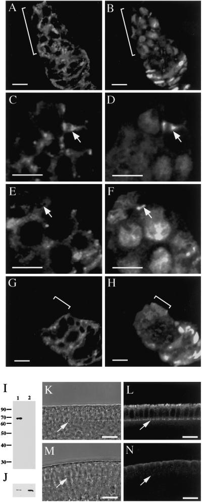
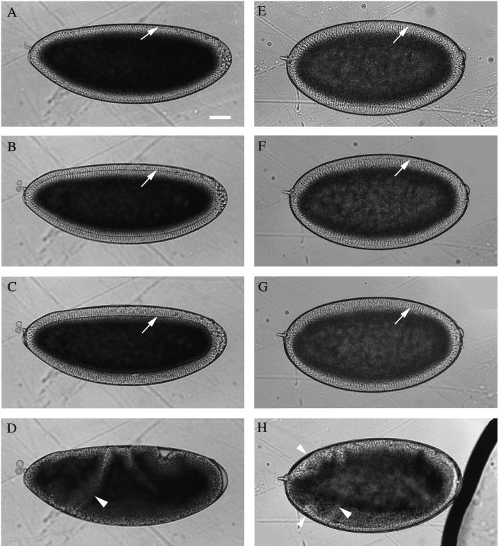
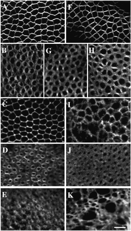
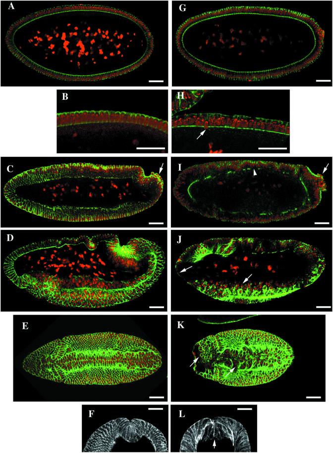
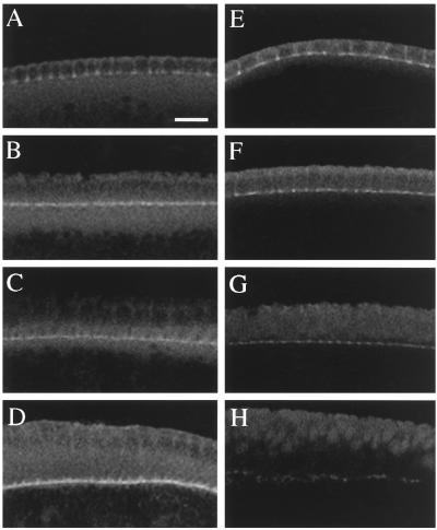
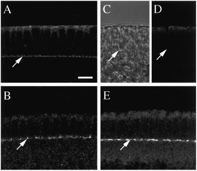
The septins are a conserved family of proteins that are involved in cytokinesis and other aspects of cell-surface organization. In Drosophila melanogaster, null mutations in the pnut septin gene are recessive lethal, but homozygous pnut mutants complete embryogenesis and survive until the pupal stage. Because the completion of cellularization and other aspects of early development seemed likely to be due to maternally contributed Pnut product, we attempted to generate embryos lacking the maternal contribution in order to explore the roles of Pnut in these processes. We used two methods, the production of germline clones homozygous for a pnut mutation and the rescue of pnut homozygous mutant flies by a pnut(+) transgene under control of the hsp70 promoter. Remarkably, the pnut germline-clone females produced eggs, indicating that stem-cell and cystoblast divisions in the female germline do not require Pnut. Moreover, the Pnut-deficient embryos obtained by either method completed early syncytial development and began cellularization of the embryo normally. However, during the later stages of cellularization, the organization of the actin cytoskeleton at the leading edge of the invaginating furrows became progressively more abnormal, and the embryos displayed widespread defects in cell and embryo morphology beginning at gastrulation. Examination of two other septins showed that Sep1 was not detectable at the cellularization front in the Pnut-deficient embryos, whereas Sep2 was still present in normal levels. Thus, it is possible that Sep2 (perhaps in conjunction with other septins such as Sep4 and Sep5) fulfills an essential septin role during the organization and initial ingression of the cellularization furrow even in the absence of Pnut and Sep1. Together, the results suggest that some cell-division events in Drosophila do not require septin function, that there is functional differentiation among the Drosophila septins, or both.


Figures

Figure 1
Figure 1
Presence of Pnut in wild-type stem cells and absence of Pnut in pnut germline clones. (A–H) Immunofluorescence micrographs of wild-type (A–F) and pnut germline-clone (G and H) ovarioles, showing germaria that were double-stained with the 4C9 anti-Pnut antibody (A, C, E, and G) and with antianillin (B, D, F, and H) antibody. (A and B) Presence of Pnut in cells throughout the region of germline proliferation (indicated by brackets) in wild-type germaria. (C and D) Presence of Pnut in stem-cell cleavage furrows (arrows). (E and F) Absence of Pnut from some stem-cell ring canals (arrows). In F, note the presence of anillin in the nuclei, showing that the cells have progressed into interphase (Field and Alberts, 1995; de Cuevas and Spradling, 1998). (G and H) Absence of Pnut from the region of germline proliferation (brackets) in germline-clone germaria. The Pnut-expressing cells in G are the somatic follicle cells that surround the germline cells after germline proliferation is complete. (I–N) Absence of Pnut from germline-clone embryos. Embryos were collected from the wild-type stock and from germline-clone females that had been allowed to mate with their siblings. (I and J) Western blots of extracts of 0–3-h embryos of wild-type (lane 1) and pnut germline clones (lane 2) stained with the KEKK anti-Pnut antibody (I) or with anti-Bicaudal D antibody as a loading control (J). (K–N) Transmitted-light (K and M) and immunofluorescence (L and N) micrographs of cellularizing wild-type (K and L) and germline-clone (M and N) embryos stained with the KEKK antibody. Arrows indicate the position of the cellularization front. A weak apical background staining is seen in germline-clone embryos with this antibody. The 4C9 anti-Pnut monoclonal antibody also showed no Pnut staining at the cellularization front but had a different background pattern (our unpublished observations). Bars, 5 μm (A–H), 10 μm (K–N).
Figure 2
Figure 2
Abnormal cuticle phenotype of pnut germline-clone embryos. In A–C, the anterior of the embryo is up. (A) Wild-type embryo. (B and C) Representative embryos from a mating of germline-clone females to their siblings. Bar, 50 μm.
Figure 3
Figure 3
Time-lapse observations on living embryos during cellularization and early gastrulation. (A–D) A wild-type embryo. (E–H) An embryo from the mating of HS-pnut+ flies. Anterior is to the left and dorsal is up. The dark area filling most of the interior of each embryo is the yolk-containing cytoplasm; the transparent layer contains the cortical nuclei and yolk-free cytoplasm; the cellularization front (arrows) is visible as a thin line running parallel to the surface. (A and E) Early cellularization; (B and F) slow phase; (C and G) fast phase; (D and H) early gastrulation. Large arrowheads, cephalic furrows; small arrowheads, an ectopic fold (see text). Bar, 50 μm.
Figure 4
Figure 4
Apparently normal F-actin organization during the slow phase of cellularization and abnormal F-actin organization during the fast phase of cellularization and early gastrulation in pnut germline-clone embryos. Wild-type (A–E) and germline-clone (F–K) embryos were stained with BODIPY FL-phallacidin and viewed at the level of the cellularization front (A, B, D–H, J, and K) or apical to the front (C and I). (A and F) Slow phase; (B, C, and G–I) early fast phase; (D and J) late fast phase; (E and K) early gastrulation. (G and J) Representatives of the majority class (∼90%) of pnut germline-clone embryos; (H and I) representatives of the minority class (∼10%) of pnut germline-clone embryos. Bar, 5 μm.
Figure 5
Figure 5
Largely normal cellularization and grossly defective gastrulation in pnut germline-clone embryos. Wild-type (A–F) and pnut germline-clone (G–L) embryos were stained with BODIPY FL-phallacidin (green) and propidium iodide (red) (A–E and G–K) or with BODIPY FL-phallacidin alone (F and L). (A–D and G–J) Sagittal optical sections; anterior is to the left and dorsal is up. (A, B, G, and H) Embryos in the fast phase of cellularization. Arrow, a nucleus positioned inappropriately with respect to the cortex. (C and I) Embryos at gastrulation onset. Arrows, pole cells; arrowhead, a disruption in the cytoskeleton at the cell bases. (D and J) Embryos during early gastrulation. Arrows, examples of nuclei not surrounded by an actin cytoskeleton at the anterior end and in the ventral furrow. (E and K) Surface sections at the time of ventral-furrow formation: anterior is to the left, ventral face is shown. Arrows, nuclei not surrounded by an actin cytoskeleton. (F and L) Cross sections showing the ventral furrow. Arrow, base of a ventral-furrow cell. Bars, 50 μm.
Figure 6
Figure 6
Progressive loss of anillin localization from the cellularization front in pnut germline clones. Wild-type embryos (A–D) and pnut germline-clone embryos (E–H) were stained with antianillin antibody. (A and E) Slow phase of cellularization; (B and F) early fast phase; (C and G) late fast phase; (D and H) early gastrulation. Bar, 10 μm.
Figure 7
Figure 7
Localization of Sep2, but not of Sep1, to the cellularization front in pnut germline-clone embryos. Wild-type (A and B) and pnut germline-clone (CE) embryos were stained with anti-Sep1 (A and D) or anti-Sep2 (B and E) antibodies. (C) Transmitted-light image of the region shown in D. Arrows indicate the cellularization fronts. Bar, 10 μm.

References

    1. Beites CL, Xie H, Bowser R, Trimble WS. The septin CDCrel-1 binds syntaxin and inhibits exocytosis. Nat Neurosci. 1999;2:434–439. - PubMed
    1. Bi E, Maddox P, Lew DJ, Salmon ED, McMillan JN, Yeh E, Pringle JR. Involvement of an actomyosin contractile ring in Saccharomyces cerevisiae cytokinesis. J Cell Biol. 1998;142:1301–1312. - PMC - PubMed
    1. Caltagarone J, Rhodes J, Honer WG, Bowser R. Localization of a novel septin protein, hCDCrel-1, in neurons of human brain. NeuroReport. 1998;9:2907–2912. - PubMed
    1. Carroll CW, Altman R, Schieltz D, Yates JR, III, Kellogg D. The septins are required for the mitosis-specific activation of the Gin4 kinase. J Cell Biol. 1998;143:709–717. - PMC - PubMed
    1. Chant J, Mischke M, Mitchell E, Herskowitz I, Pringle JR. Role of Bud3p in producing the axial budding pattern of yeast. J Cell Biol. 1995;129:767–778. - PMC - PubMed

Publication types

MeSH terms

LinkOut - more resources