. 2000 Dec 1;275(48):37565-71.
doi: 10.1074/jbc.M005211200.

Structure and activity of ClpB from Escherichia coli. Role of the amino-and -carboxyl-terminal domains

Affiliations

Structure and activity of ClpB from Escherichia coli. Role of the amino-and -carboxyl-terminal domains

M E Barnett et al. J Biol Chem. .

Abstract

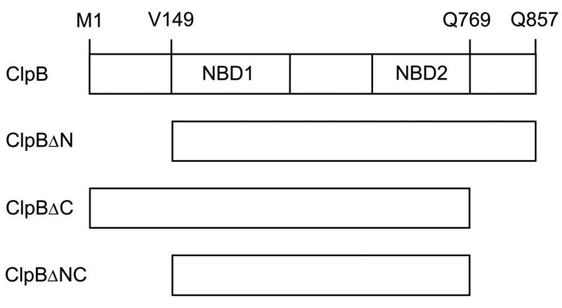
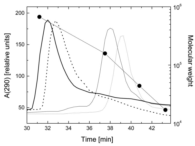
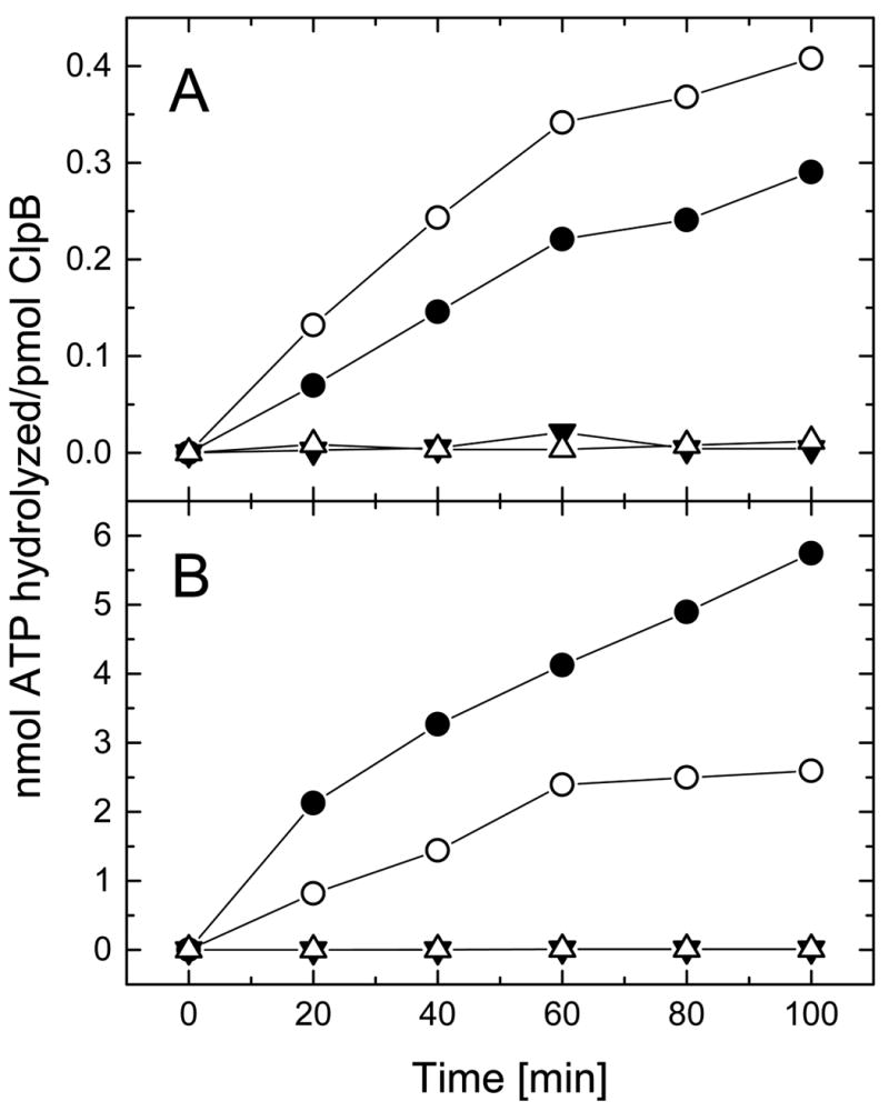
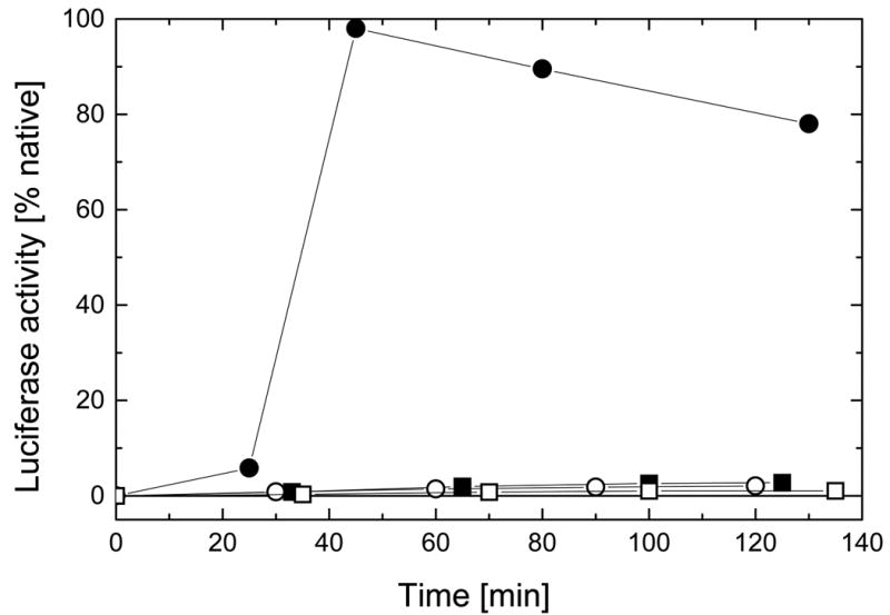
ClpB is a member of a protein-disaggregating multi-chaperone system in Escherichia coli. The mechanism of protein-folding reactions mediated by ClpB is currently unknown, and the functional role of different sequence regions in ClpB is under discussion. We have expressed and purified the full-length ClpB and three truncated variants with the N-terminal, C-terminal, and a double N- and C-terminal deletion. We studied the protein concentration-dependent and ATP-induced oligomerization of ClpB, casein-induced activation of ClpB ATPase, and ClpB-assisted reactivation of denatured firefly luciferase. We found that both the N- and C-terminal truncation of ClpB strongly inhibited its chaperone activity. The reasons for such inhibition were different, however, for the N- and C-terminal truncation. Deletion of the C-terminal domain inhibited the self-association of ClpB, which led to decreased affinity for ATP and to decreased ATPase and chaperone activity of the C-terminally truncated variants. In contrast, deletion of the N-terminal domain did not inhibit the self-association of ClpB and its basal ATPase activity but decreased the ability of casein to activate ClpB ATPase. These results indicate that the N-terminal region of ClpB may contain a functionally significant protein-binding site, whereas the main role of the C-terminal region is to support oligomerization of ClpB.


Figures

Figure 1
Figure 1. Diagram of the ClpB variants produced in this study
Shown is the full-length ClpB with its two nucleotide-binding domains (NBD1, NBD2) and three truncated forms of ClpB. In ClpBΔN, the amino acids 1 – 148 (the N-terminal domain) have been deleted. In ClpBΔC, the amino acids 770 – 857 (the C-terminal domain) have been deleted. In ClpBΔNC, both the N- and C-terminal domains have been deleted. In ClpBΔN and ClpBΔNC, the first valine residue (corresponding to V149 in the full-length ClpB) has been replaced with methionine.
Figure 2
Figure 2. Secondary structure and thermal stability of ClpB and its truncated variants
A, Far-UV circular dichroism spectra expressed as mean molar residue ellipticity (θ) of the full-length ClpB (thick solid line), ClpBΔN (thick dashed line), ClpBΔC (thin solid line), and ClpBΔNC (thin dashed line) at 37 °C. B, Temperature-induced changes in the circular dichroism signal at 222 nm for the full-length ClpB (solid circles), ClpBΔN (crosses), and ClpBΔC (open circles).
Figure 3
Figure 3. Sedimentation velocity of ClpB and its truncated variants at low and high protein concentration
Ultracentrifugation was performed at 50,000 rpm (A, C, E) or 40,000 rpm (B, D, F) at 20 °C. The protein concentration was 0.2 mg/ml (A, C, E) or 4.0 mg/ml (B, D, F) in 50 mM Hepes-KOH, pH 7.5, 0.2 M KCl, 20 mM MgCl2, 1 mM EDTA, 2 mM β-mercaptoethanol. Shown are the series of radial scans of the centrifuge cell at 5 min intervals (A, C, E) or 3.5 min intervals (B, D, F) for the full-length ClpB (A, B), ClpBΔN (C, D), and ClpBΔC (E, F). Protein concentration profiles were measured using absorption at 236 nm (A, C, E) or 290 nm (B, D, F). The direction of sedimentation is to the right and the positions of solution menisci are indicated by the light scattering peaks at ~6.4-6.5 cm.
Figure 4
Figure 4. Apparent sedimentation coefficient distributions for ClpB and its truncated variants
Shown are the results of the time-derivative analysis (19) of four late protein concentration profiles (see Fig. 3) for the full-length ClpB (A), ClpBΔN (B), and ClpBΔC (C) at 0.2 mg/ml (solid lines) and 4.0 mg/ml (dotted lines). The lines show apparent distribution functions g(s*) vs. the sedimentation coefficient s* in Svedberg units (S).
Figure 5
Figure 5. Gel filtration analysis of ClpB and its truncated variants in the presence of ATP
Aliquots of ClpB (thick solid line), ClpBΔN (thick dotted line), ClpBΔC (thin solid line), and ClpBΔNC (thin dotted line) (10 μl of ~2 mg/ml) were injected onto a Superose 6 column. Protein elution profiles were obtained with a 0.06 ml/min flow rate (running buffer: 50 mM Tris/HCl, pH 7.5, 0.2 M KCl, 20 mM MgCl2, 1 mM EDTA, 1 mM DTT, 2 mM ATP) by monitoring absorption at 290 nm. Circles correspond to elution times of thyroglobulin (Mr 670,000), gamma globulin (Mr 158,000), ovalbumin (Mr 44,000), and myoglobin (Mr 17,000).
Figure 6
Figure 6. ATP hydrolysis by ClpB and its truncated variants
ATPase activity has been measured by incubating the full-length ClpB (filled circles), ClpBΔN (open circles), ClpBΔC (filled triangles), or ClpBΔNC (open triangles) for the indicated time at 37 °C in the assay buffer (see Experimental Procedures). In panel A, 2.5 μg protein was used for each reaction. In panel B, the assay buffer contained 0.1 mg/ml κ-casein and 0.25 μg ClpB or ClpBΔN, or 2.5 μg ClpBΔC or ClpBΔNC. Notice the difference between vertical scales in panels A and B.
Figure 7
Figure 7. Calorimetric titrations of ClpB with ATP
A, calorimeter output for series of 10-μl injections of buffer B with 5.8 mM ATP into buffer B (upper trace), into 2 mg/ml full-length ClpB (lower trace), or 2 mg/ml ClpBΔC (middle trace) at 30 °C. Positive peaks correspond to endothermic effects and negative – to exothermic effects. The data traces were offset for clarity. B, Cumulative heat effect of titrating ATP into ClpBΔC (from the middle trace in panel A).
Figure 8
Figure 8. Reactivation of luciferase by ClpB and its truncated variants in the presence of DnaK, DnaJ, and GrpE
Unfolded luciferase was diluted at room temperature into the refolding buffer (see Experimental Procedures) containing DnaK, DnaJ, GrpE and the full-length ClpB (filled circles), ClpBΔN (open circles), ClpBΔC (filled squares), or ClpBΔNC (open squares). Luciferase activity was measured in aliquots withdrawn after the times indicated. Protein concentrations in the refolding solutions were: 25 nM luciferase, 0.3 μM (hexamer) ClpB or its variants, 1.0 μM DnaK, 1.1 μM DnaJ, 1.2 μM GrpE.

References

    1. Wickner S, Maurizi MR, Gottesman S. Science. 1999;286:1888–1893. - PubMed
    1. Schirmer EC, Glover JR, Singer MA, Lindquist S. Trends Biochem Sci. 1996;21:289–296. - PubMed
    1. Weber-Ban EU, Reid BG, Miranker AD, Horwich AL. Nature. 1999;401:90–93. - PubMed
    1. Zolkiewski M. J Biol Chem. 1999;274:28083–28086. - PubMed
    1. Goloubinoff P, Mogk A, Ben Zvi AP, Tomoyasu T, Bukau B. Proc Natl Acad Sci U S A. 1999;96:13732–13737. - PMC - PubMed

Publication types

MeSH terms

LinkOut - more resources