. 2000 Oct;20(19):7068-79.
doi: 10.1128/MCB.20.19.7068-7079.2000.

Induction of postmitotic neuroretina cell proliferation by distinct Ras downstream signaling pathways

Affiliations

Induction of postmitotic neuroretina cell proliferation by distinct Ras downstream signaling pathways

C Peyssonnaux et al. Mol Cell Biol. 2000 Oct.

Abstract

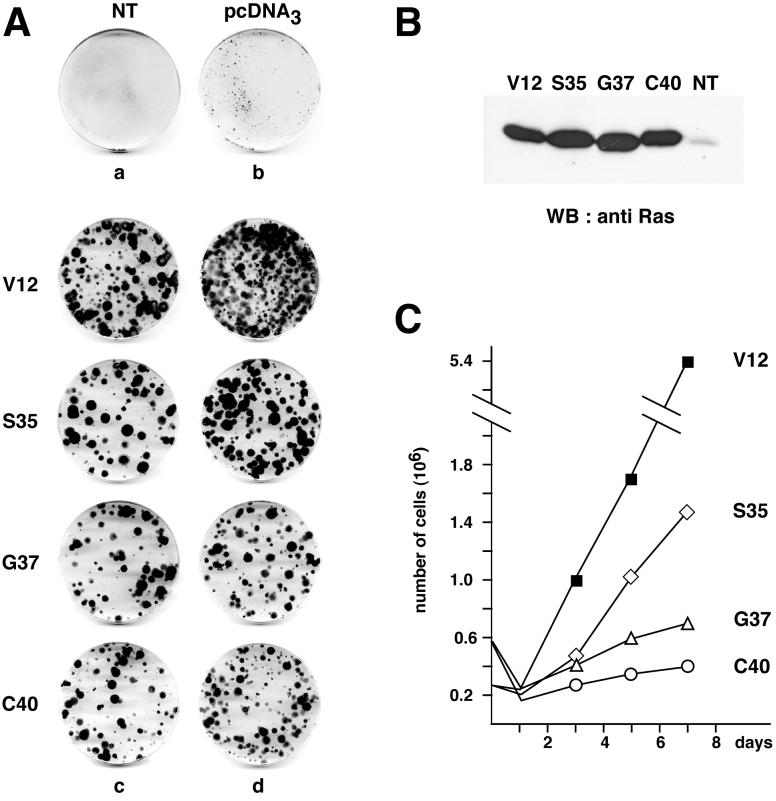
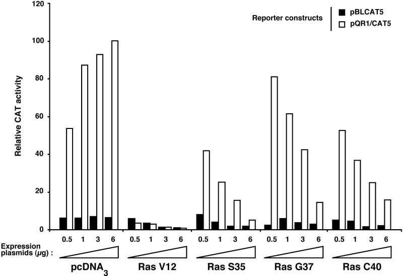
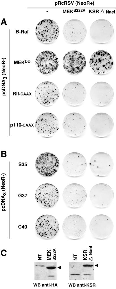
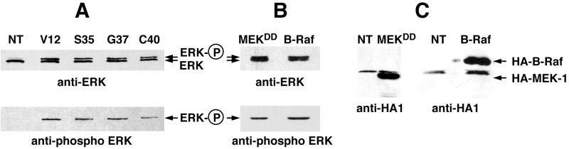
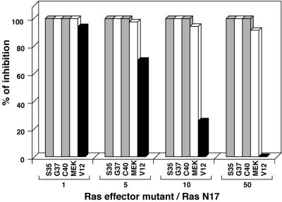
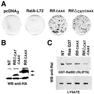
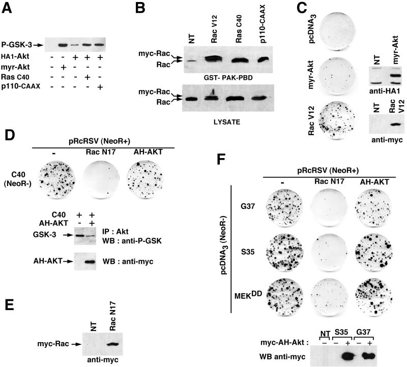
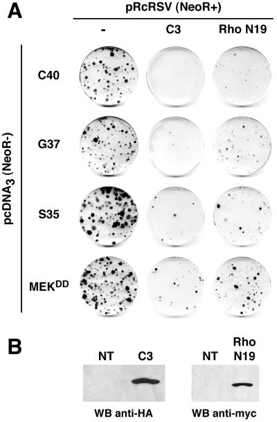
Ras-induced cell transformation is mediated through distinct downstream signaling pathways, including Raf, Ral-GEFs-, and phosphatidylinositol 3-kinase (PI 3-kinase)-dependent pathways. In some cell types, strong activation of the Ras-Raf-MEK-extracellular signal-regulated kinase (ERK) cascade leads to cell cycle arrest rather than cell division. We previously reported that constitutive activation of this pathway induces sustained proliferation of primary cultures of postmitotic chicken neuroretina (NR) cells. We used this model system to investigate the respective contributions of Ras downstream signaling pathways in Ras-induced cell proliferation. Three RasV12 mutants (S35, G37, and C40) which differ by their ability to bind to Ras effectors (Raf, Ral-GEFs, and the p110 subunit of PI 3-kinase, respectively) were able to induce sustained NR cell proliferation, although none of these mutants was reported to transform NIH 3T3 cells. Furthermore, they all repressed the promoter of QR1, a neuroretina growth arrest-specific gene. Overexpression of B-Raf or activated versions of Ras effectors Rlf-CAAX and p110-CAAX also induced NR cell division. The mitogenic effect of the RasC40-PI 3-kinase pathway appears to involve Rac and RhoA GTPases but not the antiapoptotic Akt (protein kinase B) signaling. Division induced by RasG37-Rlf appears to be independent of Ral GTPase activation and presumably requires an unidentified mechanism. Activation of either Ras downstream pathway resulted in ERK activation, and coexpression of a dominant negative MEK mutant or mKsr-1 kinase domain strongly inhibited proliferation induced by the three Ras mutants or by their effectors. Similar effects were observed with dominant negative mutants of Rac and Rho. Thus, both the Raf-MEK-ERK and Rac-Rho pathways are absolutely required for Ras-induced NR cell division. Activation of these two pathways by the three distinct Ras downstream effectors possibly relies on an autocrine or paracrine loop, implicating endogenous Ras, since the mitogenic effect of each Ras effector mutant was inhibited by RasN17.


Figures

FIG. 1
FIG. 1
The three Ras effector mutants induce sustained NR cell proliferation. (A) Primary cultures of chicken embryonic NR cells were transfected with 5 μg (c) or 10 μg (d) of pcDNA3-derived constructs encoding the RasV12 single mutant (V12) or Ras double mutants containing a second mutation (S35, G37, or C40) in addition to the V12 mutation, as indicated. Controls were nontransfected NR cell cultures (NT) (a) and NR cell cultures transfected with 10 μg of the empty vector pcDNA3 (b). After selection for G418-resistant cells, foci of proliferating NR cells were stained with crystal violet. The data presented are representative of eight independent experiments. (B) Western blot analysis of Ras protein expression in NR cell cultures induced to proliferate by the different Ras mutants. Equal amounts of protein extracts from cultures obtained as described for panel A were resolved by SDS-polyacrylamide gel electrophoresis (PAGE), transferred to Immobilon-P membranes, and probed with an anti-Ras monoclonal antibody. Nontransfected NR cells maintained in culture in the absence of G418 were used as a control (NT). (C) Growth curves of NR cell cultures induced to proliferate by the different Ras mutants. Proliferating G418-resistant cells obtained as in panel A were pooled, seeded at a density of 2.5 × 10⁵ cells (for V12 and S35 Ras mutants) or 5 × 10⁵ cells (for G37 and C40 Ras mutants) in 60-mm dishes, and counted at the indicated intervals.
FIG. 2
FIG. 2
The mitogenic capacity of Ras effector mutants correlates with their ability to repress the quiescence-inducible promoter of QR1. tsNY68-infected QNR cells were cotransfected with the QR1 reporter construct (CAT5/QR1) or the CAT5-negative control and increasing amounts of either pcDNA3 or pcDNA3-derived constructs expressing Ras mutants described for Fig. 1. CAT activity was determined after a 48-h incubation at the nonpermissive temperature (41°C), as described in Materials and Methods. Relative activities are given with respect to the effect of 6 μg of pcDNA3 on CAT5/QR1 (100%). The results of a representative experiment are shown; similar results were reproducibly obtained in three independent cotransfections.
FIG. 3
FIG. 3
The MEK-ERK pathway is required for NR cell proliferation induced by Ras effectors and Ras double mutants. (A) NR cells were cotransfected with 10 μg of pcDNA3(NeoR−)-derived constructs expressing activated MEK-1 (MEKDD) or the different Ras direct effectors: B-Raf and activated versions of Rlf (Rlf-CAAX) and PI 3-kinase (p110-CAAX), and 10 μg of pRcRSV vector or pRcRSV-derived constructs expressing an HA1-tagged dominant negative mutant of MEK-1 (MEKS222A) or the kinase domain of mKsr-1 (KSRΔNaeI). The G418 resistance is provided by the pRcRSV-derived constructs (NeoR+), whereas the pcDNA3-derived constructs alone do not allow G418 selection (NeoR−). After selection for G418-resistant cells, foci of proliferating NR cells were stained with crystal violet. (B) Same as in panel A except that the Ras double mutants (S35, G37, and C40) were used instead of the Ras effectors. Data presented in panels A and B are representative of three independent experiments. (C) Expression of MEK and KSR mutants in NR cells. NR cells were transfected with pRcRSV/HA1-MEKS222A and pRcRSV/KSRΔNaeI constructs, and protein expression was analyzed 48 h later by Western blotting (WB), as described in Materials and Methods.
FIG. 4
FIG. 4
ERK activation in NR cell cultures induced to proliferate by the Ras double mutants. (A) Western blot analysis of ERK phosphorylation in NR cell cultures induced to proliferate by the different Ras mutants. Equal amounts of protein extracts from cultures obtained as described in Fig. 1A were resolved by SDS-PAGE, transferred to Immobilon-P membranes, and probed with an anti-ERK polyclonal antibody. The membranes were then stripped and reprobed with an anti-phospho-ERK polyclonal antibody. Nontransfected NR cells maintained in culture in the absence of G418 were used as a control (NT). (B) Same as in panel A for the pcDNA3(NeoR−)-derived constructs encoding HA1-tagged MEKDD and B-Raf used in the experiments shown in Fig. 3. (C) Expression of these constructs in NR cells was controlled by Western blotting using an anti-HA1 antibody.
FIG. 5
FIG. 5
Endogenous Ras activation is required for NR cell proliferation induced by Ras effector mutants and MEK. NR cells were cotransfected with pcDNA3(NeoR−)-derived constructs expressing the different Ras mutants (V12, S35, G37, or C40) or MEKDD and with either the pRcRSV vector or the pRcRSV/RasN17 construct expressing a dominant negative mutant of Ras. The different ratios of pcDNA3- to pRcRSV-derived plasmid DNAs used are indicated (Ras effector mutant/RasN17). As in the experiment shown in Fig. 3, G418 resistance is provided by the pRcRSV-derived constructs. After selection for G418-resistant cells, foci of proliferating NR cells were stained with crystal violet and counted. Results are presented as the percent inhibition obtained from the number of foci in cultures cotransfected with the pRcRSV empty vector compared to that obtained in cultures transfected with pRcRSV/RasN17 for each Ras mutant or MEKDD. Data presented are representative of seven independent experiments.
FIG. 6
FIG. 6
NR cell proliferation induced by the G37-Rlf pathway is mediated by a Ral-independent mechanism. (A) Cultures of NR cells were transfected with 20 μg of pcDNA3 or pcDNA3-derived constructs encoding an activated RalA mutant (RalA-L72), an activated Rlf mutant (Rlf-CAAX), or an Rlf-CAAX mutant with a deletion in the scr-1 region of the catalytic domain responsible for the exchange factor activity on Ral (RlfΔCAT-CAAX), as indicated. After selection for G418-resistant cells, foci of proliferating NR cells were stained with crystal violet. Data presented are representative of five independent experiments. (B) Expression of HA1-tagged Rlf mutants in NR cells was controlled by Western blotting (WB) using an anti-HA1 antibody as indicated. (C) Endogenous Ral GTP loading in nontransfected NR cells and in NR cells induced to proliferate by the G37-Rlf pathway. Equal amounts of cell extracts from nontransfected NR cell cultures or from G418-resistant foci of NR cells induced to proliferate upon transfection with RasG37, Rlf-CAAX, and RlfΔCAT-CAAX were incubated with a GST-RalBD fusion protein containing the Ral binding domain of RLIP76, precoupled to glutathione beads, to recover GTP-bound Ral. The beads were washed four times, and collected Ral was identified by Western blotting analysis with a monoclonal anti-Ral antibody (top panel). The level of total Ral in whole lysates (50 μg of protein extracts) is also shown (bottom panel).
FIG. 7
FIG. 7
Ras-induced NR cell proliferation requires Rac but not Akt activation. (A) Activation of Akt by the C40–PI 3-kinase pathway in NR cells. NR cell cultures were cotransfected with HA1-tagged wild-type Akt and either RasC40 or constitutively activated PI 3-kinase (p110-CAAX), as indicated. NR cultures that were not transfected, transfected with HA1-tagged myristylated Akt (myr-Akt), or cotransfected with HA1-Akt and the empty pcDNA3 vector were used as controls. Akt proteins were immunoprecipitated with a monoclonal anti-HA1 antibody, and immune complexes were incubated with a GSK-3α/β peptide as a substrate. Phosphorylation of GSK-3 (P-GSK-3) was then analyzed by Western blotting using an anti-phospho-GSK-3 polyclonal antibody. (B) Rac GTP loading in NR cells induced to proliferate by the C40–PI 3-kinase pathway. Equal amounts of cell extracts from G418-resistant foci of NR cells induced to proliferate upon transfection with RasC40, p110-CAAX, and Myc-tagged RacV12 were incubated with the p21-binding domain of PAK-1 as a GST-PAK-PBD fusion protein coupled to agarose beads, to recover GTP-bound Rac. The beads were washed three times, and collected Rac was identified by Western blot analysis with an anti-Rac monoclonal antibody (top panel). The level of total Rac in the whole lysates (50 μg of protein extracts) is also shown (bottom panel). Nontransfected NR cells maintained in culture in the absence of G418 were used as a control (NT). (C) Cultures of NR cells were transfected with 20 μg of pcDNA3, pcDNA3/myr-Akt (HA1-tagged myristylated Akt), or pcDNA3/myc-RacV12 (encoding an activated mutant of Rac1), as indicated. After selection for G418-resistant cells, foci of proliferating NR cells were stained with crystal violet. Expression of these constructs in NR cells was analyzed by Western blotting using anti-HA1 and anti-Myc monoclonal antibodies in transient-transfection experiments. Data presented are representative of five independent experiments for RacV12 and two independent experiments for myr-Akt. (D) NR cells were cotransfected with 10 μg of pcDNA3(NeoR−)/RasC40 and 10 μg of pRcRSV vector or pRcRSV-derived constructs expressing a Myc-tagged dominant negative mutant of Rac1 (RacN17) or the Myc-tagged PH domain of Akt (PKB) (AH-AKT). The G418 resistance is provided by the pRcRSV-derived constructs (NeoR+), whereas the RasC40 construct alone do not allow G418 selection (NeoR−). After selection for G418-resistant cells, foci of proliferating NR cells were stained with crystal violet. Data presented are representative of four independent experiments. Inhibition of endogenous Akt kinase activity by the Akt dominant negative mutant was analyzed in G418-resistant foci of NR cells induced to proliferate upon cotransfection with both pcDNA3/RasC40 and pRcRSV/AH-Akt compared with those obtained in the absence of AH-Akt (empty pRcRSV vector), using the same experimental procedure as described for panel A, except that endogenous Akt was immunoprecipitated with an anti-Akt monoclonal antibody (IP). (E) Expression of the Myc-tagged dominant negative mutant of Rac1 (RacN17) in NR cells was analyzed by Western blotting using an anti-Myc monoclonal antibody in a transient-transfection experiment. (F) NR cells were cotransfected with 10 μg of pcDNA3(NeoR−)-derived constructs expressing the Ras double mutants (S35 and G37) or activated MEK-1 (MEKDD) and 10 μg of pRcRSV vector, pRcRSV/RacN17, or pRcRSV/AH-AKT, as described for panel D. After selection for G418-resistant cells, foci of proliferating NR cells were stained with crystal violet. Data presented are representative of four independent experiments. Expression of the Myc-AH-Akt protein was analyzed by Western blot analysis. Equal amounts of protein extracts from NR cell cultures were resolved on SDS-PAGE, transferred to Immobilon-P membranes, and probed with an anti-Myc monoclonal antibody. Nontransfected NR cells maintained in culture in the absence of G418 were used as a control (NT).
FIG. 8
FIG. 8
Inhibition of the Rho pathway prevents Ras-induced NR cell proliferation. NR cells were cotransfected with 10 μg of pcDNA3(NeoR−)-derived constructs expressing the different Ras double mutants (S35, G37, and C40) or activated MEK-1 (MEKDD) and 10 μg of pRcRSV vector or pRcRSV-derived constructs expressing a HA1-tagged C. botulinum C3 transferase (C3) or a Myc-tagged dominant negative mutant of RhoA (RhoN19), as indicated. The G418 resistance is provided by the pRcRSV-derived constructs (NeoR+), whereas the pcDNA3-derived constructs do not allow G418 selection (NeoR−). After selection for G418-resistant cells, foci of proliferating NR cells were stained with crystal violet. Data presented are representative of three independent experiments. (B) Expression of C3 transferase and RhoN19 proteins in NR cells was analyzed by Western blotting using anti-HA1 and anti-Myc monoclonal antibodies in transient-transfection experiments. NT, control.

Similar articles

Cited by

References

    1. Aktas H, Cai H, Cooper G M. Ras links growth factor signaling to the cell cycle machinery via regulation of cyclin D1 and the Cdk inhibitor p27KIP1. Mol Cell Biol. 1997;17:3850–3857. - PMC - PubMed
    1. Aoki M, Batista O, Bellacosa A, Tsichlis P, Vogt P K. The akt kinase: molecular determinants of oncogenicity. Proc Natl Acad Sci USA. 1998;95:14950–14955. - PMC - PubMed
    1. Béchade C, Calothy G, Pessac B, Martin P, Coll J, Denhez F, Saule S, Ghysdael J, Stehelin D. Induction of proliferation or transformation of neuroretina cells by the mil and myc viral oncogenes. Nature (London) 1985;316:559–562. - PubMed
    1. Benard V, Bohl B P, Bokoch G M. Characterization of Rac and Cdc42 activation in chemoattractant-stimulated human neutrophils using a novel assay for active GTPases. J Biol Chem. 1999;274:13198–13204. - PubMed
    1. Brunet A, Pages G, Pouyssegur J. Constitutively active mutants of MAP kinase kinase (MEK1) induce growth factor-relaxation and oncogenicity when expressed in fibroblasts. Oncogene. 1994;9:3379–3387. - PubMed

Publication types

MeSH terms