Comparative Study
. 2000 Oct;20(19):7183-91.
doi: 10.1128/MCB.20.19.7183-7191.2000.

Physical and functional interaction between the eukaryotic orthologs of prokaryotic translation initiation factors IF1 and IF2

Affiliations
Comparative Study

Physical and functional interaction between the eukaryotic orthologs of prokaryotic translation initiation factors IF1 and IF2

S K Choi et al. Mol Cell Biol. 2000 Oct.

Abstract

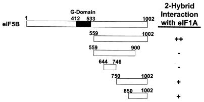
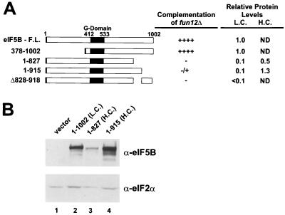
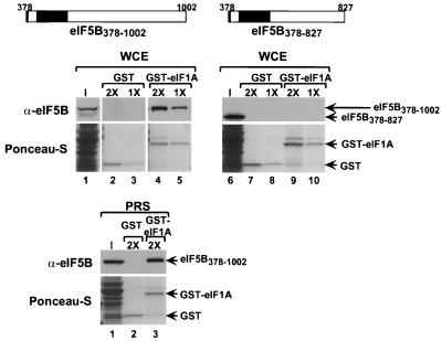
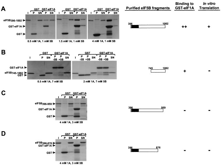
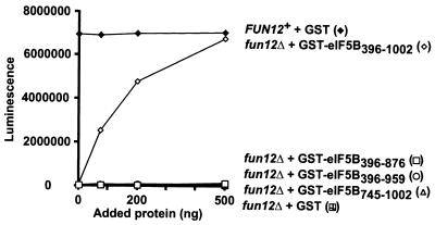
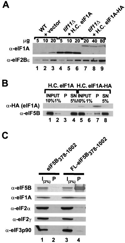
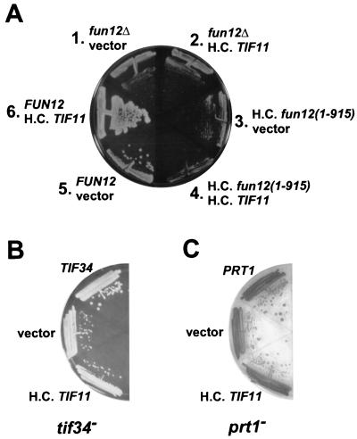
To initiate protein synthesis, a ribosome with bound initiator methionyl-tRNA must be assembled at the start codon of an mRNA. This process requires the coordinated activities of three translation initiation factors (IF) in prokaryotes and at least 12 translation initiation factors in eukaryotes (eIF). The factors eIF1A and eIF5B from eukaryotes show extensive amino acid sequence similarity to the factors IF1 and IF2 from prokaryotes. By a combination of two-hybrid, coimmunoprecipitation, and in vitro binding assays eIF1A and eIF5B were found to interact directly, and the eIF1A binding site was mapped to the C-terminal region of eIF5B. This portion of eIF5B was found to be critical for growth in vivo and for translation in vitro. Overexpression of eIF1A exacerbated the slow-growth phenotype of yeast strains expressing C-terminally truncated eIF5B. These findings indicate that the physical interaction between the evolutionarily conserved factors eIF1A and eIF5B plays an important role in translation initiation, perhaps to direct or stabilize the binding of methionyl-tRNA to the ribosomal P site.


Figures

FIG. 1
FIG. 1
eIF1A interacts with the C terminus of eIF5B in the yeast two-hybrid assay. The schematic at the top depicts full-length yeast eIF5B. The remaining schematics depict the segments of eIF5B tested in the two-hybrid assays, with the numbers referring to the amino acid positions at the termini of the eIF5B fragments. The DNA fragments encoding the indicated portions of yeast eIF5B and full-length yeast eIF1A were inserted in the yeast two-hybrid activation domain vector pGAD424 and the DNA binding domain vector pGBT9. Yeast strain Y187 bearing pGAD424 derivatives was mated with Y190 bearing pGBT9 derivatives, and diploids were isolated on synthetic complete (SC)-Trp-Leu medium. The strength of the protein-protein interactions was measured by stimulation of the HIS3 reporter present in the diploids as assayed by growth on SC-Trp-Leu-His medium containing 30 mM 3-aminotriazole after 9 days at 30°C (++, robust growth; +, weak growth, but above background levels; −, background growth equivalent to that empty-vector controls that lack eIF5B and eIF1A sequences). Equivalent results were obtained when the eIF5B fragments were fused to the GAL4 DNA binding domain or to the activation domain. G-domain, GTP-binding domain.
FIG. 2
FIG. 2
The C terminus of yeast eIF5B is required for activity in vivo. (A) Schematic of yeast eIF5B. Shown are the full-length (F.L.) form and various truncated forms or a form with or an internal deletion. The numbers refer to the amino acid positions in the proteins. Large black box, conserved GTP-binding domain; small black box (left end), N-terminal FLAG epitope tag. The various eIF5B proteins were expressed under the control of the native FUN12 promoter on low-copy-number (L.C.) or high-copy-number (H.C.) plasmids in fun12Δ strain J111 and tested for the ability to complement the slow-growth phenotype of this strain. Growth rates were assessed by streaking transformants for single colonies on minimal medium. ++++, wild-type growth with visible colonies in streak-outs after 2 days; −, fun12Δ growth with visible colonies in streak-outs after 5 days; ±, slightly larger-sized colonies in streak-outs than observed with fun12Δ strains after 5 days. The relative expression levels of the eIF5B constructs expressed from low- or high-copy-number plasmids are indicated (ND, not determined). (B) Immunoblot analysis of eIF5B expression. WCEs of transformants of strain J111 expressing the indicated eIF5B construct or vector alone were prepared and subjected to immunoblot analysis using polyclonal antiserum raised against a GST-eIF5B396–1002 fusion protein (shown) as well as monoclonal anti-FLAG antibodies. To control for protein loading, the lower half of the blot was probed with antiserum specific for yeast eIF2α. Immune complexes were visualized by ECL.
FIG. 3
FIG. 3
The C terminus of eIF5B is required for interaction with GST-eIF1A in a yeast WCE. (Top) Schematics of FLAG epitope-tagged eIF5B378–1002 and eIF5B378–827. Small black box (left end), FLAG epitope tag; large black box, GTP-binding domain. (Middle) The indicated GST (lanes 2, 3, 7, and 8) and GST-eIF1A (lanes 4, 5, 9, and 10) fusions attached to glutathione-Sepharose beads were incubated with WCEs from fun12Δ strain J111 expressing either N-terminally truncated eIF5B378–1002 (pC1043) (lanes 1 to 5) or N- and C-terminally truncated eIF5B378–827 (pC1058) (lanes 6 to 10) from low-copy-number plasmids. Both eIF5B proteins contain an N-terminal FLAG epitope tag. The 1× concentrations for GST and GST-eIF1A were 0.8 and 0.5 mM, respectively. The input (I) lanes represent 10% of the yeast extracts used for the binding assays. Following binding, the beads were pelleted and washed, and the bound proteins were analyzed by SDS-PAGE followed by electroblotting to nitrocellulose membranes. The GST fusion proteins were visualized by Ponceau-S staining (lower panels), and the eIF5B proteins were detected using anti-FLAG peptide antiserum and ECL (upper panels). (Bottom) The indicated GST (lane 2) and GST-eIF1A (lane 3) fusions attached to glutathione-Sepharose beads were incubated with PRSs from fun12Δ strain J111 expressing FLAG-tagged N-terminally truncated eIF5B378–1002 (pC1043). The concentrations of GST and GST-eIF1A proteins in the binding assays were 1.6 and 1.0 mM, respectively. The input lane represents 10% of the PRS used for the binding assays. The analysis of the binding reactions and visualization of the results were as described above.
FIG. 4
FIG. 4
eIF5B interacts directly with eIF1A. The indicated GST and GST-eIF1A fusion proteins expressed in E. coli were purified on glutathione-Sepharose beads and incubated with the following purified recombinant eIF5B fragments: eIF5B396–1002 (A), eIF5B745–1002 (B), eIF5B396–959 (C), and eIF5B396–876 (D). Following binding and washing, 10% of the supernatant fractions (SN) and 100% of the pellet fractions (P) were resolved by SDS–4 to 20% PAGE and visualized by Coomassie staining (left panels). The input (I) lanes contain 10% of the eIF5B fragments used in the binding assays. The concentrations of eIF5B and GST or GST-eIF1A proteins in the binding reaction mixtures are indicated below the results. Because breakdown products of the GST-eIF1A fusion protein comigrated with eIF5B745–1002, the pellet fractions of binding reaction mixtures lacking (P −5B) and containing (P +5B) eIF5B are presented (B). Schematic depictions of the eIF5B constructs and a qualitative summary of the results of these binding assays and of the in vitro translation assays shown in Fig. 5 are presented on the right.
FIG. 5
FIG. 5
The C terminus of eIF5B is required for function in an in vitro translation system. Translation extracts were prepared as described previously (11) from fun12Δ strain J133 carrying low-copy-number FUN12 plasmid pC479 (FUN12+) or empty vector pRS316 (fun12Δ). Extracts were incubated with 200 ng of luciferase mRNA and the indicated amounts of highly purified recombinant GST or GST-eIF5B fusions. Translational activity was determined, as described previously (11), by measuring luminescence after a 15-min incubation at 26°C. Results are representative of at least two independent experiments.
FIG. 6
FIG. 6
eIF5B and eIF1A interact in vivo. (A) Immunoblot analysis of eIF1A and eIF1A-HA expression. WCEs were prepared from the wild-type (WT) strain H1895 and derivatives of the isogenic tif11Δ strain H2809 containing the high-copy-number (H.C.) plasmid pDSO23 (TIF11 LEU2) encoding eIF1A or pDSO46 (TIF11-HA LEU2) encoding eIF1A-HA. The indicated amounts of WCEs were subjected to SDS-PAGE and analyzed by immunoblotting using polyclonal antisera raised against yeast eIF2Bε (GCD6) or eIF1A. Immune complexes were visualized by ECL. (B) Coimmunoprecipitation of eIF5B with epitope-tagged eIF1A. WCEs were prepared from the tif11Δ strains described for panel A, which express either eIF1A or HA epitope-tagged eIF1A (eIF1A-HA) from high-copy-number plasmids. Aliquots containing 800 μg of protein were incubated with monoclonal anti-HA antibodies (HA.11; Babco) prebound to protein A-Sepharose beads, and, after being washed, the bound proteins were analyzed by immunoblotting using anti-HA and anti-eIF5B antisera, as indicated. Input lanes contain 1 or 10% of the starting amount of WCE, the pellet lanes (P) containing 100% of the immunoprecipitated fraction, and the supernatant lanes (SN) contain 5% of the reaction mixtures following removal of the pellet. (C) Coimmunoprecipitation of eIF1A with epitope-tagged eIF5B. WCEs were prepared from fun12Δ strain J111 expressing, from high-copy-number plasmids, eIF1A and either an untagged (pC1037) or FLAG epitope-tagged (FL; pC1007) form of N-terminally truncated eIF5B378–1002, as indicated. Aliquots containing 800 μg of protein were incubated with anti-FLAG affinity resin, and after being washed the bound proteins were analyzed by SDS-PAGE and immunoblotting using antisera specific for the proteins indicated at the left. The input (I) lanes containing 2% of the starting amount of WCE, and the pellet lanes contain the entire immunoprecipitated fraction.
FIG. 7
FIG. 7
Overexpression of eIF1A strongly exacerbates the growth defect of a strain expressing C-terminally truncated eIF5B. (A) fun12Δ strain J130 was cotransformed with the high-copy-number (H.C.) LEU2 plasmid pDSO23 bearing the TIF11 gene encoding eIF1A (sectors 2, 4, and 6) or the empty vector pRS425 (sectors 1, 3, and 5) and either the low-copy-number URA3 plasmid pC1005 bearing a FUN12 gene encoding FLAG-tagged eIF5B (sectors 5 and 6), the empty vector pRS316 (sectors 1 and 2), or the high-copy-number plasmid pC1008 expressing the FLAG-tagged, C-terminally truncated eIF5B1–915 (sectors 3 and 4). Transformants were streaked on synthetic dextrose (SD) minimal medium and incubated for 5 days at 30°C. (B and C) Overexpression of eIF1A does not exacerbate eIF3 mutant strains. The temperature-sensitive tif34 (H2769; B) and prt1-1 (TP11B-4-1; C) strains were transformed with the high-copy-number TIF11 plasmid pDSO23, the empty vector pRS425, or the low-copy-number TIF34 (eIF3-p39, YCpU-TIF34 [2]) or PRT1 (eIF3-p90, pJA100 [34]) plasmid, as indicated. The transformants were streaked on SD minimal media with essential supplements and incubated for 5 days at 30°C.

Similar articles

Cited by

References

    1. Aevarsson A, Brazhnikov E, Garber M, Zheltonosova J, Chirgadze Y, al-Karadaghi S, Svensson L A, Liljas A. Three-dimensional structure of the ribosomal translocase: elongation factor G from Thermus thermophilus. EMBO J. 1994;13:3669–3677. - PMC - PubMed
    1. Asano K, Phan L, Anderson J, Hinnebusch A G. Complex formation by all five homologues of mammalian translation initiation factor 3 subunits from yeast Saccharomyces cerevisiae. J Biol Chem. 1998;273:18573–18585. - PubMed
    1. Battiste J B, Pestova T V, Hellen C U T, Wagner G. The eIF1A solution structure reveals a large RNA-binding surface important for scanning function. Mol Cell. 2000;5:109–119. - PubMed
    1. Benne R, Hershey J W B. The mechanism of action of protein synthesis initiation factors from rabbit reticulocytes. J Biol Chem. 1978;253:3078–3087. - PubMed
    1. Benne R, Naaktgeboren N, Gubbens J, Voorma H O. Recycling of initiation factors IF-1, IF-2 and IF-3. Eur J Biochem. 1973;32:372–380. - PubMed

Publication types

MeSH terms