Comparative Study
. 2000 Oct;20(19):7332-41.
doi: 10.1128/MCB.20.19.7332-7341.2000.

Two conserved amino acid motifs mediate protein targeting to the micronemes of the apicomplexan parasite Toxoplasma gondii

Affiliations
Comparative Study

Two conserved amino acid motifs mediate protein targeting to the micronemes of the apicomplexan parasite Toxoplasma gondii

M Di Cristina et al. Mol Cell Biol. 2000 Oct.

Abstract

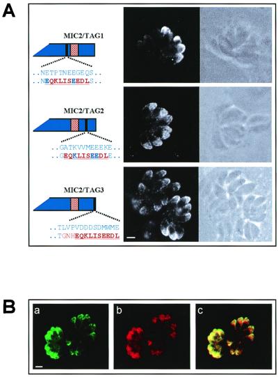
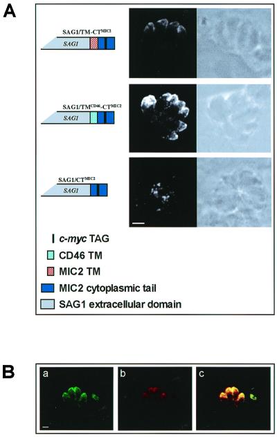
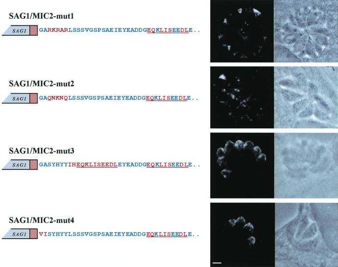
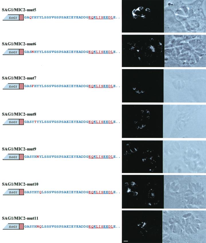
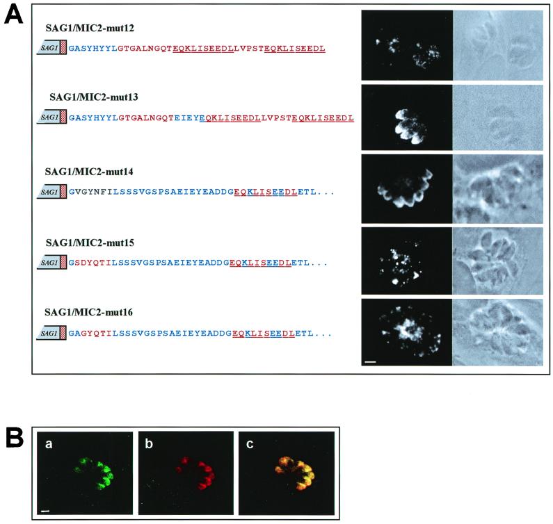
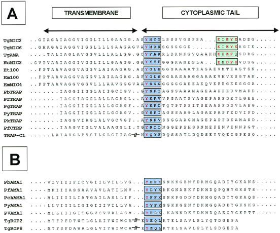
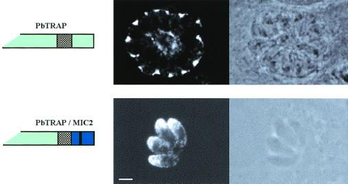
The micronemal protein 2 (MIC2) of Toxoplasma gondii shares sequence and structural similarities with a series of adhesive molecules of different apicomplexan parasites. These molecules accumulate, through a yet unknown mechanism, in secretory vesicles (micronemes), which together with tubular and membrane structures form the locomotion and invasion machinery of apicomplexan parasites. Our findings indicated that two conserved motifs placed within the cytoplasmic domain of MIC2 are both necessary and sufficient for targeting proteins to T. gondii micronemes. The first motif is based around the amino acid sequence SYHYY. Database analysis revealed that a similar sequence is present in the cytoplasmic tail of all transmembrane micronemal proteins identified so far in different apicomplexan species. The second signal consists of a stretch of acidic residues, EIEYE. The creation of an artificial tail containing only the two motifs SYHYY and EIEYE in a preserved spacing configuration is sufficient to target the surface protein SAG1 to the micronemes of T. gondii. These findings shed new light on the molecular mechanisms that control the formation of the microneme content and the functional relationship that links these organelles with the endoplasmic reticulum of the parasite.


Figures

FIG. 1
FIG. 1
(A) Immunolocalization of c-Myc-tagged MIC2 proteins, shown in confocal fluorescence (dark-field) and transmission (bright-field) photomicrographs of T. gondii tachyzoites transfected with constructs pMIC2/Tag1, pMIC2/Tag2, and pMIC2/Tag3. Intracellular parasites were incubated with MAb 9E10 directed against the c-Myc epitope and FITC-conjugated secondary antibody. The chimeric proteins encoded by the constructs are schematically shown. The blue and the red stippled boxes indicate the MIC2 sequence and the position of its TM region, respectively. The c-Myc epitope is represented as a black bar. The amino acid substitutions introduced by the insertion of the c-Myc epitope are indicated in red below the wild-type amino acid residues shown as blue letters. The amino acid sequence of the c-Myc epitope is underlined. Magnification of ×630, plus zoom factor of 2 for image acquisition; scale bar = 2 μm. (B) Confocal fluorescence photomicrographs showing the colocalization of MIC1 and the MIC2/Tag2. Intracellular pMIC2/Tag2-transfected tachyzoites were sequentially incubated with MAb T10 1F7 directed against the micronemal protein MIC1 and FITC-labeled secondary antibody (a) and biotin-labeled MAb 9E10 and rhodamine-labeled streptavidin (b). To precisely visualize the localization of MIC1 and MIC2/Tag2 within the tachyzoites, the two fluorescence photomicrographs were merged (c) using the software CoMOS version 7.0a (confocal microscope operating software) from Bio-Rad. Magnification of ×630, plus zoom factor of 2 for image acquisition; scale bar = 2 μm.
FIG. 2
FIG. 2
Immunoblot analysis of T. gondii parasites expressing different c-Myc-tagged MIC2 proteins. Protein lysates corresponding to 107 parasites were separated by SDS-PAGE on a 10% gel under reducing conditions and immunoblotted with MAb 9E10 directed against the c-Myc epitope and AP-conjugated secondary antibody. Lanes 1 and lane 2 contain T. gondii lysates from pMIC2/TAG1- and pMIC2/TAG2-transfected parasites, respectively; lane 3 contains nontransfected tachyzoites as a control. Migration positions of the high-molecular-weight standards (Sigma) are indicated in kilodaltons.
FIG. 3
FIG. 3
(A) Immunolocalization of a SAG1-MIC2 chimeric protein and variants in which the MIC2 TM region was either deleted or replaced with the TM region of human CD46. The structure of the chimeric proteins is schematically shown. Confocal fluorescence (dark-field) and transmission (bright-field) photomicrographs show intracellular tachyzoites transfected with the constructs pSAG1/TM-CTMIC2, pSAG1/TMCD46-CTMIC2, and pSAG1/CTMIC2. Intracellular parasites were incubated with MAb 9E10 and FITC-conjugated secondary antibody. Magnification of ×630, plus zoom factor of 2 for image acquisition; scale bar = 2 μm. (B) Confocal fluorescence photomicrographs showing the colocalization of MIC1 and the chimeric protein SAG1/TMCD46-CTMIC2. Magnification of ×630, plus zoom factor of 2 for image acquisition; scale bar = 2 μm.
FIG. 4
FIG. 4
Confocal fluorescence and transmission photomicrographs showing the immunolocalization of SAG1/MIC2 chimeric proteins containing amino acid substitutions in the cytoplasmic tail of MIC2. Intracellular tachyzoites, transfected with the constructs pSAG1/MIC2-mut1, pSAG1/MIC2-mut2, pSAG1/MIC2-mut3, and pSAG1/MIC2-mut4, were incubated with MAb 9E10 and FITC-conjugated secondary antibody. The red stippled box represents the MIC2 TM domain. The wild-type MIC2 amino acid residues are indicated as blue letters in the schematic representation of the constructs. Amino acid substitutions are shown in red letters, and the c-Myc epitope is underlined. Magnification of ×630, plus zoom factor of 2 for image acquisition; scale bar = 2 μm.
FIG. 5
FIG. 5
Confocal fluorescence and transmission photomicrographs showing the immunolocalization of SAG1/MIC2 chimeric proteins containing amino acid substitutions in the tyrosine-based motif of the MIC2 cytoplasmic tail. Wild-type MIC2 amino acids and mutated residues are indicated in the schematic representation of the constructs as blue and red letters, respectively. The red stippled box represents the MIC2 TM domain. The sequence of the c-Myc epitope is underlined. Magnification of ×630, plus zoom factor of 2 for image acquisition; scale bar = 2 μm.
FIG. 6
FIG. 6
(A) Confocal fluorescence and transmission photomicrographs showing the immunolocalization of chimeric proteins containing the MIC2 tyrosine-based motif alone (SAG1/MIC2-mut12) or in combination with a short glutamate-rich conserved sequence of the MIC2 cytoplasmic tail (SAG1/MIC2-mut13). The parasites were also transfected with constructs in which the MIC2 tyrosine motif had been replaced with the homologous region of PbTRAP (SAG1/MIC2-mut14) or with the endocytic targeting signals of rat TGN38 and human LAMP1 (SAG1/MIC2-mut15 and SAG1/MIC2-mut16). Wild-type MIC2 amino acids and mutated residues are indicated in the schematic representation of the constructs as blue and red letters, respectively. The red stippled box represents the MIC2 TM domain. The c-Myc epitope is underlined. PbTRAP-derived residues are indicated in green. Magnification of ×630, plus zoom factor of 2 for image acquisition; scale bar = 2 μm. (B) Confocal fluorescence photomicrographs showing the colocalization of MIC1 and the chimeric protein SAG1/MIC2-mut13. Magnification of ×630, plus zoom factor of 2 for image acquisition; scale bar, = 2 μm.
FIG. 7
FIG. 7
Sequence alignment of the putative TM sequences and cytoplasmic domains of micronemal proteins of different apicomplexan parasites. (A) Micronemal proteins. The tyrosine and glutamate motifs are indicated by light blue and green boxes, respectively. Identical or conserved residues are indicated in red. (B) Rhoptry proteins. A tyrosine-based motif is also found in the cytoplasmic tail of membrane-spanning proteins localized in the rhoptries of different Plasmodium species and T. gondii. Tg, T. gondii; Nc, N. caninum; Et, E. tenella; Em, E. maxima; C, C. parvum; Pb, P. berghei; Pf, P. falciparum; Pg, P. gallinaceum; Py, P. yoelii; Pk, P. knowlesi; Pch, P. chabaudi; Pv, P. vivax. GenBank accession numbers: TgMIC2, U62660; TgMIC6, AF110270; TgAMA, AF010264; NcMIC2, AFO61273; Et100, AF032905; Em100, M99058; PbTRAP, U67763; PfTRAP, X13022; PgTRAP, U64899; PyTRAP, M84732; PkTRAP, U64900; PfCTRP, U34363; TRAP-C1, AF017267; PbAMA1, AAC47192; PfAMA1, AF061332; PchAMA1, M25248; PyAMA1, AAC47193; PvAMA1, AAC16731; TgROP2, Z36906; TgROP8, AF011377.
FIG. 8
FIG. 8
Immunolocalization of PbTRAP and PbTRAP/MIC2. Confocal fluorescence (dark-field) and transmission (bright-field) photomicrographs show T. gondii tachyzoites transfected with the constructs pPbTRAP and pPbTRAP/MIC2. Intracellular parasites were incubated with MAb 7E4 directed against PbTRAP and FITC-conjugated secondary antibody. The proteins encoded by the constructs are schematically shown. Magnification of ×630, plus zoom factor of 2 for image acquisition; scale bar = 2 μm.

References

    1. Achbarou A, Mercereau-Puijalon O, Autheman J M, Fortier B, Camus D, Dubremetz J F. Characterization of microneme proteins of Toxoplasma gondii. Mol Biochem Parasitol. 1991;47:223–233. - PubMed
    1. Adams J H, Hudson D E, Torii M, Ward G E, Wellems T E, Aikawa M, Miller L H. The Duffy receptor family of Plasmodium knowlesiis located within the micronemes of invasive malaria merozoites. Cell. 1990;63:141–153. - PubMed
    1. Andersson A M, Melin L, Bean A, Pettersson R F. A retention signal necessary and sufficient for Golgi localization maps to the cytoplasmic tail of a Bunyaviridae(Uukuniemi virus) membrane glycoprotein. J Virol. 1997;71:4717–4727. - PMC - PubMed
    1. Aroeti B, Okhrimenko H, Reich V, Orzech E. Polarized trafficking of plasma membrane proteins: emerging roles for coats, SNAREs, GTPases and their link to the cytoskeleton. Biochim Biophys Acta. 1998;1376:57–90. - PubMed
    1. Bos K, Wraight C, Stanley K K. TGN38 is maintained in the trans-Golgi network by a tyrosine-containing motif in the cytoplasmic domain. EMBO J. 1993;12:2219–2228. - PMC - PubMed

Publication types

MeSH terms

LinkOut - more resources