. 2000 Oct;20(19):7353-62.
doi: 10.1128/MCB.20.19.7353-7362.2000.

Control of hnRNP A1 alternative splicing: an intron element represses use of the common 3' splice site

Affiliations

Control of hnRNP A1 alternative splicing: an intron element represses use of the common 3' splice site

M J Simard et al. Mol Cell Biol. 2000 Oct.

Abstract

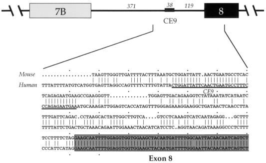
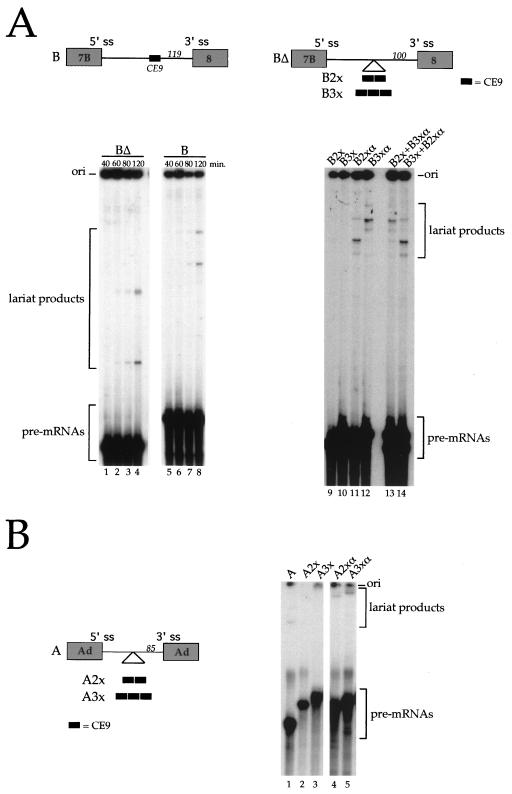
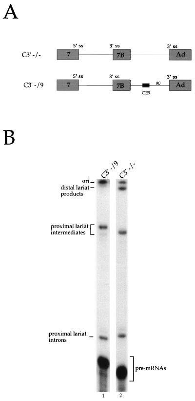
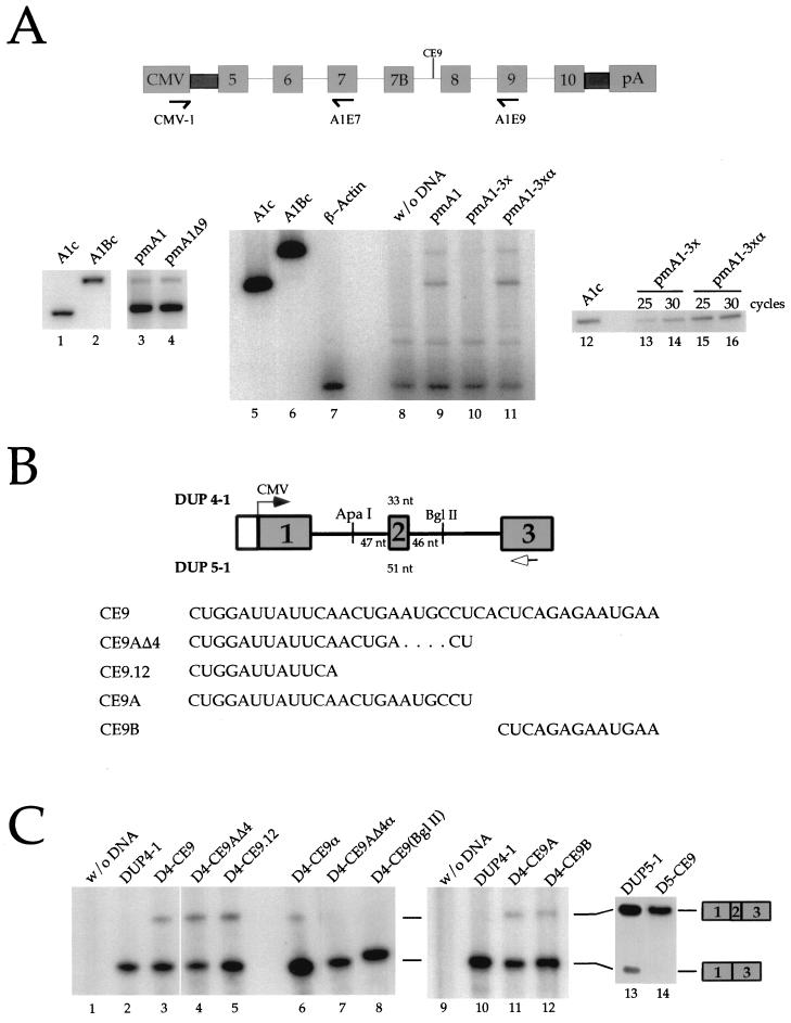
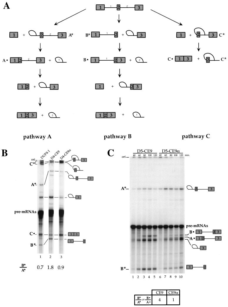
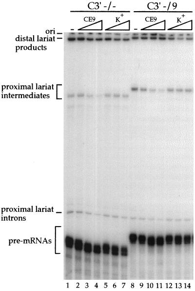
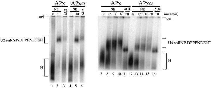
Alternative splicing of exon 7B in the hnRNP A1 pre-mRNA produces mRNAs encoding two proteins: hnRNP A1 and the less abundant A1B. We have reported the identification of several intron elements that contribute to exon 7B skipping. In this study, we report the activity of a novel element, conserved element 9 (CE9), located in the intron downstream of exon 7B. We show that multiple copies of CE9 inhibit exon 7B-exon 8 splicing in vitro. When CE9 is inserted between two competing 3' splice sites, a single copy of CE9 decreases splicing to the distal 3' splice site. Our in vivo results also support the conclusion that CE9 is a splicing modulator. First, inserting multiple copies of CE9 into an A1 minigene compromises the production of fully spliced products. Second, one copy of CE9 stimulates the inclusion of a short internal exon in a derivative of the human beta-globin gene. In this case, in vitro splicing assays suggest that CE9 decreases splicing of intron 1, an event that improves splicing of intron 2 and decreases skipping of the short internal exon. The ability of CE9 to act on heterologous substrates, combined with the results of a competition assay, suggest that the activity of CE9 is mediated by a trans-acting factor. Our results indicate that CE9 represses the use of the common 3' splice site in the hnRNP A1 alternative splicing unit.


Figures

FIG. 1
FIG. 1
Schematic representation of the downstream portion of the hnRNP A1 alternative splicing unit, with the length indicated in nucleotides. An alignment between the mouse and the human sequences from the middle of the intron to a portion of exon 8 is shown. The sequence of CE9 is underlined.
FIG. 2
FIG. 2
CE9 inhibits in vitro splicing. (A) The structures of pre-mRNA B and BΔ, as well as derivatives containing multiple copies of CE9 or complementary sequences, are shown at the top. The distance of the insertion point to the 3′ splice site (ss) is indicated in nucleotides. Note that the CE6 element is absent from all pre-mRNAs. Labeled pre-mRNAs were incubated in HeLa nuclear extracts for the times indicated for BΔ and B RNAs or for 2 h for multiple inserts. Splicing products were run on an 11% acrylamide–8 M urea gel. Mixtures containing two different RNAs were analyzed to rule out the presence of a nonspecific inhibitor in the B2x and B3x RNA preparations (lanes 13 and 14). (B) Copies of CE9 or complementary sequences were inserted into a pre-mRNA substrate derived from the adenovirus (Ad) major late transcription unit (A RNA). The labeled splicing products were resolved on a 7% acrylamide–8 M urea gel. Because mRNA products were obscured by the degradation of the pre-mRNAs, only the portion of the gels that indicates the pre-mRNAs and lariat products is shown.
FIG. 3
FIG. 3
CE9 represses the utilization of a downstream 3′ splice site (ss). (A) Schematic representation of pre-mRNAs containing competing 3′ splice sites. The position of CE9 is indicated, as is its distance from the 3′ splice site, in nucleotides. Ad, adenovirus. (B) Labeled pre-mRNAs were incubated in a HeLa nuclear extract for 2 h. Splicing products were fractionated on an 11% acrylamide–8 M urea gel. The positions of the lariat products generated from the use of the distal (Ad) or the proximal (7B) 3′ splice site are indicated.
FIG. 4
FIG. 4
CE9 affects splicing in vivo. (A) CE9 prevents the production of fully spliced mRNAs. A genomic portion of the murine A1 gene was expressed in HeLa cells using the CMV-1 promoter. The relative position of CE9 is indicated, as well as the position of the oligonucleotides used for the RT-PCR assays performed on total RNA isolated 48 h posttransfection. A derivative carrying a deletion of CE9 was used (pmA1Δ9 [lane 4]). Three copies of CE9 or three copies of the complementary sequence of CE9 were inserted into pmA1Δ9 (pmA1-3x or pmA1-3xα, respectively). Reconstructed A1 or A1B cDNAs were used as controls in PCR assays (lanes 1, 2, 5, 6, and 12). RT-PCR amplification of the endogenous β-actin mRNA was performed independently on a mock transfection (lane 7) or simultaneously with the A1 minigene analysis with oligonucleotides CMV-1 and A1E9 (lanes 8 to 11). A separate RT-PCR assay was carried out with the CMV-1 and A1E7 oligonucleotides (lanes 12 to 16). The number of cycles used in the amplification rounds is indicated above the lanes. (B) Schematic representation of the DUP constructs and derivatives. The length of the central exon for DUP 4-1 and DUP 5-1 is indicated. The transcription start site is indicated by an arrow. The distance between the different cloning sites and the central exon is indicated. The arrow below exon 3 represents the oligonucleotide used for primer extension analysis. The RNA versions of the different oligonucleotides cloned into the DUP plasmids are shown at the bottom. (C) CE9 promotes the inclusion of artificial globin exon in vivo. DUP expression was analyzed by primer extension. Plasmid names are indicated above each lane. Each DUP 4-1, DUP 5-1, or derivative was generated by inserting oligonucleotides at the ApaI site, except for D4-CE9(BglII), for which BglII in the downstream intron was used (lane 8). Extension products were loaded onto a 5% acrylamide–8 M urea. The slightly abnormal migration of the 1-3 product in lane 8 is a gel artifact.
FIG. 5
FIG. 5
Effect of CE9 on DUP 4-1 and DUP 5-1 splicing in vitro. (A) Representation of different pathways used for the splicing of DUP pre-mRNAs. Pathway A occurs when intron 1 is the first intron to be removed, while pathway B takes place when intron 2 is spliced first. Pathway C corresponds to skipping of the internal exon. The splicing products, A*, A•, B*, B•, C*, and C•, indicate molecules that are unique to each pathway and correspond to the bands shown in the splicing gels (panels B and C). (B) CE9 shifts splicing in favor of pathway B. Labeled pre-mRNAs were incubated in HeLa extracts for 1 h. Splicing products were fractionated on an 11% acrylamide–8 M urea gel. Selected products are indicated. (C) CE9 also improves pathway B in DUP 5-1. Products specific to pathway A or B are indicated. Note that the band immediately below product A• corresponds to the intron 2-exon 3 lariat intermediate, a product common to pathways A and B. Pre-mRNAs were incubated under splicing conditions for the times indicated. Splicing products were loaded onto an 8% acrylamide–8 M urea gel. The ratio of product B* to product A* (or B•/A•) is indicated and is based on the 2-h time point. The structure of DUP pre-mRNA and derivatives is as shown in Fig. 4A, except that the labeled pre-mRNAs were synthesized in vitro using T7 RNA polymerase.
FIG. 6
FIG. 6
A trans-acting factor mediates the repression by CE9 on a downstream 3′ splice site. Splicing was performed in a HeLa nuclear extract preincubated for 10 min with increasing amounts of the CE9 RNA as a competitor (lanes 2 to 4 and 9 to 11) or with a control RNA derived from pBluescript K(+) (lanes 5 to 7 and 12 to 14). Each set of the competition was performed with 0.5 fmol of pre-mRNA and 50, 250, or 500 fmol of unlabeled competitor RNA.
FIG. 7
FIG. 7
CE9 does not block the assembly of snRNP-containing complexes. The time course of splicing complex assembly was determined by using adenovirus pre-mRNAs containing two copies of CE9 or two copies of the complementary sequences (A2x or A2xα, respectively) (see Fig. 2B for a schematic diagram of the pre-mRNAs). Nuclear extracts were pretreated for 1 h at 30°C in the presence of RNase H alone (NE) or RNase H and oligonucleotides complementary to U2 snRNA (ΔU2) or U4 snRNA (ΔU4). The reaction mixtures were loaded on a 2% low-melting-point agarose gel. The origins of the gels and the identities of the complexes are indicated.

Similar articles

Cited by

References

    1. Amendt B A, Hesslein D, Chang L-J, Stoltzfus C M. Presence of negative and positive cis-acting RNA splicing elements within and flanking the first tat coding exon of human immunodeficiency virus type 1. Mol Cell Biol. 1994;14:3960–3970. - PMC - PubMed
    1. Amendt B A, Si Z-H, Stoltzfus C M. Presence of exon splicing silencers within human immunodeficiency virus type 1 tat exon 2 and tat-rev exon 3: evidence for inhibition mediated by cellular factors. Mol Cell Biol. 1995;15:4606–4615. . (Erratum, 15:6480.) - PMC - PubMed
    1. Ashiya M, Grabowski P J. A neuron-specific splicing switch mediated by an array of pre-mRNA repressor sites: evidence of a regulatory role for the polypyrimidine tract binding protein and a brain-specific PTB counterpart. RNA. 1997;3:996–1015. - PMC - PubMed
    1. Black D L. Activation of c-src neuron-specific splicing by an unusual RNA element in vivo and in vitro. Cell. 1992;69:795–807. - PubMed
    1. Black D L. Finding splice sites within a wilderness of RNA. RNA. 1995;1:763–771. - PMC - PubMed

Publication types

MeSH terms

Substances

LinkOut - more resources