Comparative Study
. 2000 Oct;20(19):7410-7.
doi: 10.1128/MCB.20.19.7410-7417.2000.

Mutations in a tRNA import signal define distinct receptors at the two membranes of Leishmania mitochondria

Affiliations
Comparative Study

Mutations in a tRNA import signal define distinct receptors at the two membranes of Leishmania mitochondria

S N Bhattacharyya et al. Mol Cell Biol. 2000 Oct.

Abstract

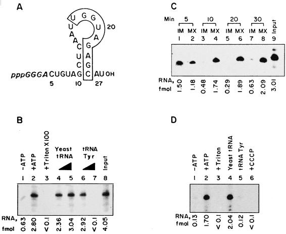
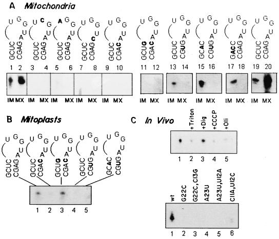
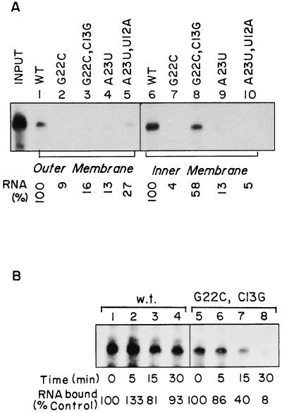
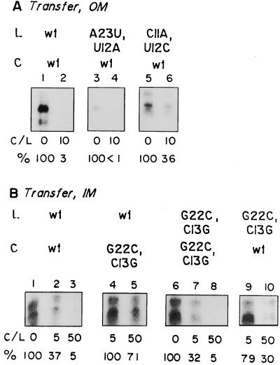
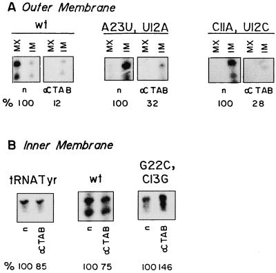
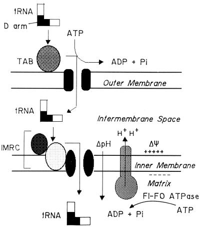
Nucleus-encoded tRNAs are selectively imported into the mitochondrion of Leishmania, a kinetoplastid protozoan. An oligoribonucleotide constituting the D stem-loop import signal of tRNA(Tyr)(GUA) was efficiently transported into the mitochondrial matrix in organello as well as in vivo. Transfer through the inner membrane could be uncoupled from that through the outer membrane and was resistant to antibody against the outer membrane receptor TAB. A number of mutations in the import signal had differential effects on outer and inner membrane transfer. Some mutants which efficiently traversed the outer membrane were unable to enter the matrix. Conversely, restoration of the loop-closing GC pair in reverse resulted in reversion of transfer through the inner, but not the outer, membrane, and binding of the RNA to the inner membrane was restored. These experiments indicate the presence at the two membranes of receptors with distinct specificities which mediate stepwise transfer into the mitochondrial matrix. The combination of oligonucleotide mutagenesis and biochemical fractionation may provide a general tool for the identification of tRNA transport factors.


Figures

FIG. 1
FIG. 1
Import of D arm minihelix in vitro. (A) Sequence of the wild-type minihelix. Numbers correspond to positions on the intact tRNATyr molecule (11). The secondary structure shown was derived by energy minimization using the FOLDRNA program (26). Bases in italics are derived from the T7 RNA polymerase initiation sequence on the template. The boxed region contains the conserved nonanucleotide motif found in importable RNAs (13). (B) Total uptake of the wild-type minihelix by intact mitochondria. Reaction mixtures were incubated without (lane 1) or with (lanes 2 through 7) 4 mM ATP. In lane 3, Triton X-100 (0.5%) was added after the import incubation. In lanes 4 through 7, unlabeled yeast tRNA (lanes 4 and 5) or low-specific-activity tRNATyr transcript (lanes 6 and 7) was added as the competitor at concentrations of 0.1 (lanes 4 and 6) or 1 (lanes 5 and 7) pmol. After RNase treatment, the total internalized RNA was analyzed. (C) Intramitochondrial distribution of D arm minihelix. Intact mitochondria were incubated with wild-type RNA and ATP at 37°C for the time intervals shown. After each incubation, the mitochondria were RNase treated and subfractionated, and the contents of RNA in the IM and MX fractions were analyzed. (D) Import of D arm minihelix into mitoplasts. Mitoplasts (100 μg of protein) were incubated with the wild-type minihelix in the absence (lane 1) or presence (lanes 2 through 6) of 4 mM ATP. In lane 3, Triton X-100 (0.5%) was added after the incubation. Reactions 4 and 5 contained 1 pmol of yeast tRNA or tRNATyr, respectively, as the competitor. In reaction 6, mitoplasts were preincubated with 50 μM CCCP before the import incubation. RNase-resistant RNA was recovered for analysis. The region of the major minihelix band in each lane was excised and counted; after background subtraction, femtomole values were computed from the specific activity.
FIG. 2
FIG. 2
Effect of mutations on intramitochondrial location of D arm minihelix. (A) 32P-labeled wild-type or mutant minihelix (100 fmol) was incubated with mitochondria (100 μg of protein). After 15 min at 37°C, RNase was added, and the washed mitochondria were fractionated into IM and MX compartments. Lanes 1 through 10 and 11 through 20 show the results of two different experiments. The RNAs used were as follows. Lanes 1 and 2, 19 and 20, wild type; lanes 3 and 4, G18→C; lanes 5 and 6, U17→A; lanes 7 and 8, A21→C; lanes 9 and 10, G22→C; lanes 11 and 12, G22→C, C13→G; lanes 13 and 14, A23→U; lanes 15 and 16, A23→U, U12→A; and lanes 17 and 18, C11→A, U12→C. (B) 32P-labeled wild-type (lane 1), G22→C (lane 2), G22→C, C13→G (lane 3), A23→U (lane 4), and A23→U, U12→A (lane 5) RNAs (100 fmol of each) were incubated with mitoplasts (100 μg of protein) in the presence of ATP for 15 min at 37°C, and the RNase-resistant RNA was analyzed. Band quantitation was performed as in Fig. 1; the smear at the bottom of lane 7 was disregarded. (C) Matrix targeting in vivo. (Upper panel) Promastigotes were transfected with 32P-labeled wild-type minihelix (1 pmol) in the absence (lanes 1 to 3) or presence of 10 μM CCCP (lane 4) or of 50 μM oligomycin (lane 5). Aliquots of the transfected cells were lysed, and mitochondrial fractions were treated with RNase and DNase in the absence (lanes 1, 4, and 5) or presence of 1% Triton X-100 (lane 2) or 320 μM digitonin (lane 3). (Lower panel) Promastigotes were transfected with the wild type (lane 1) or the G22→C (lane 2), G22→C, C13→G (lane 3), A23→U (lane 4), A23+→U, U12→A (lane 5), and C11→A, U12→C (lane 6) mutants, and the RNase-resistant RNA associated with the mitochondrial fraction was analyzed. Quantitation was performed by densitometry.
FIG. 3
FIG. 3
Binding of minihelices to the OM and IM. (A) The following 32P-labeled RNAs were incubated with mitochondria (lanes 1 to 5) or mitoplasts (lanes 6 to 10): lanes 1 and 6, wild type; lanes 2 and 7, G22→C; lanes 3 and 8, G22→C, C13→G; lanes 4 and 9, A23→U; and lanes 5 and 10, A23→U, U12→A. After washing with STE, the membrane-bound RNA was recovered and analyzed as in Fig. 1. The amounts of wild-type RNA (taken as 100%) bound to mitochondria (lane 1) and mitoplasts (lane 6) were 0.64 and 1.12 fmol, respectively. (B) Stability of inner membrane complexes of wild-type (lanes 1 to 4) and G22→C, C13→G mutant (lanes 5 to 8) minihelices. Binding reactions with mitoplasts were performed with the incubation intervals shown. Control reactions without incubation (lanes 1 and 5) yielded 1.80 and 1.01 fmol, respectively. Quantitations were performed by densitometric scanning of the major band in each lane.
FIG. 4
FIG. 4
Cross competition between wild-type and mutant minihelices for transfer across OM and IM. Mitochondria (A) or mitoplasts (B) were incubated with high-specific-activity 32P-labeled RNA (L) in the absence or presence of low-specific-activity competitor (C; C/L, ratio of competitor to substrate), and total uptake was assayed with RNase protection. Panel A, lanes 1 to 2, 3 to 4, and 5 to 6, contained high-specific-activity wild-type, A23→U, U12→A, and C11→A, A12→U RNA (100 fmol), respectively. Wild-type competitor (1 pmol) was included in reactions 2, 4, and 6. In panel B, high-specific-activity wild-type (lanes 1 to 5) or G12→C, C13→G (lanes 6 to 10) RNA (100 fmol) was incubated without competitor (lanes 1 and 6), with 0.5 pmol (lanes 2 and 9) or 5 pmol (lanes 3 and 10) of wild-type competitor, or with 0.5 pmol (lanes 4 and 7) or 5 pmol (lanes 5 and 8) of G22→C, C13→G competitor. Quantitations were performed by densitometric scanning of the major band in each lane.
FIG. 5
FIG. 5
Effect of anti-TAB antibody on transfer of RNA across the OM and IM. (A) Intact mitochondria (100 μg of protein) preincubated with normal (n) or anti-TAB (α) IgG were incubated with 100 fmol of wild-type D arm minihelix (left), A23→U, U12→A mutant (center), or C11→A, U12→C mutant (right) in the presence of ATP. The amounts of total internalized RNA (IM + MX) are expressed as percentages of the control values obtained in the presence of normal IgG. (B) Mitoplasts were incubated with normal (n) or anti-TAB (α) IgG and then with 100 fmol of tRNATyr(GUA) transcript (left), wild-type D arm minihelix (center), or G22→C, C13→G mutant (right) for 15 min at 37°C and treated with RNase, and the internalized RNA was recovered. IM transfer is expressed as the percentage of the control value obtained with normal IgG. Quantitations were performed as for Fig. 4. In panel B, middle, both bands are of nearly equal intensity and yield similar values for extent of inhibition.
FIG. 6
FIG. 6
Stepwise transfer of tRNA through the two mitochondrial membranes. For details see the text. TAB, outer membrane receptor; IMRC, inner membrane receptor complex components (two components are shown); ΔΨ, membrane potential; ΔpH, proton gradient; F1-F0 ATPase, oligomycin-sensitive proton pump.

Similar articles

Cited by

References

    1. Adhya S, Ghosh T, Das A, Bera S K, Mahapatra S. Role of an RNA binding protein in import of tRNA into Leishmania mitochondria. J Biol Chem. 1997;272:21396–21402. - PubMed
    1. Chiu N, Chiu A, Suyama Y. Native and imported transfer RNA in mitochondria. J Mol Biol. 1975;94:37–50. - PubMed
    1. Das S, Adhya S. Organization and chromosomal localization of β-tubulin genes in Leishmania donovani. J Biosci. 1990;15:239–248.
    1. Ghosh A, Ghosh T, Ghosh S, Das S, Adhya S. Interaction of small ribosomal and transfer RNAs with a protein from Leishmania donovani. Nucleic Acids Res. 1994;22:1663–1669. - PMC - PubMed
    1. Gray M W, Burger G, Lang B F. Mitochondrial evolution. Science. 1999;283:1476–1481. - PubMed

Publication types

MeSH terms

LinkOut - more resources