. 2000 Sep 15;28(18):3542-50.
doi: 10.1093/nar/28.18.3542.

Transcriptional activation by the PHD finger is inhibited through an adjacent leucine zipper that binds 14-3-3 proteins

Affiliations

Transcriptional activation by the PHD finger is inhibited through an adjacent leucine zipper that binds 14-3-3 proteins

T Halbach et al. Nucleic Acids Res. .

Abstract

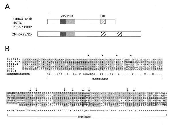
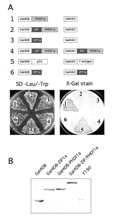
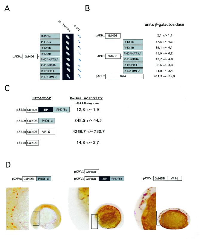
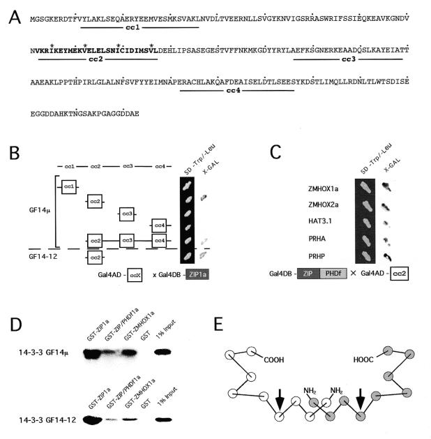
The PHD finger, a Cys(4)-His-Cys(3) zinc finger, is found in many regulatory proteins from plants or animals which are frequently associated with chromatin-mediated transcriptional regulation. We show here that the PHD finger activates transcription in yeast, plant and animal cells. In plant homeodomain transcription factors the PHD finger is combined with an upstream leucine zipper. Both domains together form a highly conserved 180 amino acid region called the ZIP/PHDf motif and transcriptional activity of the PHD finger is masked when embedded in this motif. Our results indicate that the ZIP/PHDf domain is a potential regulatory domain of PHDf-HD proteins. The leucine zipper upstream of the PHD finger interacts with 14-3-3GF14 mu from Arabidopsis thaliana and 14-3-3GF14-12 from maize via a leucine zipper conserved in helix 4 of various 14-3-3 proteins from plants and animals. PHD-type plant homeodomain proteins consequently may represent potential targets of 14-3-3 signalling.


Figures

Figure 1
Figure 1
Position and conservation of the ZIP/PHDf domain in plant HD proteins. (A) Position of the conserved 180 amino acid region of the ZIP/PHDf domain in different PHD-HD proteins. (B) Alignment of five plant PHDf and the second PHD finger in Drosophila dMI-2 protein. Asterisks indicate leucine residues of the leucine zipper; arrows mark the cysteine residues in the PHD finger. Shading indicates amino acid conservation in at least three genes. The identity between the five plant sequences exceeds 60%.
Figure 2
Figure 2
Dissectional analysis of the ZMHOX1a ZIP/PHDf domain in the yeast two-hybrid system. (A) Combinations of constructs 1–6 as indicated were used for co-transformation of Y190 yeast cells. Staining for lacZ activity was performed after 3 days at 30°C on minimal selection medium devoid of leucine and tryptophan. Galactosidase activity with the ZMHOX1a PHD finger (1), as for the positive control (5) comprising the p53–T antigen interaction, was detectable after 3 h. Note that the ZIP/PHDf domain (2) and the isolated leucine zipper (3) do not exert lacZ activity and show no homodimerisation (4 and 6). Identical results were observed with constructs 1–3 on single selection medium (leu). See also Figure 3A. (B) Western blot analysis of protein extracts prepared from Y190 cells transformed with constructs as indicated above each lane. Equal amounts of protein extract were analysed with a monoclonal antibody against the Gal4DB domain and subsequent chemoluminiscent detection. Polypeptides exhibited the predicted sizes: Gal4DB, 22 kDa; Gal4DB-ZIP1a, 29 kDa; Gal4DB-PHDf1a, 31 kDa; Gal4DB-ZIP/PHDf1a, 40 kDa. No reacting polypeptide was detectable in untransformed Y190 cells.
Figure 3
Figure 3
Transcriptional activation is a common feature of PHD fingers in yeast, plant and animal cells. (A) Galactosidase staining obtained after transformation of Y190 yeast cells with various GAL4DB-PHDf fusions as schematically indicated to the left. All signals appeared within 2 h incubation at 37°C. (B) Quantification of lacZ activity [O-nitrophenyl-β-d-galactopyranoside (ONPG) test] in yeast protein extracts transformed with different PHD finger constructs. The values are calculated from three independent transformation experiments. (C) Relative GUS activities calculated from transient gene expression experiments performed in Arabidopsis protoplasts. Effector construct Gal4DB-PHDf1a, Gal4DB-ZIP/PHDf1a or Gal4DB-VP16, as a strong transcriptional activation domain, was combined with the UAS core::GUS reporter and a 35S::luciferase marker as internal reference. The relative GUS activities represent data from eight independent transfection experiments and were normalised to the luciferase standard. (D) Transcriptional activation by PHDf1a observed after injection of zebrafish embryos. The Gal4DB-PHDf1a, Gal4DB-ZIP/PHDf1a and Gal4DB effector proteins were expressed under the constitutive CMV promoter and monitored by the UAS::myc-notch:intra reporter. Antibody staining of a myc-tagged notch-intra marker monitors reporter gene activation. Note that staining is observed with Gal4DB-PHDf1a (left) and Gal4DB-VP16 (right) but not with Gal4DB-ZIP/PHDf1 (middle) or Gal4DB (data not shown)
Figure 4
Figure 4
In vivo and in vitro analysis of the 14-3-3 protein interactions. (A) Coiled-coil predictions of 14-3-3 proteins. The highly conserved leucine zipper in cc2 is bold; asterisks mark its heptad repeat spacing. (B) Interactions between cc motifs of 14-3-3GF14µ and 14-3-3GF14-12 fused to the activation domain (Gal4AD-ccX) and the ZMHOX1a leucine zipper bait (Gal4DB-ZIP1a). The cc2 motif of 14-3-3GF14µ and 14-3-3GF14-12 is sufficient to mediate interactions in the yeast two-hybrid system. The N-terminally truncated peptide (Δ1–75) of 14-3-3GF14µ lacking region cc1 binds to the ZIP1a motif. Expression of full-length maize and Arabidopsis 14-3-3 proteins in yeast unfortunately failed. (C) Interactions between Gal4AD-cc2 of Arabidopsis 14-3-3GF14µ and different ZIP/PHDf domains as bait (Gal4DB-ZIP/PHDf). Individual ZIP/PHDf domains are indicated in the margin. (D) Co-precipitation of full-length 14-3-3GF14µ and 14-3-3 GF14-12 proteins by different ZMHOX1a peptides. The radioactively labelled 14-3-3GF14µ and 14-3-3GF14-12 proteins of maize and Arabidopsis bind to ZIP1a, ZIP/PHDf1a and the complete ZMHOX1a protein, but not to GST. (E) Helical structure of the 14-3-3 dimer. Circles indicate antiparallel α-helices, nine in each monomer. The two arrows mark α-helix 4 and the orientation of the leucine zipper interaction surface.

References

    1. Bellmann R. and Werr,W. (1992) EMBO J., 11, 3367–3374. - PMC - PubMed
    1. Schindler U., Beckmann,H. and Cashmore,A.R. (1993) Plant J., 4, 137–150. - PubMed
    1. Aasland R., Gibson,T.J. and Stewart,A.F. (1995) Trends Biochem. Sci., 20, 56–59. - PubMed
    1. Saha V., Chaplin,T., Gregorini,A., Ayton,P. and Young,B.D. (1995) Proc. Natl Acad. Sci. USA, 92, 9737–9741. - PMC - PubMed
    1. Bochar D.A., Savard,J., Wang,W., Lafleur,D.W., Moore,P., Cote,J. and Shiekhattar,R. (2000) Proc. Natl Acad. Sci. USA, 97, 1038–1043. - PMC - PubMed

Publication types

MeSH terms