. 2000 Sep 15;28(18):3605-14.
doi: 10.1093/nar/28.18.3605.

Stable expression and cell-specific chromatin structure of human alpha1-antitrypsin cosmid transgenes in rat hepatoma cells

Affiliations

Stable expression and cell-specific chromatin structure of human alpha1-antitrypsin cosmid transgenes in rat hepatoma cells

P Rollini et al. Nucleic Acids Res. .

Abstract

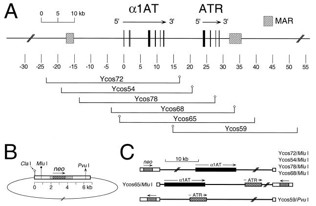
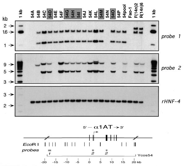
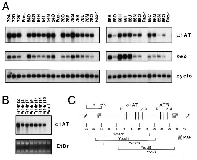
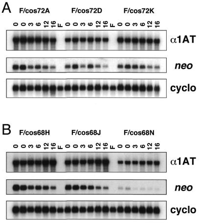
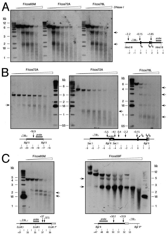
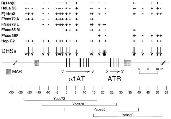
The human gene encoding alpha1-antitrypsin (alpha1AT, gene symbol PI) resides in a cluster of serine protease inhibitor (serpin) genes on chromosome 14q32.1. alpha1AT is highly expressed in the liver and in cultured hepatoma cells. We recently reported the chromatin structure of a >100 kb region around the gene, as defined by DNase I-hypersensitive sites (DHSs) and matrix-attachment regions, in expressing and non-expressing cells. Transfer of human chromosome 14 by microcell fusion from non-expressing fibroblasts to rat hepatoma cells resulted in activation of alpha1AT transcription and chromatin reorganization of the entire region. In the present study, we stably introduced cosmids containing alpha1AT with various amounts of flanking sequence and a linked neo selectable marker into rat hepatoma cells. All single-copy transfectants with >14 kb of 5' flanking sequence expressed wild-type levels of alpha1AT mRNA in a position-independent manner. In contrast, expression of transgenes containing only approximately 1.5-4 kb of flanking sequence was highly variable. Long-term culture of transfectant clones in the absence of selection resulted in gradual loss of neo expression, but expression of the linked alpha1AT gene remained constant. DHS mapping of cosmid transgenes integrated at ectopic sites revealed a hepatoma-specific chromatin structure in each transfectant clone. The implications of these findings are discussed.


Figures

Figure 1
Figure 1
Genomic map of the α1AT region and cosmids used in this study. (A) Physical map of the α1AT-ATR region. The map is drawn to scale, with position zero defined as an EcoRI site in macrophage-specific exon IA of α1AT (5,35). Exons are indicated by black boxes, and MARs as hatched boxes. The positions and names of relevant cosmids used in this study are shown below the map. Small diamonds on either side of the genomic inserts represent the diagnostic ClaI site of the cosmid vector. (B) Schematic representation of the SuperCos1 vector ligated to a genomic insert. White boxes indicate λ or plasmid sequences. The bacterial aminoglycoside phosphotransferase (neo) gene (hatched box) is under the control of SV40 promoter and polyadenylation signals (gray boxes). Relevant restriction sites used to determine insert orientations and/or to linearize constructs for electroporation are indicated. (C) Diagrammatic representation of various linearized cosmid clones, showing the presumptive genomic organization of single-copy, intact integrants. The relative positions and orientations of neo (gray boxes) and α1AT (black boxes) and/or ATR (hatched boxes) in the various linearized templates are indicated.
Figure 2
Figure 2
Genotypic analysis of Ycos54 transfectants. Genomic DNAs from individual cosmid transfectants (54A, 54B, etc.) and from a polyclonal mixture of approximately 30 clones (54pool) were prepared, and 5 µg of each were digested with EcoRI. The restriction digests were analyzed by Southern hybridization using various probes in and around the human α1AT gene, as well as with a control rat probe (rHNF-4). Control DNAs were prepared from untransfected rat hepatoma cells (Fao-1) and from rat hepatoma [F(14n)2] or fibroblast [R(14n)6] microcell hybrids containing human chromosome 14. The map at the bottom of the figure shows the human α1AT gene with the hepatocyte-specific transcription start site (arrow) and relevant flanking sequences, including genomic EcoRI sites and the locations of probes 1 and 2. These probes are described in Materials and Methods. The rHNF-4 probe was an ∼1.1 kb SphI–BamHI fragment from pLEN4S (36) containing 3′ untranslated sequences of rat HNF-4 cDNA. Shaded boxes over individual transfectants indicate clones that contained single copy, intact transgenes. Arrows beside the gels show the positions (in kb) of size markers (1 kb ladder).
Figure 3
Figure 3
Expression of human α1AT mRNA in single-copy cosmid transfectants. (A) Cytoplasmic RNAs (5 µg/lane) from the indicated single copy Ycos72, Ycos54, Ycos78, Ycos68 and Ycos65 transfectant clones or untransfected rat hepatoma cells (Fao-1) were prepared, and steady-state levels of human α1AT mRNA were analyzed by northern hybridization. The α1AT probe was a human-specific exon II DNA fragment that did not cross-hybridize with rat α1AT sequences. Aminoglycoside phosphotransferase (neo) mRNA levels from the SuperCos1 vector were also assayed. As a loading control, the filters were stripped and rehybridized with a rat cyclophilin (cyclo) probe. (B) Seven rat hepatoma microcell hybrids containing an apparently intact human chromosome 14 (31) were analyzed for human α1AT mRNA levels as above. As loading controls, the intensities of the ethidium bromide-stained ribosomal RNA bands (EtBr) are shown. These images are composites made from two different gels. (C) The map shows the extent of each cosmid insert, together with the position of the serpin genes and the MARs, as described in the legend to Figure 1.
Figure 4
Figure 4
Expression of neo and human α1AT mRNAs in the absence of selection. Six single-copy, intact cosmid transfectant clones (F/cos72A, F/cos72D, F/cos72K, F/cos68H, F/cos68J and F/cos68N) were grown in the absence of G418 selection for more than 30 passages, and cytoplasmic RNAs (5 µg/lane) were prepared after 3, 6, 12 and 16 weeks (lanes marked 3, 6, 12 and 16) and analyzed for neo, human α1AT and rat cyclophilin mRNA expression as described in the legend to Figure 3. Lanes marked ‘0’ are two separate RNA samples from the cell lines prior to removal of G418. Lanes marked F contained RNA from parental Fao-1 rat hepatoma cells.
Figure 5
Figure 5
DNase I-hypersensitive site mapping in cosmid transfectants. Nuclei from the indicated single-copy, intact cosmid transfectants were treated with increasing concentrations of DNase I. DNA was purified, digested with HindIII (A), BglII (B, left and right; C, right), SacI (B, center), or EcoRI (C, left) and analyzed by Southern hybridization using specific probes around α1AT described in Materials and Methods. The diagrams indicate the positions of relevant restriction sites, DHSs, probes and α1AT exons, using as coordinate zero an EcoRI site in the middle of exon IA of α1AT. Light gray boxes in (C) indicate a fragment of the SuperCos1 vector at the 3′ end of the transgene, and restriction sites with an asterisk are either in the vector or at the integration site of the cosmid in the rat genome. The ∼4.1 kb EcoRI fragment without DHSs (C, left; approximately position +31 to +35.1 kb) only hybridized weakly to the probe used, and is barely visible below the stronger ∼4.3 kb fragment recognized by the probe. Arrows beside the autoradiographs indicate the positions of sub-fragments generated by DNase I cleavage at specific DHSs. A 1 kb ladder was used as a size standard, and the positions of diagnostic bands are indicated beside the autoradiographs.
Figure 6
Figure 6
Long-range DHS map of α1AT-ATR alleles. The map is drawn to scale, with position zero defined as an EcoRI site in exon IA of α1AT. Exons are indicated as black boxes, with arrows showing the transcriptional orientations of the genes. MARs are shown as hatched boxes. DHSs are depicted above the map as vertical arrows. Long arrows indicate strong DHSs, and short arrows weak sites in the HepG2 cells. The presence or absence of specific DHSs in various microcell hybrids containing human chromosome 14 and single-copy cosmid transfectants [HepG2, expressing human hepatoma allele; HeLa S3, non-expressing human allele; F(14n)2, activated allele in rat hepatoma cells; R(14n)6, extinguished allele in rat fibroblasts; F/cos72A, F/cos78L, F/cos65M and F/cos59F, cosmid transfectants; see Figs 1 and 3] is indicated above the map by + or – signs. Small + indicates a DHS weaker than that of the expressed HepG2 allele. The positions of the genomic inserts of the four transfectants analyzed are indicated below the map.

Similar articles

Cited by

References

    1. Potempa J., Korzus,E. and Travis,J. (1994) J. Biol. Chem., 269, 15957–15960. - PubMed
    1. Bao J.J., Reed-Fourquet,L., Sifers,R.N., Kidd,V.J. and Woo,S.L. (1988) Genomics, 2, 165–173. - PubMed
    1. Hofker M.H., Nelen,M., Klasen,E.C., Nukiwa,T., Curiel,D., Crystal,R.G. and Frants,R.R. (1988) Biochem. Biophys. Res. Commun., 155, 634–642. - PubMed
    1. Kelsey G.D., Parkar,M. and Povey,S. (1988) Ann. Hum. Genet., 52, 151–160. - PubMed
    1. Rollini P. and Fournier,R.E.K. (1997) Genomics, 46, 409–415. - PubMed

Publication types