. 2000 Sep 15;28(18):3684-93.
doi: 10.1093/nar/28.18.3684.

Two novel human and mouse DNA polymerases of the polX family

Affiliations

Two novel human and mouse DNA polymerases of the polX family

S Aoufouchi et al. Nucleic Acids Res. .

Abstract

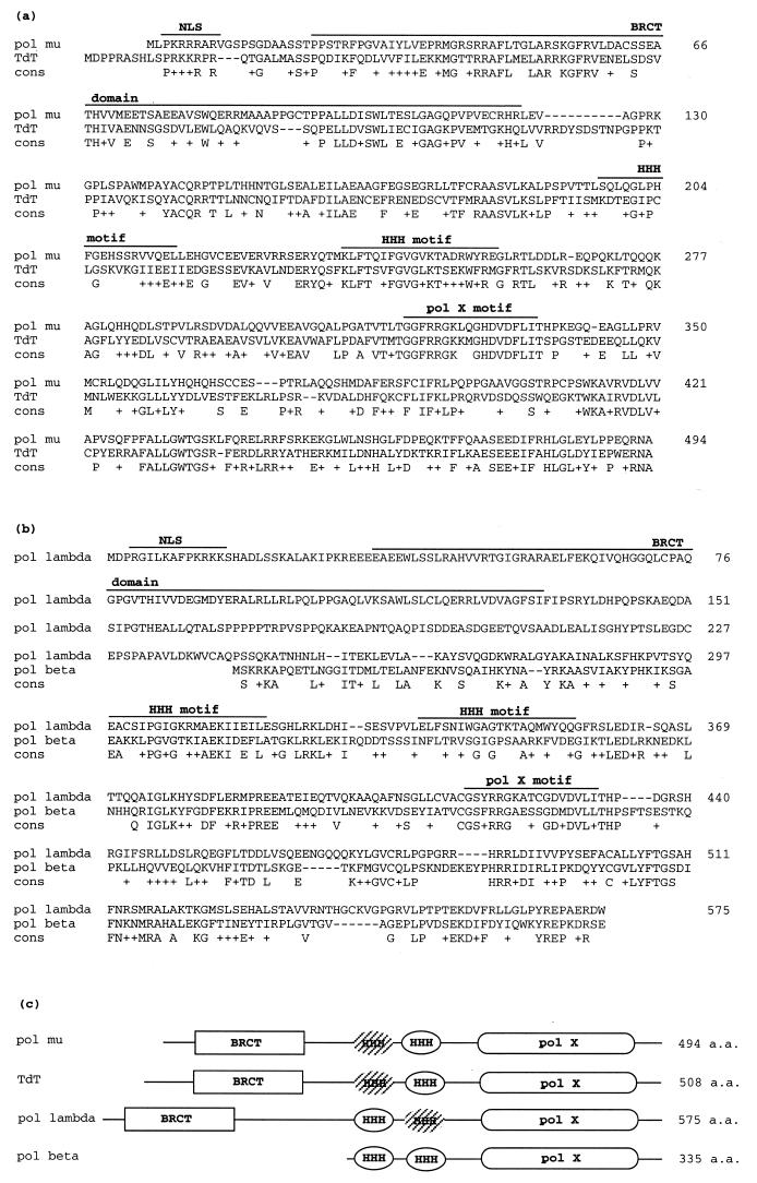
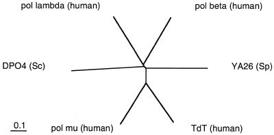
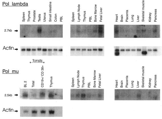
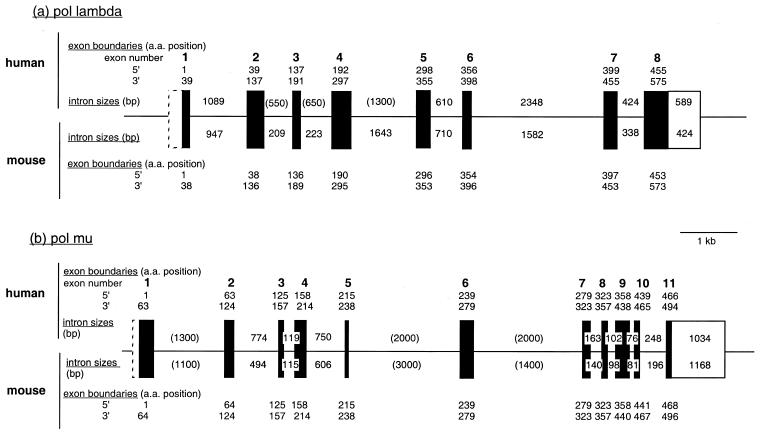
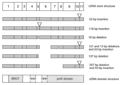
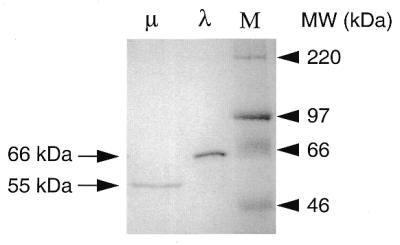
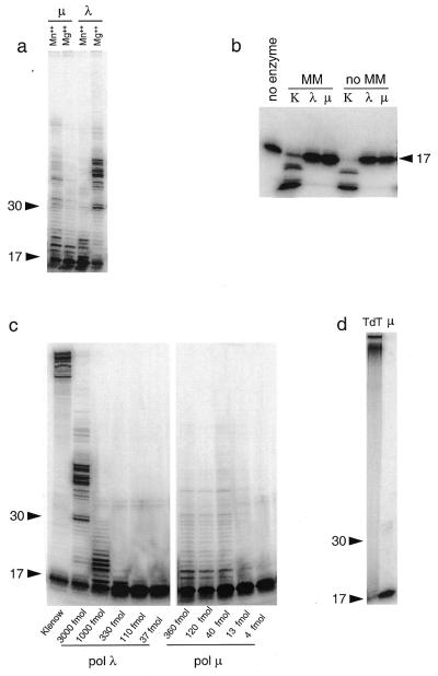
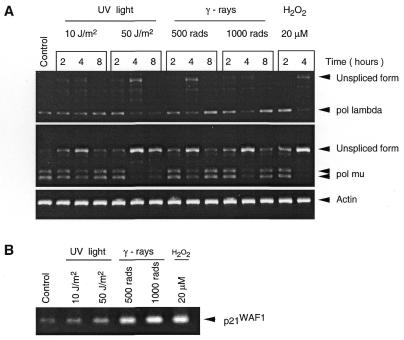
We describe here two novel mouse and human DNA polymerases: one (pol lambda) has homology with DNA polymerase beta while the other one (pol mu) is closer to terminal deoxynucleotidyltransferase. However both have DNA polymerase activity in vitro and share similar structural organization, including a BRCT domain, helix-loop-helix DNA-binding motifs and polymerase X domain. mRNA expression of pol lambda is highest in testis and fetal liver, while expression of pol mu is more lymphoid, with highest expression both in thymus and tonsillar B cells. An unusually large number of splice variants is observed for the pol mu gene, most of which affect the polymerase domain. Expression of mRNA of both polymerases is down-regulated upon treatment by DNA damaging agents (UV light, gamma-rays or H(2)O(2)). This suggests that their biological function may differ from DNA translesion synthesis, for which several DNA polymerase activities have been recently described. Possible functions are discussed.


Figures

Figure 1
Figure 1
Human pol µ and pol λ DNA polymerase protein sequences. (a) Comparison of human pol µ and TdT proteins performed using the advanced BLAST program. Identical residues are indicated below the alignment with (+) for conservative changes. Nuclear localization signal (NLS), BRCT domain (17), HHH motifs (16) and polX motif (Prosite database) are marked. Amino acid positions of the pol µ sequence are indicated. (b) Same comparison performed between human pol λ and DNA polymerase β. (c) Schematic domain organization of pol µ, TdT, pol λ and DNA polymerase β. Hatched HHH motifs contain residues divergent from consensus at the Gly-(hydrophobe)-Gly core positions. Amino acid (aa) length of the human proteins is indicated.
Figure 2
Figure 2
Phylogenetic comparison of yeast and human proteins of the polX family. Alignment of sequences was performed with the CLUSTALW program using the default settings. The bar corresponds to 0.1 amino acid substitution per residue. The protein accession numbers are as follows: yeast (S.cerevisiae), DP04, P25615; yeast (S.pombe), YA26, Q09693; human DNA polymerase β, P06746; human TdT, P04053.
Figure 3
Figure 3
Tissue-specific expression of human pol µ and pol λ genes. Multi-tissue northern blots from Clontech were hybridized with full-length human pol µ and pol λ cDNA probes. An additional blot was hybridized, containing 0.5 µg of poly(A)+ RNA from the BL2 cell line (18), from total human tonsils and CD19+CD38+ tonsillar cells and from thymus. Control hybridization with actin is shown once for each blot. Exposure times are 3 days for pol µ and pol λ hybridizations, and 75 min for actin.
Figure 4
Figure 4
Structure of mouse and human pol λ and pol µ genes. Exon–intron organization of human pol λ and pol µ is drawn on scale with mouse genes represented below. Intron sizes are indicated (in bp). Values in parentheses are size estimates, others correspond to sequenced regions. The mouse pol λ gene has been completely sequenced (chromosome 19, accession number AC003694), and sequencing of the human pol λ gene is in progress (chromosome 10, AC008027). The amino acid positions of exon boundaries are indicated. Coding exons are represented as black boxes, and non-coding exons as open boxes (dashed boxes for 5′-untranslated regions whose exact length is not known).
Figure 5
Figure 5
Splice variants for the human pol µ gene. Six representative examples of splice variants, isolated by RT–PCR amplification of poly(A)+ mRNA from the Ramos or the BL2 cell line with primers framing the coding sequence of pol µ, are depicted. The positions of the specific domains, BRCT, HHH and polX are indicated. Multiple events of alternate splicing are frequently observed within a cDNA (see last three examples).
Figure 6
Figure 6
SDS–PAGE profile of purified pol λ and pol µ. The fraction purified after heparin–Sepharose chromatography was analyzed by electrophoresis on 10% polyacrylamide gel and visualized by Coomassie blue staining. Pol µ (500 ng) and pol λ (2 µg) were loaded. M indicates molecular weight markers (Rainbow marker, Amersham).
Figure 7
Figure 7
Both purified pol λ and pol µ proteins exhibit a template-dependent DNA polymerase activity in vitro. (a) In vitro assay for template-dependent DNA polymerase activity. DNA polymerase activity was assayed by extension of a 5′-radiolabeled 17mer primer annealed to single-stranded circular M13mp18, as described in Materials and Methods, in the presence of either Mg++ or Mn++ metal activators. For each reaction, 75 fmol of substrate and 75 fmol of purified protein (either pol λ or pol µ) were used. Migration positions of 17mer and 30mer oligonucleotides are marked. (b) 3′→5′ exonuclease assay. The 3′→5′ exonuclease activity was investigated in the absence of dNTP on either 3′-mismatched (MM) or 3′-matched (no MM) primer-template substrate. Klenow fragment (0.5 U) was used as positive control. (c) Processivity assay. The processivity of pol λ and pol µ was assayed with a fixed amount of substrate (25 fmol of M13 template hybridized with the 17mer primer) incubated with decreasing amounts of polymerase as specified. Klenow fragment (2 × 10⁻³ U) was used as control. The weak doublet observed above the 30 nt marker originates from the labeled primer (see also below). (d) Assay for terminal transferase activity. TdT activity was assessed using 0.3 pmol of 5′-labeled single-stranded 17mer primer and 75 fmol of purified pol µ, in the presence of 250 µM of the four dNTPs. Calf thymus TdT (1 U) was used as positive control.
Figure 8
Figure 8
Pol µ and pol λ are down-regulated upon exposure to DNA damaging agents. (A) Following the DNA damaging treatment, Ramos cells were collected, total RNA extracted and semi quantitative RT–PCR performed, using the actin gene as control. The results of non-saturating PCR amplification conditions are illustrated. The various treatments are indicated. The size of the expected cDNA fragments, and of the unspliced forms is marked. For pol µ, the 5′ and 3′ primers are located in exons 9 and 11, respectively. The amplified doublet (246 and 299 bp) corresponds to the spliced mRNA with or without the 53 bp addition (see splicing variants). The unspliced form (570 bp) contains introns 9–10 and 10–11. For pol λ, the 5′ and 3′ primers located in exons 4 and 6, respectively, amplify a 360 bp fragment. The unspliced form (∼1 kb) corresponds to the presence of the sole intron 5–6, the fully unspliced species with both introns present being too large (∼2.3 kb) to be efficiently amplified in this assay. (B) Expression level of p21WAF1 gene was estimated by PCR amplification of the cDNAs from the 4 h time point of the different treatments.

Similar articles

Cited by

References

    1. Diaz M., Velez,J., Singh,M., Cerny,J. and Flajnik,M.F. (1999) Int. Immunol., 11, 825–833. - PubMed
    1. Masutani C., Kusumoto,R., Yamada,A., Dohmae,N., Yokoi,M., Yuasa,M., Araki,M., Iwai,S., Takio,K. and Hanaoka,F. (1999) Nature, 399, 700–704. - PubMed
    1. Gerlach V.L., Aravind,L., Gotway,G., Schultz,R.A., Koonin,E.V. and Friedberg,E.C. (1999) Proc. Natl Acad. Sci. USA, 96, 11922–11929. - PMC - PubMed
    1. McDonald J.P., Rapic-Otrin,V., Epstein,J.A., Broughton,B.C., Wang,X., Lehmann,A.R., Wolgemuth,D.J. and Woodgate,R. (1999) Genomics, 60, 20–30. - PubMed
    1. Ogi T., Kato,T.,Jr, Kato,T. and Ohmori,H. (1999) Genes Cells, 4, 607–618. - PubMed

Publication types

MeSH terms

Associated data