. 2000 Sep 26;97(20):10808-13.
doi: 10.1073/pnas.190230097.

Femtosecond dynamics of the forbidden carotenoid S1 state in light-harvesting complexes of purple bacteria observed after two-photon excitation

Affiliations

Femtosecond dynamics of the forbidden carotenoid S1 state in light-harvesting complexes of purple bacteria observed after two-photon excitation

P J Walla et al. Proc Natl Acad Sci U S A. .

Abstract

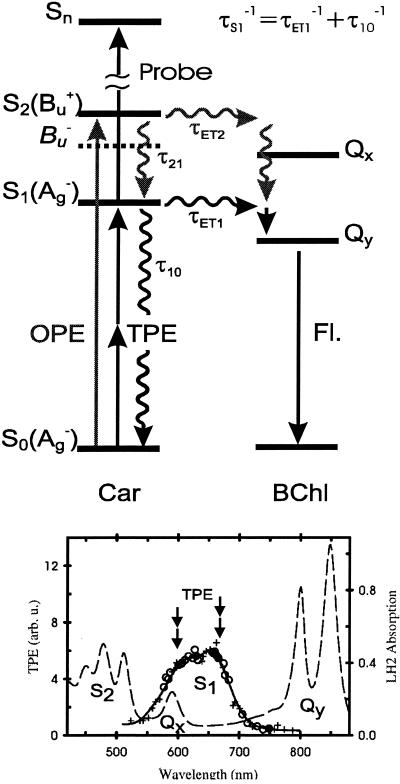
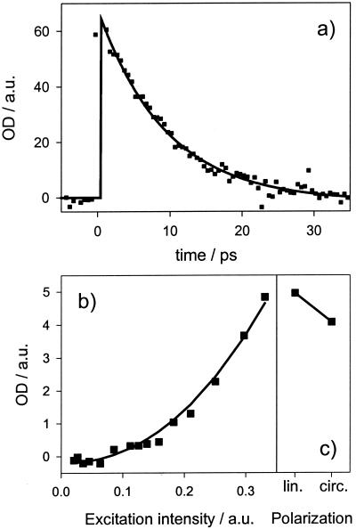
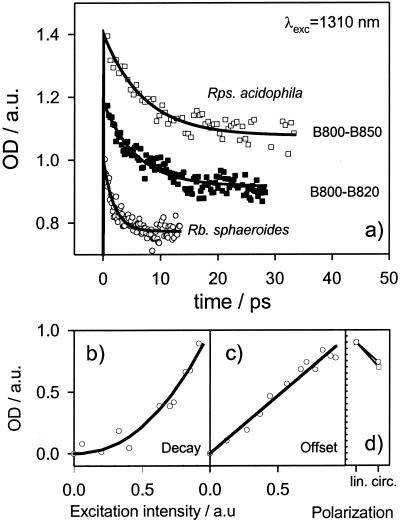
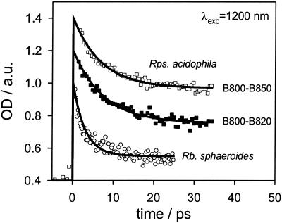
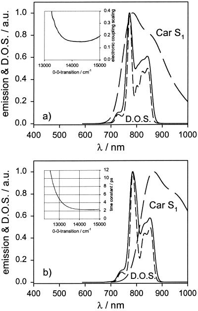
Time-resolved excited-state absorption intensities after direct two-photon excitation of the carotenoid S(1) state are reported for light-harvesting complexes of purple bacteria. Direct excitation of the carotenoid S(1) state enables the measurement of subsequent dynamics on a fs time scale without interference from higher excited states, such as the optically allowed S(2) state or the recently discovered dark state situated between S(1) and S(2). The lifetimes of the carotenoid S(1) states in the B800-B850 complex and B800-B820 complex of Rhodopseudomonas acidophila are 7+/-0.5 ps and 6+/-0.5 ps, respectively, and in the light-harvesting complex 2 of Rhodobacter sphaeroides approximately 1.9+/-0.5 ps. These results explain the differences in the carotenoid to bacteriochlorophyll energy transfer efficiency after S(2) excitation. In Rps. acidophila the carotenoid S(1) to bacteriochlorophyll energy transfer is found to be quite inefficient (phi(ET1) <28%) whereas in Rb. sphaeroides this energy transfer is very efficient (phi(ET1) approximately 80%). The results are rationalized by calculations of the ensemble averaged time constants. We find that the Car S(1) --> B800 electronic energy transfer (EET) pathway ( approximately 85%) dominates over Car S(1) --> B850 EET ( approximately 15%) in Rb. sphaeroides, whereas in Rps. acidophila the Car S(1) --> B850 EET ( approximately 60%) is more efficient than the Car S(1) --> B800 EET ( approximately 40%). The individual electronic couplings for the Car S(1) --> BChl energy transfer are estimated to be approximately 5-26 cm(-1). A major contribution to the difference between the energy transfer efficiencies can be explained by different Car S(1) energy gaps in the two species.


Figures

Figure 1
Figure 1
(Upper) Energy diagram for LH2. Black arrows indicate energy pathways occurring in the TPE pump probe experiment. Gray arrows indicate additional energy pathways in conventional one-photon experiments. OPE, One-photon excitation. Fl., Fluorescence. τET2, Time constant for Car S2 → BChl EET [≈50 fs (30)]. τ21, Time constant for Car S2 → S1 IC [≈135 fs (30)]. τET1, Time constant for Car S1 → BChl EET. τ10, Time constant for Car S1 → S0 IC. τS1, (τET1−1 + τ10−1)−1 lifetime of the Car S1 state measured in this work. (Lower) TPE spectrum of LH2 of Rb. sphaeroides (line with symbols), one-photon absorption spectrum (dashed line). Data taken from ref. . TPE-wavelengths for the TPE pump probe experiment are indicated with double arrows.
Figure 2
Figure 2
(a) Time dependence of ESA (λdet = 550 nm) of β-carotene in octane (squares) observed after TPE (λexc = 1,310 nm). Monoexponential fit: 9 ± 0.2 ps (solid line). (b) Power dependence of ESA. Power fit: Exponent = 2.2 ± 0.3 (solid line). (c) Relative intensity of ESA excited with linear and circular polarized light (Ω = 0.84 ± 0.07). The “coherence spike” is very large in the two-photon experiments (off scale) and has a quadratic dependence on the excitation power and a linear dependence on the Car concentration. No spike was seen with a pure buffer sample. In experiments with chlorophyll a, which has no two-photon allowed states, the coherence spike was found to be substantially smaller in magnitude (data not shown).
Figure 3
Figure 3
(a) Time dependence of ESA (λdet = 550 nm) along with monoexponential fits with constant offset of the B800-B850 complex (□, τS1 = 7 ± 0.5 ps) and the B800-B820 complex (▪, τS1 = 6 ± 0.5 ps) of Rps. acidophila and LH2 of Rb. sphaeroides (○, τS1 = 1.9 ± 0.5 ps) observed after TPE (λexc = 1,310 nm). The traces have been normalized and offset from each other by 0.2. (b) Power dependence of the prefactor in the monoexponential fit to the data of Rb. sphaeroides (○). Power fit: Exponent = 2.2 ± 0.3 (solid line). (c) Power dependence of the constant offset in the fit to the data of Rb. sphaeroides (○). Solid line: Linear fit. (d) Relative intensity of the fluorescence obtained after TPE (λexc = 1,310 nm) with linear and circular polarized light of B800-B850 of Rps. acidophila (□, Ω = 0.80 ± 0.04) and LH2 of Rb. sphaeroides (○, Ω = 0.84 ± 0.04).
Figure 4
Figure 4
Time dependence of ESA (λdet = 550 nm) along with monoexponential fits with constant offset of the B800-B850 complex (□, τS1 = 7 ± 0.5 ps) and the B800-B820 complex (■, τS1 = 7.5 ± 0.5 ps) of Rps. acidophila and LH2 of Rb. sphaeroides (○, τS1 = 3 ± 0.5 ps) observed after TPE (λexc = 1,200 nm). The traces have been normalized and offset from each other by 0.2.
Figure 5
Figure 5
(a) Acceptor DOS in LH2 of Rb. sphaeroides calculated with (solid line) and without introducing disorder (short dashed line) before solving the eigenvalue problem for the acceptor aggregate along with Car S1 emission (long dashed line). Note that the shown DOS is not weighed by electronic coupling. For details see text. (Inset) Electronic coupling scaling factor (see text) needed to reproduce the experimentally observed time constant τET1 as a function of the 0–0 transition of the Car S1 emission. (b) Same results for the B800–B850 complex of Rps. acidophila. (Inset) Calculated time constants τET1 as a function of the Car S1 state 0–0 transition.

Similar articles

Cited by

References

    1. McDermott G, Prince S M, Freer A A, Hawthornthwaitelawless A M, Papiz M Z, Cogdell R J, Isaacs N W. Nature (London) 1995;374:517–521.
    1. McDermott G, Prince S M, Freer A A, Isaacs N W, Papiz M Z, Hawthornthwaitelawless A M, Cogdell R J. Protein Eng. 1995;8:43–43.
    1. Koepke J, Hu X C, Muenke C, Schulten K, Michel H. Structure (London) 1996;4:581–597. - PubMed
    1. Sundstrom V, Pullerits T, van Grondelle R. J Phys Chem B. 1999;103:2327–2346.
    1. Nagarajan V, Johnson E T, Williams J C, Parson W W. J Phys Chem B. 1999;103:2297–2309.

Publication types

LinkOut - more resources