

Measuring the thickness of the human cerebral cortex from magnetic resonance images
- PMID: 10984517
- PMCID: PMC27146
- DOI: 10.1073/pnas.200033797
Measuring the thickness of the human cerebral cortex from magnetic resonance images
Abstract
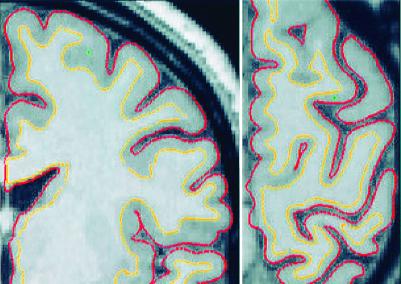
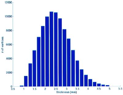
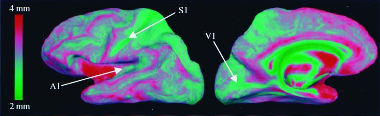
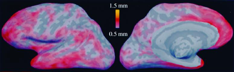
Accurate and automated methods for measuring the thickness of human cerebral cortex could provide powerful tools for diagnosing and studying a variety of neurodegenerative and psychiatric disorders. Manual methods for estimating cortical thickness from neuroimaging data are labor intensive, requiring several days of effort by a trained anatomist. Furthermore, the highly folded nature of the cortex is problematic for manual techniques, frequently resulting in measurement errors in regions in which the cortical surface is not perpendicular to any of the cardinal axes. As a consequence, it has been impractical to obtain accurate thickness estimates for the entire cortex in individual subjects, or group statistics for patient or control populations. Here, we present an automated method for accurately measuring the thickness of the cerebral cortex across the entire brain and for generating cross-subject statistics in a coordinate system based on cortical anatomy. The intersubject standard deviation of the thickness measures is shown to be less than 0.5 mm, implying the ability to detect focal atrophy in small populations or even individual subjects. The reliability and accuracy of this new method are assessed by within-subject test-retest studies, as well as by comparison of cross-subject regional thickness measures with published values.
Figures

References
-
- Zilles K. In: The Human Nervous System. Paxinos G, editor. San Diego: Academic; 1990. pp. 757–802.
-
- von Economo C. The Cytoarchitectonics of the Human Cerebral Cortex. London: Oxford Univ. Press; 1929.
-
- Brodmann K. Vergleichende Lokalisationslehre der Großhirnrinde in ihren Prinzipien dargestellt auf Grund des Zellenbaues. Leipzig, Germany: Barth; 1909.
-
- De Leon M J, George A E, Golomb J, Tarshish C, Convit A, Kluger A, De Santi S, McRae T, Ferris S H, Reisberg B, et al. Neurobiol Aging. 1997;18:1–11. - PubMed
Publication types
MeSH terms
Grants and funding
LinkOut - more resources
Full Text Sources
Other Literature Sources
Medical
Research Materials
