. 2000 Sep 12;97(19):10395-9.
doi: 10.1073/pnas.97.19.10395.

Molecular basis for CD40 signaling mediated by TRAF3

Affiliations

Molecular basis for CD40 signaling mediated by TRAF3

C Z Ni et al. Proc Natl Acad Sci U S A. .

Abstract

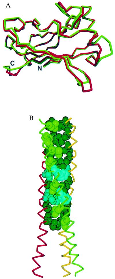
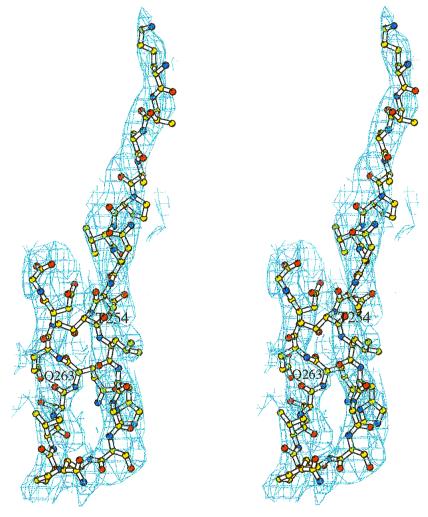
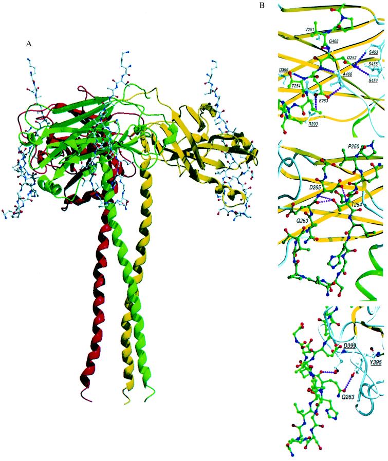
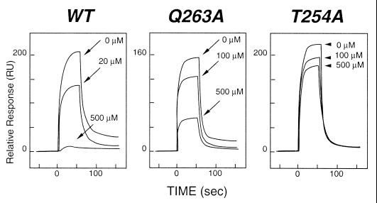
Tumor necrosis factor receptors (TNFR) are single transmembrane-spanning glycoproteins that bind cytokines and trigger multiple signal transduction pathways. Many of these TNFRs rely on interactions with TRAF proteins that bind to the intracellular domain of the receptors. CD40 is a member of the TNFR family that binds to several different TRAF proteins. We have determined the crystal structure of a 20-residue fragment from the cytoplasmic domain of CD40 in complex with the TRAF domain of TRAF3. The CD40 fragment binds as a hairpin loop across the surface of the TRAF domain. Residues shown by mutagenesis and deletion analysis to be critical for TRAF3 binding are involved either in direct contact with TRAF3 or in intramolecular interactions that stabilize the hairpin. Comparison of the interactions of CD40 with TRAF3 vs. TRAF2 suggests that CD40 may assume different conformations when bound to different TRAF family members. This molecular adaptation may influence binding affinity and specific cellular triggers.


Figures

Figure 1
Figure 1
The overall structure of the TRAF3 subunit is composed of an elongated helix followed by an eight-stranded β-sandwich (TRAF domain). (A) Superimposition of the polypeptide backbone of the C-terminal TRAF domain of TRAF3 (red) and TRAF2 (24). The rms deviation between corresponding α-carbons is 1 Å. Strong homology (59% identical) exists between TRAF3 and TRAF2 in this domain. The sandwich is formed by two layers of β-sheet, each with four antiparallel strands. The topology of this β-sandwich is found thus far only in TRAF domains. (B) The TRAF-N domain of TRAF3 is a long amphipathic α-helix that forms a coiled-coil when TRAF3 trimerizes (see Fig. 3). Residues 300–347 were ordered in the electron density map. The coiled-coil interactions are stabilized by nine heptad repeats of hydrophobic residues. This hydrophobic pattern is interrupted at residues 324 and 331 where three histidines are found in the interior of the coiled-coil. The side chains of these histidines extend out of the coiled-coil. The TRAF3 fragment is considerably longer at the N terminus than the TRAF2 fragment, where residues from the N terminus were missing or disordered (24, 33). This model provides structural details for most of the helical TRAF-N region.
Figure 2
Figure 2
Model for the CD40 fragment displayed in the electron density map. The 3.5-Å 2FoFc map was contoured at 0.75σ and is displayed in stereo. For modeling, the peptide was fitted to FoFc difference maps and omitmaps (36). Clear electron density was visible for the backbone to position the bound peptide; however, density was weak for some side chains. The reverse turn configuration was clearly defined.
Figure 3
Figure 3
TRAF3/CD40 interactions. (A) Schematic drawing of the TRAF3 trimer with the polypeptide backbone of TRAF3 presented as a ribbon model and the CD40 peptide shown as a ball and stick model. One CD40 fragment binds to each TRAF3 monomer at the edge of the TRAF3 domain, crossing one β-sheet. No conformational changes were seen when comparing TRAF3 alone or bound to CD40. (B) Close-up view of the CD40 fragment bound to TRAF2 and TRAF3. Residues in CD40 are labeled, and critical contact residues in TRAF3 are also marked and underlined for identification. Interactions within 3.0 Å that are proposed to dictate specific recognition of CD40 and TRAF3 or TRAF2 are shown as dotted lines. The images show intramolecular hydrogen bonds within the CD40 fragment that stabilize the reverse turn (Middle) and direct contacts between CD40 and TRAF3 (Bottom). These images can be contrasted with the contacts in TRAF2 (Top; 1CZZ-; based on figure 3 of Ye et al. in ref. 34).
Figure 4
Figure 4
Inhibition of TRAF3 binding to CD40 by synthetic peptides. Binding was measured by using surface plasmon resonance with a BIACORE 3000 instrument (Biacore AB, Uppsala). A recombinant fragment representing the entire cytoplasmic domain of CD40 was cloned as a glutathione S-transferase fusion protein, expressed in Escherichia coli, and purified by affinity chromatography on a glutathione-Sepharose (Amersham Pharmacia) column. The fusion partner was removed by thrombin digestion, and the CD40 fragment was purified by ion-exchange chromatography. The CD40 domain was immobilized on a Biacore CM5 sensor chip, and the TRAF3 TRAF domain was injected at 3.5 μM. The Left sensorgram shows the relative response for binding interactions between immobilized CD40 and TRAF3 and inhibition of this binding by a synthetic peptide from the cytoplasmic domain of CD40 (250PVQETLHGCQPVTQEDG266). A series of peptides with alanine substituted singly for each residue in the fragment was tested for inhibition of binding of TRAF3 to CD40. Mutant peptides were injected at concentrations ranging from 20 to 500 μM. As can be seen from the Center and Right sensorgrams, striking changes in inhibition were observed when alanine was substituted for Q263 or T254, suggesting reduced binding of the mutant fragments. Substitution of alanine for other residues in the peptide did not significantly alter levels of inhibition observed with the wild-type (WT) peptide.

Similar articles

Cited by

References

    1. Foy T M, Aruffo A, Bajorath J, Buhlmann J E, Noelle R J. Annu Rev Immunol. 1996;14:591–617. - PubMed
    1. Banchereau J, Bazan F, Blanchard D, Briere F, Galizzi J P, van Kooten C, Liu Y J, Rousset F, Saeland S. Annu Rev Immunol. 1994;12:881–922. - PubMed
    1. Clark E A, Ledbetter J A. Nature (London) 1994;367:425–428. - PubMed
    1. Eliopoulos A G, Dawson C W, Mosialos G, Floettmann J E, Rowe M, Armitage R J, Dawson J, Zapata J M, Kerr D J, Wakelam M J, et al. Oncogene. 1996;13:2243–2254. - PubMed
    1. Eliopoulos A G, Stack M, Dawson C W, Kaye K M, Hodgkin L, Sihota S, Rowe M, Young L S. Oncogene. 1997;14:2899–2916. - PubMed

Publication types

LinkOut - more resources