. 2000 Sep 12;97(19):10595-600.
doi: 10.1073/pnas.97.19.10595.

A cGMP-signaling pathway in a subset of olfactory sensory neurons

Affiliations

A cGMP-signaling pathway in a subset of olfactory sensory neurons

M R Meyer et al. Proc Natl Acad Sci U S A. .

Abstract

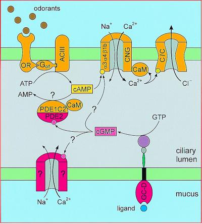
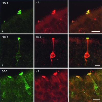
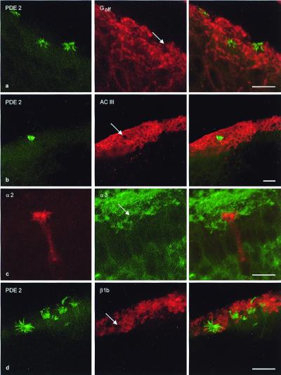
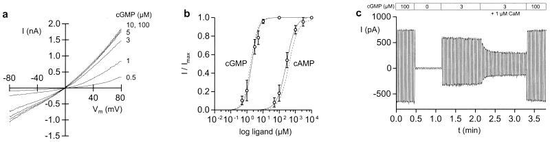
It is well established that signal transduction in sensory neurons of the rat olfactory epithelium involves a cAMP-signaling pathway. However, a small number of olfactory neurons specifically express cGMP-signaling components, namely a guanylyl cyclase (GC-D) and a cGMP-stimulated phosphodiesterase (PDE2). Here, we show that this subset of olfactory neurons expressing GC-D and PDE2 does also express the subunit of a cGMP-selective cyclic nucleotide-gated (CNG) channel that has been previously identified in cone photoreceptors. Further, components of the prototypical cAMP-signaling pathway could not be detected in this subpopulation of cells. These results imply that these neurons use an alternative signaling pathway, with cGMP as the intracellular messenger, and that, in these cells, the receptor current is initiated by the opening of cGMP-gated channels.


Figures

Figure 1
Figure 1
Two alternative hypotheses of cGMP signaling in a subset of OSNs. (Upper) An odorant receptor (OR) activates a cAMP-signaling pathway involving a G protein (Golf), an adenylyl cyclase (ACIII), a cyclic nucleotide-gated (CNG) channel (α3α4β1b), and a chloride channel (ClC). The cAMP is degraded by a CaM-dependent phosphodiesterase (PDE1C2). (Lower) Components of a cGMP-signaling pathway and putative targets of cGMP: receptor guanylyl cyclase GC-D; cGMP-regulated PDE2; an unknown cGMP-regulated ion channel; and the known CNG channel of the cAMP-signaling pathway.
Figure 2
Figure 2
Primary structure of the rat α2b channel. (A) The exon, which is missing in the cDNA of α2a, is highlighted by gray background. The transmembrane segments S1–S6, the pore region, and the cyclic-nucleotide (cNMP)-binding domain are indicated. Established intron positions (18) are indicated (filled triangles). Open triangles indicate intron positions that were not determined by genomic sequencing, but could be deduced because of the intron-exon-structure of α2 orthologs (23, 25) and the existence of consensus sites for exon-exon boundaries. (B) Comparison of the putative CaM-binding sites of α2a and α2b. Because of alternative usage of exon 3, rat α2a and α2b possess different putative CaM-binding sites. The position of the amino acids is indicated on the right (for α2b). The 1–5-8–14 motif (26, 51) of the putative CaM-binding sites is indicated (numbers at the top).
Figure 3
Figure 3
Immunohistochemical localization of the α2 subunit. (a) Section through the rat retina. (Left) Differential interference contrast; (Right) anti-α2 immunofluorescence in cone outer segments. ROS, rod outer segments; IS, inner segments; ONL, outer nuclear layer; OPL, outer plexiform layer; INL, inner nuclear layer; IPL, inner plexiform layer; GCL, ganglion cell layer. (b) Coronal sections through the rat olfactory epithelium. (Left) Anti-α2 immunoreactivity shown by dark diaminobenzidine reaction product. Cilia of a single cell are strongly stained; weaker staining is found throughout the cell body. (Right) Labeled cells are preferentially found in recesses of the olfactory turbinates close to the cribriform plate. C, ciliary layer; SC, supporting cell layer; OSN, somata of olfactory sensory neurons. Scale bars = 50 μm in a and b Right; and 10 μm in b Left.
Figure 4
Figure 4
The α2 subunit is coexpressed with PDE2 and GC-D. (a Left) Immunofluorescent detection of PDE2 in cilia of two cells; (Middle) anti-α2 immunofluorescence (antibody FPc66K); (Right) merged image showing colocalization of PDE2 and α2. (b Left) anti-PDE2 immunofluorescence; (Middle) anti-GC-D immunofluorescence; (Right) merged image; both proteins are colocalized. (c Left) anti-GC-D immunofluorescence; (Middle) anti-α2 immunofluorescence (antibody CNC9C1); (Right) merged image; both proteins are colocalized. Scale bars = 10 μm.
Figure 5
Figure 5
Components of the olfactory cAMP-signaling pathway are absent from α2-positive neurons. (a Left) Cilia of three PDE2-positive cells; plane of section is slightly oblique. (Middle) Golf/Gs-positive cilia form a continuous layer; arrow indicates the position of one PDE2-positive ciliary bundle in the red “lawn.” (Right) Merged image, no colocalization is observed. (b Left) Cilia of a PDE2-positive cell; (Middle) ACIII-positive cilia form a continuous layer with a hole (arrow); (Right) merged image; no colocalization. (c Left) Anti-α2 immunofluorescence. (Middle) α3-positive cilia; note the hole (arrow). (Right) Merged image, no colocalization. (d Left) PDE2-positive cilia. (Middle) β1 is found throughout the ciliary layer; arrow indicates the position of a PDE2-positive ciliary bundle. (Right) Merged image, no colocalization. Scale bars = 10 μm.
Figure 6
Figure 6
Properties of heterologously expressed α2 channels. (a) Current-voltage (I/Vm) relations of α2a channels in the presence of different cGMP concentrations. (b) Dose-response curves for α2a (solid lines) and α2b (dashed lines) at Vm = +60 mV. All currents were normalized to currents at saturating cGMP. For clarity, data points and error bars were omitted from dashed traces. (c) α2 channels are modulated by CaM. Vm was switched from −40 mV to +40 mV in 1-s intervals. The inside-out patch (α2b) was superfused with 100 μM cGMP to record saturating currents, followed by superfusion in 0 μM cGMP. During superfusion with 3 μM cGMP for 1 min, the current stayed constant. With 3 μM cGMP and 1 μM CaM, the current decreased by roughly 50% within 30 s. The current at saturating cGMP (100 μM) was not altered by CaM.

Similar articles

Cited by

References

    1. Gold G H. Annu Rev Physiol. 1999;61:857–871. - PubMed
    1. Buck L, Axel R. Cell. 1991;65:175–187. - PubMed
    1. Mombaerts P. Nat Neurosci. 1999;2:686–687. - PubMed
    1. Bakalyar H A, Reed R R. Science. 1990;250:1403–1406. - PubMed
    1. Jones D T, Reed R R. Science. 1989;244:790–795. - PubMed

Publication types

Associated data

LinkOut - more resources