. 2000 Sep 12;97(19):10613-8.
doi: 10.1073/pnas.180223197.

A common polymorphism associated with antibiotic-induced cardiac arrhythmia

Affiliations

A common polymorphism associated with antibiotic-induced cardiac arrhythmia

F Sesti et al. Proc Natl Acad Sci U S A. .

Abstract

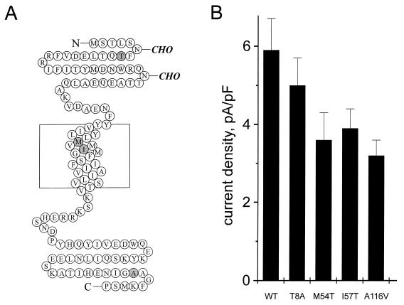
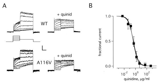
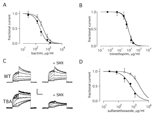
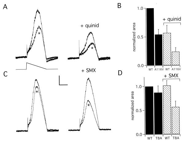
Drug-induced long QT syndrome (LQTS) is a prevalent disorder of uncertain etiology that predisposes to sudden death. KCNE2 encodes MinK-related peptide 1 (MiRP1), a subunit of the cardiac potassium channel I(Kr) that has been associated previously with inherited LQTS. Here, we examine KCNE2 in 98 patients with drug-induced LQTS, identifying three individuals with sporadic mutations and a patient with sulfamethoxazole-associated LQTS who carried a single-nucleotide polymorphism (SNP) found in approximately 1.6% of the general population. While mutant channels showed diminished potassium flux at baseline and wild-type drug sensitivity, channels with the SNP were normal at baseline but inhibited by sulfamethoxazole at therapeutic levels that did not affect wild-type channels. We conclude that allelic variants of MiRP1 contribute to a significant fraction of cases of drug-induced LQTS through multiple mechanisms and that common sequence variations that increase the risk of life-threatening drug reactions can be clinically silent before drug exposure.


Figures

Figure 1
Figure 1
KCNE2 missense mutations diminish IKr current density. (A) KCNE2 polymorphisms identified in patients with acquired LQTS. Schematic of the predicted primary sequence and membrane topology of human MiRP1: a single transmembrane segment is based on the predicted sequence of MiRP1and its homology to MinK; an externally-oriented amino-terminal domain is based on antibody localization (9). Residues that were altered in patients in this study are indicated. (B) Current density assessed by normalizing steady-state tail currents at −40 mV to the cell capacitance for CHO cells expressing the indicated MiRP1 variants and HERG and studied by the standard protocol (Materials and Methods). Each bar represents the mean ± SEM for a group of 34–40 cells; the mean decrease in current density compared with wild-type MiRP1 was 15, 39, 34, and 47% for T8A-MiRP1, M54T-MiRP1, I57T-, and A116V-MiRP1, respectively, with P < 10−4 for each variant compared with wild type (unpaired t test).
Figure 2
Figure 2
IKr channels formed with A116V-MiRP1 show wild-type blockade by quinidine. CHO cells expressing wild-type MiRP1 or A116V-MiRP1 and HERG were studied by whole-cell clamp. (A) Whole-cell currents recorded from channels formed with wild-type MiRP1 (WT) and A116V-MiRP1 (A116V) in the absence (left column) or presence of 0.5 μg/ml of quinidine (+quinid). Scale bars represent 50 and 30 pA and 0.7 s for wild type and A116V-MiRP1, respectively. (Inset) Protocol: holding −80-mV, 1-s pulses from −60 to 20 mV in 10-mV steps, 2 s at −40 mV, 1-s interpulse interval. (B) Current–dose relationship for channels formed with wild-type MiRP1 (open circles) and A116V-MiRP1 (filled squares) in the presence of increasing amounts of quinidine. Data from peak currents at −40 mV after a prepulse to +20 mV by using the standard protocol. Theoretical lines were constructed to the function: 1/(1 + H) were H = ([drug]/Ki)n. Ki and n represent the equilibrium dissociation constant and the Hill coefficient, respectively. Fits give values for Ki and n of 0.41 ± 0.04 μg/ml and 1.1 ± 0.1 for wild-type MiRP1 and 0.43 ± 0.06 μg/ml and 1.1 ± 0.2 for A116V-MiRP1; 6 cells in each case.
Figure 3
Figure 3
Channels formed with T8A-MiRP1 are more sensitive to SMX. Currents and fits as in Fig. 2. (A) Current–dose relationship for channels formed with wild-type MiRP1 (open circles) and T8A-MiRP1 (filled circles) in the presence of increasing amounts of TMP/SMX. Data from peak currents at −40 mV after a prepulse to +20 mV by using the standard protocol. Fits give values for wild-type MiRP1 of Ki = 380 ± 40 μg/ml and n = 1.8 ± 0.3 and for T8A-MiRP1 of Ki = 210 ± 30 μg/ml and n = 1.9 ± 0.2; 13 cells in each case. (B) Current–dose relationships in the presence of TMP for wild-type MiRP1 (open circles) and T8A-MiRP1 (filled circles). Fits give values for Ki and n of 71 ± 12 μg/ml and 1.8 ± 0.4 for wild-type MiRP1 and 75 ± 9 μg/ml and 1.9 ± 0.3 for T8A-MiRP1; 5 cells each case. The data thus suggest inhibition was cooperative and two TMP molecules were required to block. (C) Whole-cell currents recorded from channels formed with wild-type MiRP1 (WT) and T8A-MiRP1 (T8A) in the absence (left column) or presence of 633 μg/ml of SMX (+SMX). Scale bars represent 50 pA and 40 pA for wild type and T8A, respectively, and 1 s. (D) Current–dose relationships in the presence of SMX for wild-type MiRP1 (open circles) and T8A-MiRP1 (filled circles). Fits give values for Ki and n of 2,600 ± 200 μg/ml and 1.6 ± 0.6 for wild-type MiRP1 and 675 ± 60 μg/ml and 1.4 ± 0.2 for T8A-MiRP1; 6 cells each case.
Figure 4
Figure 4
Missense mutations and drug inhibition combine to diminish potassium flux. Simulated cardiac action potentials were used to study channels formed with wild-type MiRP1, A116V-MiRP1 and T8A-MiRP1 with HERG in CHO cells. (A) Whole-cell currents for simulated action potentials with wild-type MiRP1 and A116V-MiRP1 (*) in the absence and presence of 0.5 μg/ml quinidine (+quinid). Scale bars represent 1.3 pA/pF and 0.8 s. (Inset) Protocol: holding −80 mV, voltage ramp from 40 to −80 mV, dV/dt = −71 mV/s. (B) Normalized potassium flux assessed by measurement of area under the curve for simulated action potentials in cells expressing wild-type MiRP1 (WT) or A116V-MiRP1 (A116V) in the presence of 0.5 μg/ml quinidine (+quinid) normalized to wild-type channels without drug. Currents were divided by cell capacitance before normalization. Each bar represents the mean ± SEM for 6–10 cells. (C) Whole-cell currents for simulated action potentials with wild-type MiRP1 and T8A-MiRP1 (*) in the absence and presence of 300 μg/ml SMX. Scale bars represent 1.6 pA/pF and 0.8 s. (D) Normalized potassium flux assessed by measurement of area under the curve for simulated action potentials in cells expressing wild-type MiRP1(WT) and T8A-MiRP1 (T8A) in the presence of 300 μg/ml SMX normalized to wild-type channels without drug. Currents were divided by cell capacitance before normalization. Each bar represents the mean ± SEM for 7–10 cells.

References

    1. Roden D M, Lazzara R, Rosen M, Schwartz P J, Towbin J, Vincent G M. Circulation. 1996;94:1996–2012. - PubMed
    1. Ackerman M J, Clapham D E. N Engl J Med. 1997;336:1575–1586. - PubMed
    1. Keating M T, Sanguinetti M C. Science. 1996;272:681–685. - PubMed
    1. Schwartz P J, Priori S G, Napolitano C. In: Cardiac Electrophysiology: From Cell to Bedside. Zipes D P, Jalife J, editors. Philadelphia: Saunders; 2000. pp. 597–615.
    1. Bennett P B, Yazawa K, Makita N, George A L., Jr Nature (London) 1995;376:683–685. - PubMed

Publication types