

Immune reaction against the cytoskeleton in coeliac disease
- PMID: 10986212
- PMCID: PMC1728086
- DOI: 10.1136/gut.47.4.520
Immune reaction against the cytoskeleton in coeliac disease
Abstract
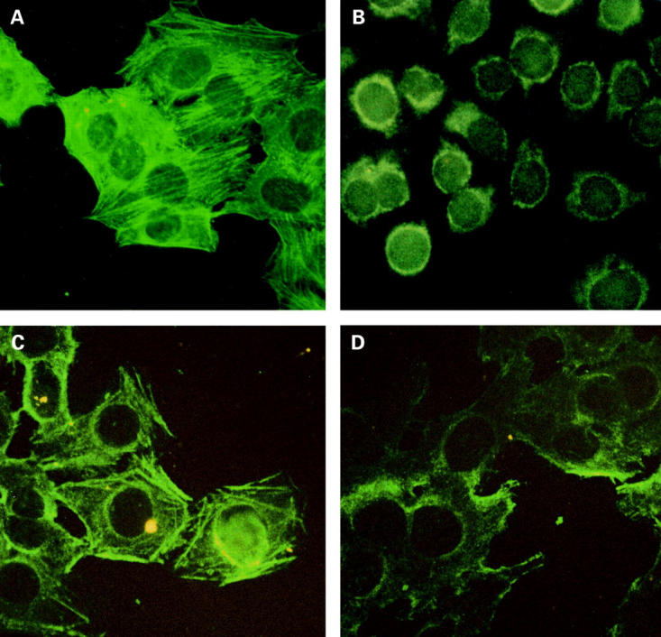
Background: The cytoskeleton actin network of intestinal microvilli has been found to be rapidly impaired after gluten challenge in coeliac disease (CD). The aim of this study was to investigate the presence of an immune reaction towards cytoskeleton structures such as actin filaments in CD.
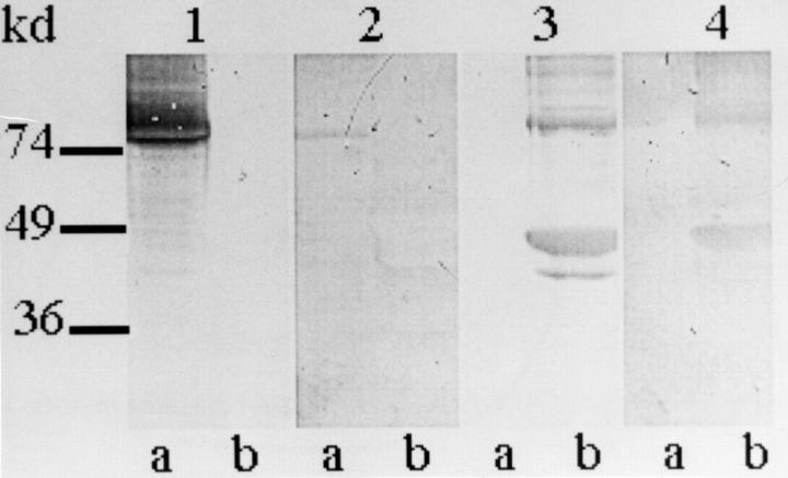
Methods: Eighty three antiendomysial antibody positive CD patients (52 children and 31 adults) were studied at our outpatient clinics from 1996 to 1998 using indirect immunofluorescence, ELISA, and western blotting for antiactin (AAA) and antitissue transglutaminase (TGA) antibodies before and after a gluten free diet (GFD). Sixteen patients with smooth muscle antibody positive autoimmune hepatitis, 21 with inflammatory bowel diseases, seven with small bowel bacterial overgrowth, and 60 healthy subjects were studied as controls.
Results: Fifty nine of 83 CD patients (28/31 adults (90.3%); 31/52 children (59.6%)) were positive for IgA and/or IgG AAA. Seventy seven (92.7%) were positive for IgA TGA. IgA AAA were strongly correlated with more severe degrees of intestinal villous atrophy (p<0.0001; relative risk 86.17). After a GFD, AAA became undetectable within five months.
Conclusions: Apart from the immune reaction against the extracellular matrix, we have described an immune reaction against the cytoskeleton in both children and adults with CD. As AAA are strongly associated with more severe degrees of villous atrophy, they may represent a useful serological marker of severe intestinal atrophy in CD.
Figures
References
Publication types
MeSH terms
Substances
LinkOut - more resources
Full Text Sources
Other Literature Sources
Medical
Research Materials
Miscellaneous