

doi: 10.1016/s0006-8993(00)02710-4.
Assessment of hindlimb gait as a powerful indicator of axonal loss in a murine model of progressive CNS demyelination
Affiliations
- PMID: 10986359
- PMCID: PMC5321677
- DOI: 10.1016/s0006-8993(00)02710-4
Item in Clipboard
Assessment of hindlimb gait as a powerful indicator of axonal loss in a murine model of progressive CNS demyelination
Brain Res.
.
Abstract
Identifying the role of axonal injury in the development of permanent, irreversible neurologic disability is important to the study of central nervous system (CNS) demyelinating diseases. Our understanding of neurologic dysfunction in demyelinating diseases and the ability to assess therapeutic interventions depends on the development of objective functional assays that can non-invasively measure axonal loss. In this study, we demonstrate in a murine model of progressive CNS demyelination that assessment of the hindlimb width of stride provides a powerful indicator of axonal loss and can dissociate between deficits induced by demyelination versus axonal loss.
Figures
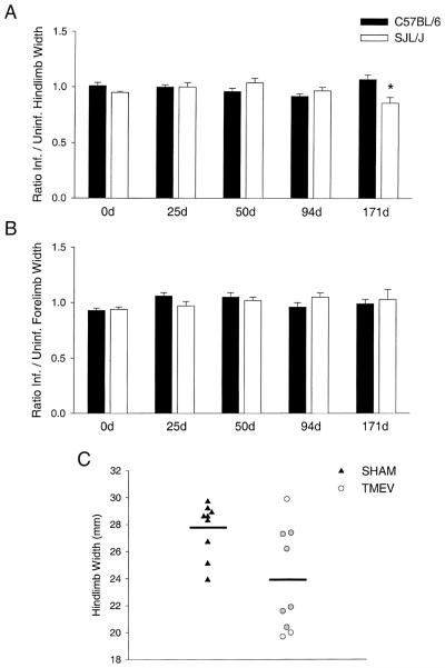
Assessment of hindlimb and forelimb stride width. Hindlimb (A) and forelimb (B) stride width were assessed in sham-infected and TMEV-infected C57BL/6 and SJL/ J mice at baseline (0d), 25, 50, 94, and 171 d.p.i. Data are represented as a ratio of infected / sham-infected hindlimb widths (mean±S.E.M.) at each time point. Statistical differences (denoted by an asterisk) were assessed by two-way repeated measures ANOVA. Pairwise comparisons were made using the Student–Newman–Keuls method (P<0.05). (C) Actual hindlimb widths (in mm) are shown for sham-infected (triangles) and TMEV-infected (circles) SJL/ J mice at 171 d.p.i. Horizontal lines represent the means for each group. The gray circles represent the six TMEV-infected mice that were randomly selected for quantification of axonal loss in the normal-appearing spinal cord white matter. (One TMEV-infected SJL/ J mouse could not be included on this graph because it was severely impaired and unable to walk at 171 d.p.i.).
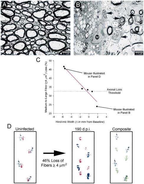
Alterations in hindlimb width as an indicator of spinal cord axonal loss. (A) An electron micrograph from the anterolateral columns of a sham-infected mouse illustrates normal myelin (thick black sheaths) surrounding intact axons. (B) In contrast, an electron micrograph from the lesion of a 190-day-infected mouse shows macrophage infiltration (M) and numerous degenerating axons (black arrows). (C) A strong negative correlation was found between the percentage of medium and large normally myelinated axon loss and the change in hindlimb width from baseline in chronically infected SJL/ J mice. Correlation coefficients were calculated using the Pearson product moment correlation (P<0.05). (D) A forelimb (red) and hindlimb (blue) stride pattern is shown for the same mouse before and after a 46% loss of medium and large axons. The composite image shows an overlay of hindlimb steps for this mouse at the baseline (blue) and 190 d.p.i. (green). Note that, after significant medium and large fiber loss, the infected mouse is one stride length short and there is a narrowing in the width of the stride.
References
-
- Trapp BD, Ransohoff RM, Rudick R. Axonal pathology in multiple sclerosis: relationship to neurologic disability. Curr. Opin. Neurol. 1999;12:295–302. - PubMed
-
- Bjartmar C, Yin XH, Trapp BD. Axonal pathology in myelin disorders. J. Neurocytol. 1999;28:383–395. - PubMed
-
- Dal Canto MC, Lipton HL. Recurrent demyelination in chronic central nervous system infection produced by Theiler’s murine encephalomyelitis virus. J. Neurol. Sci. 1979;42:391–405. - PubMed
-
- Lipton HL, Dal Canto MC. Chronic neurologic disease in Theiler’s virus infection of SJL / J mice. J. Neurol. Sci. 1976;30:201–207. - PubMed
Publication types
MeSH terms
Substances
Grants and funding
LinkOut - more resources
Full Text Sources
