. 2000 Sep 15;527 Pt 3(Pt 3):479-92.
doi: 10.1111/j.1469-7793.2000.00479.x.

Inhibition of a mammalian large conductance, calcium-sensitive K+ channel by calmodulin-binding peptides

Affiliations

Inhibition of a mammalian large conductance, calcium-sensitive K+ channel by calmodulin-binding peptides

A P Braun et al. J Physiol. .

Abstract

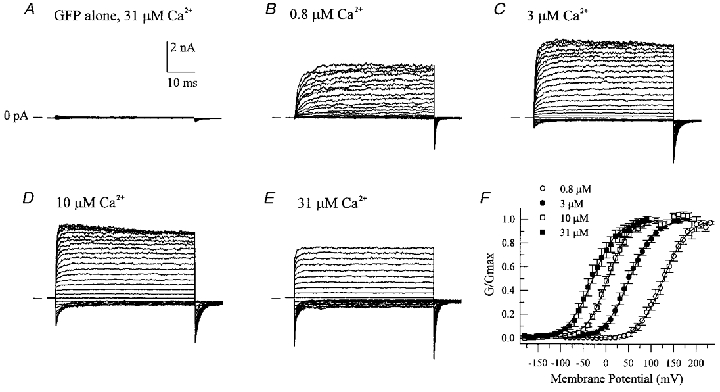
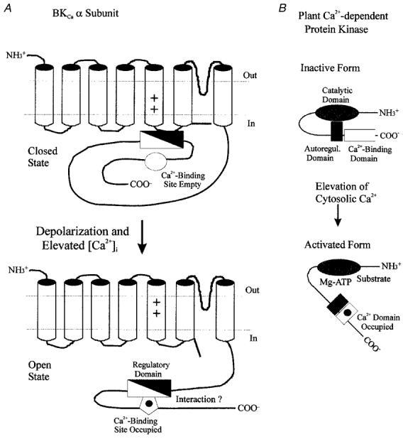
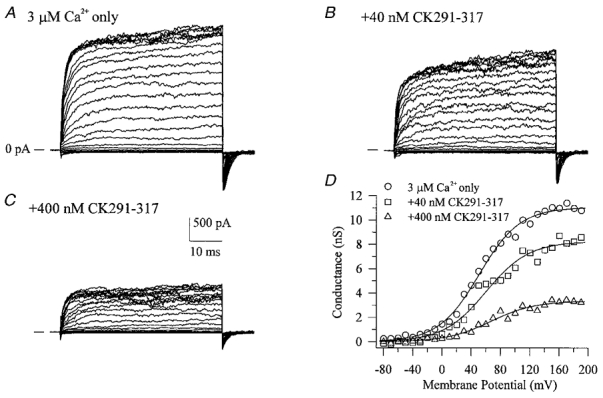
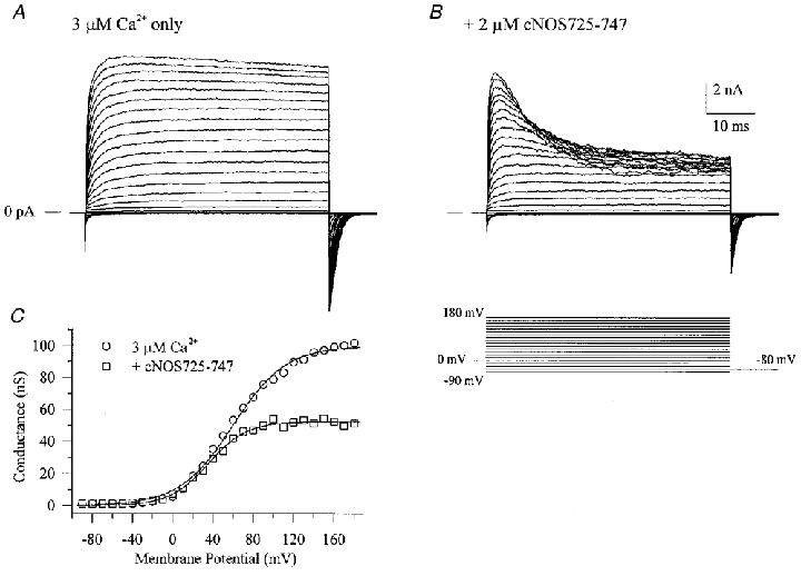
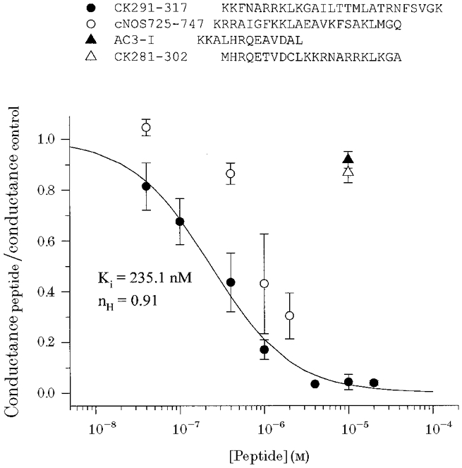
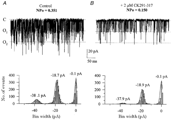
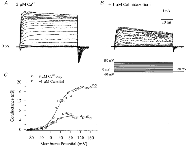
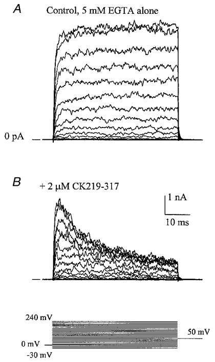
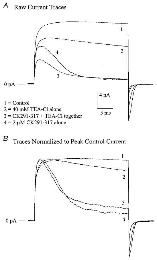
The large conductance, calcium-sensitive K+ channel (BKCa channel) is a voltage-activated ion channel in which direct calcium binding shifts gating to more negative cellular membrane potentials. We hypothesized that the calcium-binding domain of BKCa channels may mimic the role played by calmodulin (CaM) in the activation of calcium-CaM-dependent enzymes, in which a tonic inhibitory constraint is removed on CaM binding. To examine such a hypothesis, we used peptides from the autoregulatory domains of CaM kinase II (CK291-317) and cNOS (the constitutive nitric oxide synthase; cNOS725-747) as probes for the calcium-dependent activation of murine BKCa channels transiently expressed in HEK 293 cells. We found that these CaM-binding peptides produced potent, time-dependent inhibition of mammalian BKCa channel current following voltage-dependent activation. Inhibition was observed in both the presence and the absence of cytosolic free calcium. Similar application of CK291-31 had no effect on either the amplitude or kinetics of voltage-dependent, macroscopic currents recorded from rabbit smooth muscle Kv1.5 potassium channels transiently expressed in HEK 293 cells. Cytosolic application of both CK291-317 and tetraethylammonium (TEA) produced an additive and non-competitive block of BKCa current. This finding suggests that the peptide-binding site is distinct (e.g. outside the pore region of the channel) from that of TEA. Our results are thus consistent with a model in which the BKCa channel's voltage-dependent gating process is under an intramolecular constraint that is relieved upon calcium binding. The intrinsic calcium sensor of the channel may thus interact with an inhibitory domain present in the BKCa channel, and by doing so, remove an inhibitory 'constraint' that permits voltage-dependent gating to occur at more negative potentials.


Figures

Figure 1
Figure 1. Transient expression of mSlo α subunit in HEK 293 cells
Macroscopic currents were recorded from inside-out patches excised from cells expressing either GFP alone (A) or GFP + mSlo α subunit (B-E). Membrane voltage was stepped from −90 to +180 mV (A-D) or −180 to +90 mV (E) in the presence of the indicated amount of cytoplasmic free calcium. Tail currents recorded at either −80 mV (A-D) or −120 mV (E) were then used to calculate normalized conductance-voltage (G-V) relations. F shows the average G-V relations (±s.e.m.) calculated from 4–8 individual membrane patches; smooth curves represent fits to single Boltzmann functions, i.e. G/Gmax = 1/(1 + exp((V½Vm)/slope)) where 1/slope =zF/RT (Vm is membrane potential, V½ is the membrane potential at which 50% of the channels are open, and z, F, R and T have their usual meanings). Derived V½ and slope parameters are as follows: 0.8 μm Ca2+, 127.4 mV and 26.5 mV per e-fold change in G; 3 μm Ca2+, 51.7 mV and 26.9 mV per e-fold change in G; 10.5 μm Ca2+, 3.9 mV and 23.2 mV per e-fold change in G; 31 μm Ca2+, −29.1 mV and 27.0 mV per e-fold change in G.
Figure 2
Figure 2. Hypothetical mechanism of calcium-dependent gating in BKCa channels
The calcium-induced leftward shift of voltage-dependent gating in BKCa channels (A) may occur by a mechanism similar to that described for the activation of Ca2+-dependent protein kinases by calcium-CaM (Kemp & Pearson, 1991; Ito et al. 1991; Knighton et al. 1992; Nairn & Picciotto, 1994). In the latter situation, an intrinsic autoregulatory domain interacts with the enzyme's catalytic site, keeping it inactive. In the plant Ca2+-dependent protein kinase (B), this inactivation is removed by calcium binding to a CaM-like domain contained within the enzyme's primary structure, thereby allowing substrate and ATP to access the catalytic site. In BKCa channels, an analogous regulatory segment (filled triangle) may also exist, that could, for example, constrain movement of the channel's voltage sensor. Binding of calcium to its intrinsic site may then allow interaction of this site with a region of the regulatory segment (open triangle), and thus remove the constraint on the channel's gating mechanism. The intrinsic calcium-binding site of the BKCa channel may thus act in a similar fashion to Ca2+-CaM in the activation of Ca2+-dependent protein kinases.
Figure 3
Figure 3. Inhibition of BKCa channels by a CaM-binding peptide
A shows macroscopic currents recorded during voltage clamp steps from −90 to +180 mV in the presence of 3 μm cytoplasmic free calcium. Addition of a CaM-binding peptide (CK219–317) derived from the autoregulatory domain of CaM kinase II (Payne et al. 1988) to the cytoplasmic face of the excised patch produced a dose-dependent decrease in both outward and inward current amplitude (B and C). Conductance-voltage relations derived from tail current data in the absence and presence of CK291–317 are shown in D.
Figure 4
Figure 4. A CaM-binding peptide from cNOS inhibits BKCa channels
Macroscopic currents were recorded in the absence (A) or presence (B) of a CaM-binding peptide (cNOS725–747) applied to the cytoplasmic surface of the patch. cNOS725–747 was derived from the CaM-binding domain of constitutive nitric oxide synthase (Bredt et al. 1991; Zhang & Vogel, 1994). C shows conductance-voltage relations calculated from tail current data that have been fitted with single Boltzmann functions.
Figure 5
Figure 5. Plot of dose-dependent inhibition of BKCa channels by the CaM-binding peptides CK291–317 and cNOS725–747
BKCa channel inhibition was calculated as the fraction of total conductance remaining in the presence of increasing concentrations of peptide compared to control in the absence of peptide. The primary amino acid sequences of the 2 CaM-binding peptides (CK291–317 and cNOS725–747; Hanley et al. 1987; Zhang & Vogel, 1994), along with 2 other peptide inhibitors of CaM kinase II (AC3-I and CK281–302; Malinow et al. 1989; Braun & Schulman, 1995a) used as controls, are shown above the plot in single letter code. Data represent the means ±s.e.m. of 3–6 individual patches for each peptide. The smooth curve represents the fit of the CK291–317 data to a Hill equation as follows:
Figure 6
Figure 6. The CaM-binding peptide CK291–317 inhibits BKCa single channel events
In the presence of ∼200 μm cytoplasmic free calcium, single channel activity was recorded at a membrane voltage of −60 mV. A shows an excised inside-out patch containing at least 3 BKCa channels under control conditions; C denotes the closed level, O1 and O2 denote the amplitude levels of a single open channel and 2 simultaneously open channels, respectively. B shows the same patch following addition of 2 μm CK291–317 to the cytoplasmic surface. The all-points amplitude histograms below the current records were constructed using bin widths of 1 pA. Binned data in A and B were then fitted with Gaussian distributions, from which the peak amplitudes of each level were derived.
Figure 7
Figure 7. The CaM antagonist calmidazolium inhibits BKCa channels
Cytoplasmic application of 1 μm calmidazolium produced inhibition of both inward and outward macroscopic currents (B) compared to control currents in the presence of 3 μm cytoplasmic free calcium only (A). C shows conductance-voltage relations of the data in A and B; data points have been fitted with single Boltzmann functions.
Figure 8
Figure 8. The CaM-binding peptide CK291–317 inhibits BKCa channels in the absence of cytoplasmic free calcium
In the presence of 5 mm EGTA and no added calcium, macroscopic BKCa currents were recorded in response to voltage clamp steps from −30 to +240 mV (A). Following cytoplasmic application of 2 μm CK291–317 for 1 min, a time-dependent inhibition of current was observed during the 50 ms voltage clamp step (B). The voltage clamp protocol is shown at the bottom of the figure.
Figure 9
Figure 9. CK291–317 does not inhibit Kv1.5 voltage-dependent potassium channels
Macroscopic currents were recorded from recombinant rabbit vascular smooth muscle Kv1.5 channels in excised, inside-out membrane patches at 35 °C from a holding potential of −70 mV. Positive-going voltage clamp steps were delivered at a rate of 0.1 Hz, in 20 mV increments, followed by repolarization to a tail potential of −40 mV (refer to protocol in C). Following a series of voltage clamp steps under control conditions (A), 2 μm CK219–317 was applied to the cytoplasmic face of the patch and the same voltage clamp protocol was repeated (B). After washout of CK219–317, voltage clamp pulses were delivered in the presence of internal 0.5 mm 4-AP (C). Recovery from inhibition of the macroscopic currents was observed following washout of 4-AP from the bath (D). The horizontal and vertical scale bars shown apply to all four panels.
Figure 10
Figure 10. Internal TEA does not affect the time course of current inactivation by CK291–317
A, in the presence of 2 μm cytoplasmic free Ca2+, macroscopic BKCa channel currents were recorded from an inside-out patch in response to voltage clamps steps from 0 to +140 mV once every 4 s. Following control recordings (trace 1), 40 mm TEA-Cl was added to the bath solution, producing a significant decrease in amplitude (trace 2). The bath solution was then switched to one containing 40 mm TEA-Cl + 2 μm CK219–317, leading to a time-dependent inactivation of current (trace 3). TEA-Cl was then washed out, leaving only 2 μm CK291–317 (trace 4). B shows the relative time courses of current decay for traces 1–4, following normalization of each current to the peak current amplitude of trace 1. CK291–317 produces a similar rate of decay in the absence and presence of internal TEA. Note that traces 1–4 represent the averages of 5–8 individual sweeps recorded under each condition.

Similar articles

Cited by

References

    1. Asano M, Masuzawa-Ito K, Matsuda T. Charybdotoxin-sensitive K+ channels regulate the myogenic tone in the resting state of arteries from spontaneously hypertensive rats. British Journal of Pharmacology. 1993;108:214–222. - PMC - PubMed
    1. Asano M, Nomura Y, Ito K, Uyama Y, Imaizumi Y, Watanabe M. Increased function of voltage-dependent Ca2+ channels and Ca2+-activated K+ channels in resting state of femoral arteries from spontaneously hypertensive rats at prehypertensive stage. Journal of Pharmacology and Experimental Therapeutics. 1995;275:775–783. - PubMed
    1. Blatz AL, Magleby KL. Calcium-activated potassium channels. Trends in Neurosciences. 1987;10:463–467.
    1. Braun AP, Schulman H. A non-selective cation current activated via the multifunctional Ca2+-calmodulin-dependent protein kinase in human epithelial cells. The Journal of Physiology. 1995a;488:37–55. - PMC - PubMed
    1. Braun AP, Schulman H. The multifunctional calcium/calmodulin-dependent protein kinase: from form to function. Annual Review of Physiology. 1995b;57:417–445. - PubMed

Publication types

MeSH terms

Substances

LinkOut - more resources