. 2000 Sep 15;527 Pt 3(Pt 3):515-28.
doi: 10.1111/j.1469-7793.2000.00515.x.

Functional and molecular characterization of neuronal nicotinic ACh receptors in rat CA1 hippocampal neurons

Affiliations

Functional and molecular characterization of neuronal nicotinic ACh receptors in rat CA1 hippocampal neurons

S N Sudweeks et al. J Physiol. .

Abstract

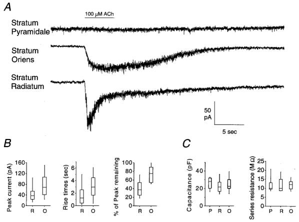
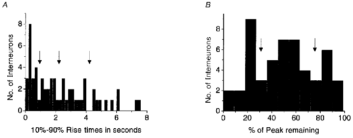
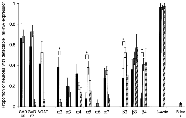
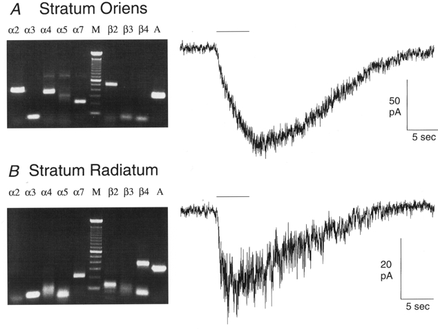
The molecular and functional properties of neuronal nicotinic acetylcholine receptors (nAChRs) were characterized from CA1 neurons in rat hippocampal slices using single-cell reverse-transcription polymerase chain reaction (RT-PCR) in conjunction with whole-cell patch-clamp recordings. We analysed the presence of the neuronal nAChR subunit mRNAs alpha2-7 and beta2-4, along with the mRNA for the GABAergic markers GAD (glutamic acid decarboxylase) 65 and 67 isoforms, and VGAT (vesicular GABA transporter) in interneurons from the stratum radiatum and stratum oriens, and in CA1 pyramidal neurons. Functional nAChR-mediated currents were detected in both interneuron populations, but not in pyramidal neurons. The neuronal nAChR subunit mRNAs detected in > 20 % of the populations examined were alpha4, alpha5, alpha7 and beta2-4 in stratum radiatum interneurons; alpha2, alpha3, alpha4, alpha7, beta2 and beta3 subunits in stratum oriens interneurons; and beta2-4 in pyramidal neurons. High levels of GABAergic marker mRNAs were detected in both interneuron populations, but not in pyramidal neurons. Significant co-expression of nAChR subunits within individual neurons was detected for alpha3 + alpha5, alpha4 + beta2, alpha4 + beta3, alpha7 + beta2, beta2 + beta3 and beta3 + beta4. The kinetics of the nAChR-mediated currents in response to the application of 100 microM ACh were significantly correlated with the expression of particular nAChR subunits. The alpha3, alpha7 and beta2 nAChR subunits were individually correlated with a fast rise time, the alpha2 nAChR subunit was correlated with a medium rise time, and the alpha4 nAChR subunit was correlated with a slow rise time. The alpha2 and alpha4 nAChR subunits were also significantly correlated with slow desensitization kinetics.


Figures

Figure 1
Figure 1. Whole-cell patch-clamp recordings of the nAChR-mediated currents
A, typical nAChR responses elicited by a 5 s application of ACh (indicated by horizontal bar) from each of the three neuronal populations examined. B, box plots showing the distributions of the peak currents, the 10–90 % rise times, and the percentage of the peak current remaining at the end of the 5 s ACh application for the two populations with functional nAChRs: stratum radiatum (R) and stratum oriens (O) interneurons. These were all significantly different between the two populations (Mann-Whitney test, P < 0.01). C, box plots representing the distributions for the capacitance and series resistance measurements from the three neuron populations examined: stratum pyramidale (P), stratum radiatum (R) and stratum oriens (O). The boxes represent the ranges of the 25th–75th percentiles (50th percentile is the middle line), and the bars represent the ranges of the 10th–90th percentiles.
Figure 4
Figure 4. Histograms of functional nAChR kinetic property distributions
These histograms were obtained from whole-cell recordings of 58 individual interneurons (53 of which were also examined by single-cell RT-PCR for nAChR subunits) in response to the application of 100 μm ACh. A, the distribution of the 10–90 % rise times. To compute correlations with nAChR subunit expression, interneurons were divided by their rise time into one of four groups, according to break-points in the histogram (indicated by arrows): 0–0.9 s, 0.91–2.20 s, 2.21–4.20 s and > 4.21 s. B, the distribution given by plotting the percentage of the peak current remaining at the end of the 5 s ACh application from each interneuron. To compute correlations between nAChR subunit expression and desensitization kinetics, interneurons were divided into one of three groups, according to break-points in the histogram (indicated by arrows): < 32 %, 32–75 % and > 75 %.
Figure 2
Figure 2. mRNA expression patterns by neuron type
The chart depicts the overall single-cell RT-PCR detection of each mRNA examined, by neuron type. To determine statistically significant expression, the proportion of neurons with detectable expression was compared with the proportion of overall false positives (shown in far right column, see Methods for calculation of false positives). There was statistically significant expression of all messages examined (in at least one neuron type), except for the α6 nAChR subunit. Stratum oriens interneurons (filled bars) had significant expression of GAD 65, GAD 67, VGAT and the α2, α3, α4, α7, β2 and β3 nAChR subunits. Stratum radiatum interneurons (open bars) had significant expression of GAD 65, GAD 67, VGAT and the α3, α4, α5, α7, β2, β3 and β4 nAChR subunits. Pyramidal neurons (hatched bars) had significant expression of the α4, α5, β2, β3 and β4 nAChR subunits. β-Actin expression was detected in over 95 % of all three neuron types. Statistically significant differences between the two populations expressing functional nAChRs (stratum oriens and stratum radiatum) are indicated by an asterisk. All statistical comparisons utilized a z test of two proportions, with P < 0.05. When comparing the detection of different subunits (i.e. α2 in stratum oriens vs.α3 in stratum oriens) it should be kept in mind that the detection limits for the subunits are not necessarily the same. The experimentally determined detection limits are reported in the Methods, and the approximated levels of relative subunit expression for each neuron population are reported in the Results.
Figure 3
Figure 3. RT-PCR products and whole-cell patch-clamp recordings
Two example agarose gels of RT-PCR products and the corresponding nAChR responses (100 μm applications of ACh for 5 s, indicated by horizontal bars) from the same interneurons. Expected RT-PCR product sizes are (in bp): α2 425, α3 305, α4 356, α5 396, α7 214, β2 507, β3 306, β4 382 and β-actin 327. The marker lane (M) shows bands at 100 bp increments. The stratum oriens interneuron (A) expressed detectable levels of the α2, α4, α7 and β2 nAChR subunits, and β-actin (A), and had a very slowly activating nAChR-mediated current. The stratum radiatum interneuron (B) expressed detectable levels of the α7 and β4 nAChR subunits, and β-actin, and had a significantly faster activating nAChR-mediated current than the stratum oriens interneuron. As these gels illustrate, non-specific bands occasionally occurred when running single-cell RT-PCR (see α4 and α5 in A, for examples), as would be expected from running multiplex PCR (in the initial round) and a total of 65 PCR cycles; spurious bands of the incorrect sizes were ignored. The subunit specificity of the PCR products from these gels was verified by direct sequencing of the DNA bands of the expected sizes after gel purification.

References

    1. Albuquerque EX, Pereira EFR, Castro NG, Alkondon M, Reinhardt S, Schroder H, Maelicke A. Nicotinic receptor function in the mammalian central nervous system. Annals of the New York Academy of Sciences. 1995;757:48–72. - PubMed
    1. Alkondon M, Albuquerque EX. Diversity of nicotinic acetylcholine receptors in rat hippocampal neurons. I. Pharmacological and functional evidence for distinct structural subtypes. Journal of Pharmacology and Experimental Therapeutics. 1993;265:1455–1473. - PubMed
    1. Alkondon M, Pereira EFR, Albuquerque EX. α-Bungarotoxin- and methyllcaconitine-sensitive nicotinic receptors mediate fast synaptic transmission in interneurons of rat hippocampal slices. Brain Research. 1998;810:257–263. - PubMed
    1. Alkondon M, Pereira EFR, Barbosa CT, Albuquerque EX. Neuronal nicotinic acetylcholine receptor activation modulates γ-aminobutyric acid release from CA1 neurons of rat hippocampal slices. Journal of Pharmacology and Experimental Therapeutics. 1997;283:1396–1411. - PubMed
    1. Anand R, Peng X, Lindstrom J. Homomeric and native α7 acetylcholine receptors exhibit remarkably similar but non-identical pharmacological properties, suggesting that the native receptor is a heteromeric protein complex. FEBS Letters. 1993;327:241–246. - PubMed

Publication types

MeSH terms

LinkOut - more resources