. 2000 Sep 15;527 Pt 3(Pt 3):611-22.
doi: 10.1111/j.1469-7793.2000.t01-1-00611.x.

Large vasodilatations in skeletal muscle of resting conscious dogs and their contribution to blood pressure variability

Affiliations

Large vasodilatations in skeletal muscle of resting conscious dogs and their contribution to blood pressure variability

A Just et al. J Physiol. .

Abstract

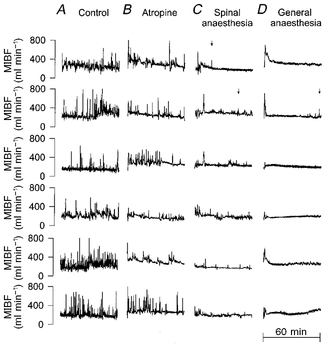
Large (up to +400 %) transient ( approximately 20 s) increases of blood flow were observed in the external iliac arteries of resting conscious dogs (n = 10) in the absence of major alerting or muscular activity. At the same time arterial pressure (AP) fell slightly while heart rate (HR) rose. The vasodilatations were resistant to atropine, ganglionic, beta-adrenergic and NO-synthase inhibition, but were suppressed by spinal or general anaesthesia. Vasodilatations of similar appearance were elicited by an alerting sound; these were abolished by atropine. The spontaneous vasodilatations occurred simultaneously and their magnitudes were well correlated between both legs, but were not correlated to the amount of concomitant activation of the surface electromyogram. The duration of this activation almost never outlasted 10 s. The reactive hyperaemia observed after a total occlusion of the artery even for 16 s was not large enough to explain the size of the spontaneous vasodilatations. Occlusion during peak flow of the vasodilatations did not affect the size of the reactive hyperaemia. Spectral analysis made separately for data segments with and without vasodilatation revealed that the vasodilatations substantially enhanced the variability of AP and HR at frequencies below approximately 0.1 Hz. In conclusion, large coordinated skeletal muscle vasodilatations were identified in resting conscious dogs, which are initiated neurally, but not by sympathetic-cholinergic or nitroxidergic fibres and which do not show any clear correlation to muscular contraction. The vasodilatations substantially affect the regulation of skeletal muscle blood flow and explain a significant portion of AP and HR variability.


Figures

Figure 1
Figure 1. Spontaneous vasodilatations in the hindlimb of conscious resting dogs
A, original trace of mean blood flow in the left external iliac artery (MIBF) recorded for 4 h. B, averaged time course of MIBF determined from all vasodilatations observed during 4 h in each dog. Mean (continuous line) ± s.e.m. (dotted lines) of the averages from 10 dogs. Data were smoothed by sliding averages over 2 s at 4 Hz. C, mean averaged time course of mean arterial blood pressure (MAP) concurrent with the vasodilatation. D, mean averaged time course of heart rate (HR). E, frequency histogram of the magnitude of all episodes expressed by the maximum MIBF reached. Error bars denote s.e.m. from 10 dogs.
Figure 3
Figure 3. Vasodilator response of mean iliac blood flow (MIBF) to a sudden loud noise
The sound was applied at time 0. Mean time course (continuous line) ± s.e.m. (dotted lines) from six dogs. A, control conditions, averaged from six responses in each dog. Note the close similarity to the time course of the spontaneous vasodilatations in Fig. 1B. B, after atropine, the vasodilator response was completely abolished.
Figure 4
Figure 4. Correlation between the vasodilatation and the concomitant surface electromyogram activity (EMG) in both legs
A, correlation between the total amount of flowed blood integrated from MIBF in the left (∫MIBF left) and the right iliac artery (∫MIBF right) during each of all individual vasodilatations detected in the 4 h recordings of four dogs. B, correlation between the integrated values of EMG and MIBF in the left leg (∫EMG left and ∫MIBF left; a.u., arbitrary units). C, correlation between integrated EMG activity recorded from the left (∫EMG left) and the right thigh (∫EMG right).
Figure 7
Figure 7. Spontaneous vasodilatations during control conditions and after ganglionic blockade
A, original traces of mean arterial pressure (MAP) and mean iliac blood flow (MIBF) during control conditions. B, original traces after ganglionic blockade. C, averaged time courses of MAP, heart rate (HR) and MIBF from all vasodilatations during control conditions. Means ± s.e.m. from six dogs. Data were smoothed by low pass filter < 3 Hz to allow spectral analysis. D, averaged time courses in the same six dogs after ganglionic blockade.
Figure 2
Figure 2. Original traces of mean iliac blood flow (MIBF) during control conditions, after atropine, spinal and general anaesthesia
Original traces from the second hour of the 2 h recordings of all six dogs (2 s mean values). Each row of traces represents the data from the same individual; each column shows experiments of the same type. A, time-control experiment, in which no drug was given. B, atropine, given at the beginning of the trace. C, spinal anaesthesia. The anaesthetic was slowly injected over the first 15 min of the trace. After that time the vasodilatations were largely suppressed. The vasodilatations that remained were smaller and less steep than usual and were always associated with movements of the forelimbs and the upper trunk. The lower three traces were recorded after additional ganglionic blockade. D, general anaesthesia. Sodium pentobarbitone was injected during the first few minutes of the trace. This probably caused the initial vasodilatation. Subsequently, the vasodilatations were completely abolished, except for the reoccurrence in one dog towards the end of the trace, when the anaesthesia waned. Arrows mark the only instances in the traces of C and D, in which a vasodilatation was detected by the detection algorithm. In A and B detection is not indicated.
Figure 5
Figure 5. Comparison between spontaneous vasodilatation and reactive hyperaemia after artificial occlusion of the iliac artery
A, mean iliac blood flow (MIBF, thick line) and integrated surface electromyogram activity (EMG, thin line) from a single vasodilatation. B, averaged response of MIBF to an occlusion of the iliac artery for a duration of 1, 2, 4, 8 or 16 s obtained in three dogs repeated at least three times in each dog. Numbers in the figure denote the duration of the occlusion. Data in A and B were smoothed by sliding averages over 2 s at 4 Hz.
Figure 6
Figure 6. Reactive hyperaemia after occlusion of the left iliac artery at the time of spontaneous vasodilatation
Time course of blood flow (MIBF) in the left (•) and in the right (○) iliac artery during single examples of spontaneous vasodilatation in three dogs. The left iliac artery was occluded as soon as a spontaneous vasodilatation could be anticipated. Data were smoothed by sliding averages over 2 s at 4 Hz.
Figure 8
Figure 8. Spectral content with and without spontaneous vasodilatation
A, power spectral density of mean arterial pressure (PSD-MAP), heart rate (PSD-HR) and mean iliac blood flow (PSD-MIBF) calculated from the data segments containing a spontaneous vasodilatation from Fig. 7C (thick line) as compared to spectra of those segments without vasodilatation (thin line). Means ± s.e.m. from six dogs. B, power density spectra from the same six dogs after ganglionic blockade.

Similar articles

Cited by

References

    1. Abrahams VC, Hilton SM, Zbrozyna AW. The role of active muscle vasodilatation in the alerting stage of the defence reaction. The Journal of Physiology. 1964;171:189–202. - PMC - PubMed
    1. Bendat JS, Piersol AG. Random Data: Analysis and Measurement Procedures. New York: Wiley; 1986.
    1. Bigger JTJ, Fleiss JL, Rolnitzky LM, Steinman RC. The ability of several short-term measures of RR variability to predict mortality after myocardial infarction. Circulation. 1993;88:927–934. - PubMed
    1. Camm AJ, Malik M, Bigger JT, Breithardt G, Cerutti S, Cohen RJ, Coumel P, Fallen EL, Kennedy HL, Kleiger RE, Lombardi F, Malliani A, Moss AJ, Rottman JN, Schmidt G, Schwartz PJ, Singer DH. Heart rate variability – standards of measurement, physiological interpretation, and clinical use (Task force of the European Society of Cardiology and the North American Society of Electrophysiology) Circulation. 1996;93:1043–1065. - PubMed
    1. Caraffa-Braga E, Granata L, Pinotti O. Changes in blood flow distribution during acute emotional stress in dogs. Pflügers Archiv. 1973;339:203–316. - PubMed

Publication types

MeSH terms

LinkOut - more resources