Clinical Trial
. 2000 Sep 15;527 Pt 3(Pt 3):633-9.
doi: 10.1111/j.1469-7793.2000.t01-1-00633.x.

Excitability changes induced in the human motor cortex by weak transcranial direct current stimulation

Affiliations
Clinical Trial

Excitability changes induced in the human motor cortex by weak transcranial direct current stimulation

M A Nitsche et al. J Physiol. .

Abstract

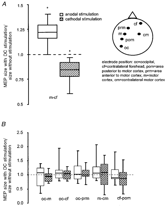
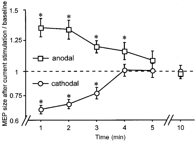
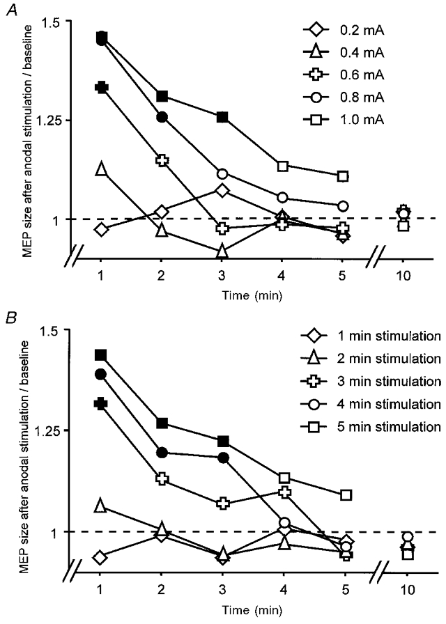
In this paper we demonstrate in the intact human the possibility of a non-invasive modulation of motor cortex excitability by the application of weak direct current through the scalp. Excitability changes of up to 40 %, revealed by transcranial magnetic stimulation, were accomplished and lasted for several minutes after the end of current stimulation. Excitation could be achieved selectively by anodal stimulation, and inhibition by cathodal stimulation. By varying the current intensity and duration, the strength and duration of the after-effects could be controlled. The effects were probably induced by modification of membrane polarisation. Functional alterations related to post-tetanic potentiation, short-term potentiation and processes similar to postexcitatory central inhibition are the likely candidates for the excitability changes after the end of stimulation. Transcranial electrical stimulation using weak current may thus be a promising tool to modulate cerebral excitability in a non-invasive, painless, reversible, selective and focal way.


Figures

Figure 1
Figure 1. Cortical excitability change during current flow
Rapidly induced effects of weak DC stimulation on the size of the motor-evoked potential (MEP) in the right abductor digiti minimi (ADM) muscle, revealed by transcranial magnetic stimulation (TMS), using the motor cortex-contralateral forehead arrangement (A), and the lack of effect using other diverse electrode positions (B). Normalised MEP amplitudes during stimulation are divided by normalised MEP amplitudes without stimulation. During DC stimulation, the MEP amplitude increased with anodal and decreased with cathodal current stimulation. Asterisks indicate significant differences between the values with and without stimulation (two-tailed t test, paired samples, P < 0.05). The boxes cover the range 25th to 75th percentiles, the error bars the 10th to 90th percentiles; the horizontal lines in the boxes indicate the median. Stimulation polarity always refers to the motor cortical electrode, respectively pre- and postmotor cortical electrode, except for the occipital-contralateral forehead condition, where it refers to the contralateral forehead electrode.
Figure 2
Figure 2. Polarity-specific after-effect of DC stimulation
Time course of polarity-specific motor cortex excitability changes outlasting stimulation duration, shown after 5 min DC stimulation at 1 mA. MEP amplitudes returned to baseline within 5 min. Asterisks indicate significant differences between MEP amplitudes after stimulation and at baseline (two-tailed t test, paired samples, P < 0.05).
Figure 3
Figure 3. Size and endurance of the DC stimulation after-effect depends on stimulation duration and current intensity
Dependency of the size of prolonged motor cortex excitability changes after anodal DC stimulation on current intensity (A) and stimulation duration (B). The MEP amplitudes relative to baseline are plotted against the time course. Filled symbols indicate significant differences (two-tailed t test, paired samples, P < 0.05) from the lowest stimulation intensity of 0.2 mA (A) or the shortest stimulation duration of 1 min (B). A minimum of 0.6 mA current intensity stimulation or a minimum stimulation duration of 3 min was needed to induce stimulation after-effects. Increasing either current intensity or stimulation duration led to prolonged and larger after-effects.

References

    1. Agnew WF, McCreery DB. Considerations for safety in the use of extracranial stimulation for motor evoked potentials. Neurosurgery. 1987;20:143–147. - PubMed
    1. Akimova IM, Novikova TA. Ultrastructural changes in the cerebral cortex following transcranial micropolarization. Biulleten Eksperimentalnoi Biologii i Meditsiny. 1978;86:737–739. - PubMed
    1. Artola A, Brocher S, Singer W. Different voltage-dependent thresholds for inducing long-term depression and long-term potentiation in slices of rat visual cortex. Nature. 1990;347:69–72. - PubMed
    1. Bindman LJ, Lippold OCJ, Redfearn JWT. Long-lasting changes in the level of the electrical activity of the cerebral cortex produced by polarizing currents. Nature. 1962;196:584–585. - PubMed
    1. Bindman LJ, Lippold OCJ, Redfearn JWT. The action of brief polarizing currents on the cerebral cortex of the rat (1) during current flow and (2) in the production of long-lasting after-effects. The Journal of Physiology. 1964;172:369–382. - PMC - PubMed

Publication types

LinkOut - more resources