. 2000 Oct;68(10):5679-89.
doi: 10.1128/IAI.68.10.5679-5689.2000.

Sequence polymorphism, predicted secondary structures, and surface-exposed conformational epitopes of Campylobacter major outer membrane protein

Affiliations

Sequence polymorphism, predicted secondary structures, and surface-exposed conformational epitopes of Campylobacter major outer membrane protein

Q Zhang et al. Infect Immun. 2000 Oct.

Abstract

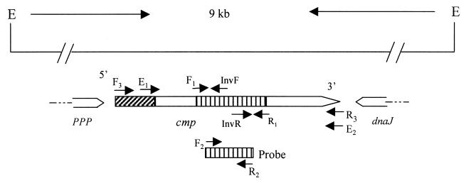
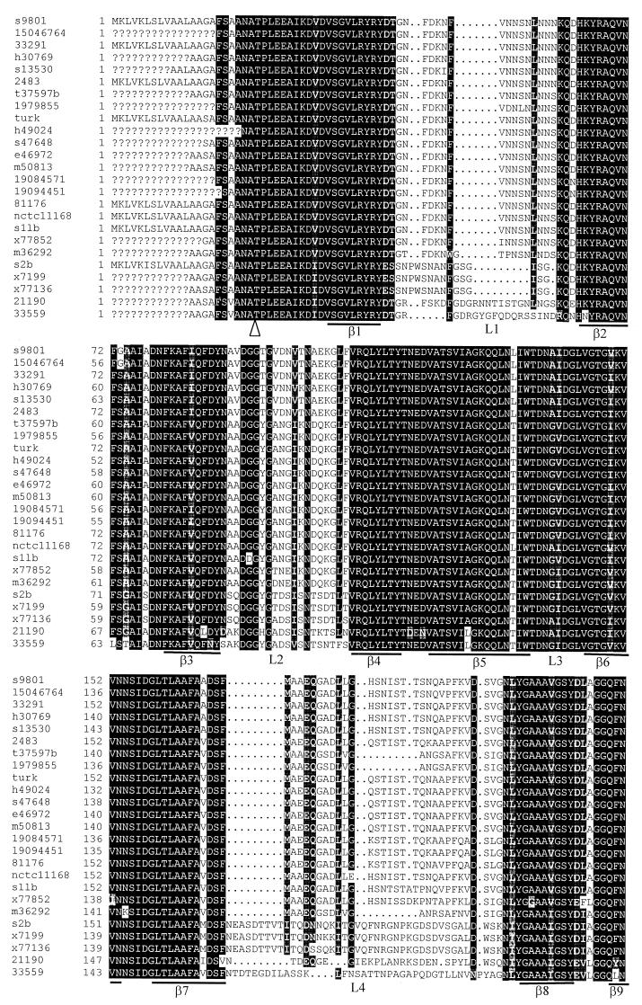
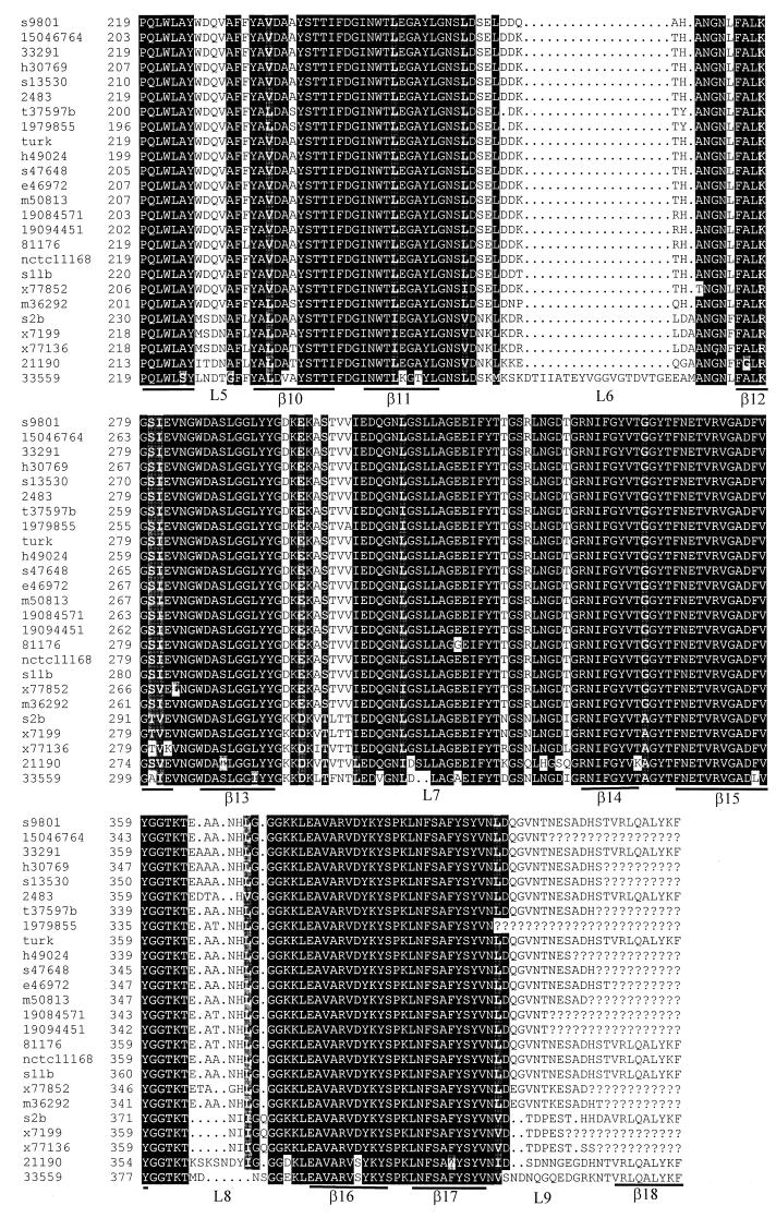
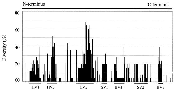
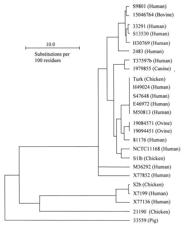
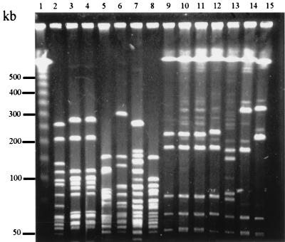
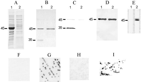
The major outer membrane protein (MOMP), a putative porin and a multifunction surface protein of Campylobacter jejuni, may play an important role in the adaptation of the organism to various host environments. To begin to dissect the biological functions and antigenic features of this protein, the gene (designated cmp) encoding MOMP was identified and characterized from 22 strains of C. jejuni and one strain of C. coli. It was shown that the single-copy cmp locus encoded a protein with characteristics of bacterial outer membrane proteins. Prediction from deduced amino acid sequences suggested that each MOMP subunit consisted of 18 beta-strands connected by short periplasmic turns and long irregular external loops. Alignment of the amino acid sequences of MOMP from different strains indicated that there were seven localized variable regions dispersed among highly conserved sequences. The variable regions were located in the putative external loop structures, while the predicted beta-strands were formed by conserved sequences. The sequence homology of cmp appeared to reflect the phylogenetic proximity of C. jejuni strains, since strains with identical cmp sequences had indistinguishable or closely related macrorestriction fragment patterns. Using recombinant MOMP and antibodies recognizing linear or conformational epitopes of the protein, it was demonstrated that the surface-exposed epitopes of MOMP were predominantly conformational in nature. These findings are instrumental in the design of MOMP-based diagnostic tools and vaccines.


Figures

FIG. 1
FIG. 1
Genomic organization and partial features of the cmp gene in C. jejuni strain 33291. The line at the top shows the 9-kb genomic fragment flanked by EcoRV (E). The location and direction of identified ORFs are indicated by boxed arrows below the line. The shaded box at the 5′ end of cmp indicates a signal sequence, while the shaded region in the middle of cmp denotes the sequence amplified by the degenerate primers designed from known peptide sequences. The locations of the key primers used in this study are labeled with arrows along the ORF of cmp. The shaded box below cmp indicates the location of the DNA probe in relation to the cmp ORF. The sequences of ppp (phosphotyrosine protein phosphatase) and dnaJ are partially determined.
FIG. 2
FIG. 2
Multiple alignments of deduced MOMP sequences and predicted secondary structures of MOMP. Question marks indicate undetermined sequences. Dots represent gaps in the alignments. The regions predicted to form β-strands are underlined and labeled β1 to β18. The putative external loops are marked L1 to L9. The arrowhead indicates the cleavage site for mature MOMP. The names of the strains are labeled on the left of the figure. The numbers indicate the amino acid numbers of MOMP in each strain. The MOMP sequences of strains NCTC 11168 and 2483 were obtained from the GenBank database (accession AL111168 and U96452, respectively).
FIG. 2
FIG. 2
Multiple alignments of deduced MOMP sequences and predicted secondary structures of MOMP. Question marks indicate undetermined sequences. Dots represent gaps in the alignments. The regions predicted to form β-strands are underlined and labeled β1 to β18. The putative external loops are marked L1 to L9. The arrowhead indicates the cleavage site for mature MOMP. The names of the strains are labeled on the left of the figure. The numbers indicate the amino acid numbers of MOMP in each strain. The MOMP sequences of strains NCTC 11168 and 2483 were obtained from the GenBank database (accession AL111168 and U96452, respectively).
FIG. 3
FIG. 3
Quantitative measurement of sequence variation in each aa position in MOMP. The percent diversity is calculated using the PsFind program(29) from the aligned sequences in Fig. 2. The N-terminal 22 amino acids and the C-terminal 24 amino acids in the alignment are omitted from the calculation because of the unavailability of the terminal sequences in some strains. The arbitrarily designated hypervariable (HV) and semivariable (SV) regions are marked by lines. The variable regions correspond to the predicted external loops L1, L2, L4, L5, L6, L7, and L8, respectively.
FIG. 4
FIG. 4
MOMP-based dendrogram constructed by the UPMGA method of Growtrees in GCG. The MOMP sequences listed in Fig. 2 were used to create a distance matrix. The names of the Campylobacter strains are shown on the right of the figure, and the sources of isolation for each strain are given in parentheses. The MOMP sequences of strains NCTC11168 and 2483 were obtained from the GenBank database (accession no. AL111168 and U96452, respectively).
FIG. 5
FIG. 5
PFGE analysis of C. jejuni using restriction enzymes KpnI (lanes 2 to 8) and SalI (lanes 9 to 15). Lane 1 contains DNA size markers (λ ladder [Promega]). The strains included in this figure are Turk (lanes 2 and 9), H49024 (lanes 3 and 10), M50813 (lanes 4 and 11), S9801 (lanes 5 and 12), 15046764 (lanes 6 and 13), 33291 (lanes 7 and 14), and S13530 (Lanes 8 and 15). The relationship of these strains as determined by cmp sequence homology is shown in Fig. 4.
FIG. 6
FIG. 6
Antigenic features of rMOMP33291 and demonstration of surface-exposed conformational epitopes of MOMP. (A) Inclusion bodies (lane 1) and supernatant (lane 2) of sonicated E. coli expressing rMOMP were separated by SDS-PAGE and stained with Coomassie blue R-250. (B) rMOMP purified with Empigen BB (lane 1) or urea (lane 2) was treated in sample buffer at 25°C and then subjected to SDS-PAGE. The gel was stained with Coomassie blue R-250. (C and D) Replica samples in panel B were transferred to nitrocellulose membrane and blotted with antibodies A-8 (C) or A-5 (D). (E) rMOMP33291 purified with urea was immunostained with A-8 in the absence (lane 1) or presence (lane 2) of Empigen BB. The positions of the denatured monomer (45 kDa) and the folded monomer (35 kDa) are indicated on the left of panels A to E. (F to I) Colonies of C. jejuni strain 33291 were immunoblotted with A-5 (F), A-8 (G), A-8 absorbed with rMOMP33291 purified with Empigen (H), or A-8 absorbed with rMOMP33291 purified with urea (I).

References

    1. Amako K, Baba N, Suzuki N, Wai S N, Umeda A. A structural analysis of the regularly arranged porin on the outer membrane of Campylobacter jejuni based on correlation averaging. Microbiol Immunol. 1997;41:855–859. - PubMed
    1. Amako K, Wai S N, Umeda A, Shigematsu M, Takade A. Electron microscopy of the major outer membrane protein of Campylobacter jejuni. Microbiol Immunol. 1996;40:749–754. - PubMed
    1. Bacon D J, Johnson W M, Rodgers F G. Identification and characterisation of a cytotoxic porin-lipopolysaccharide complex from Campylobacter jejuni. J Med Microbiol. 1999;48:139–148. - PubMed
    1. Black R E, Levine M M, Clements M L, Hughes T P, Blaser M J. Experimental Campylobacter jejuni infection in humans. J Infect Dis. 1988;157:472–479. - PubMed
    1. Blaser M J, Hopkins J A, Berka R M, Vasil M L, Wang W L. Identification and characterization of Campylobacter jejuni outer membrane proteins. Infect Immun. 1983;42:276–284. - PMC - PubMed

Publication types

MeSH terms