. 2000 Sep 18;192(6):789-99.
doi: 10.1084/jem.192.6.789.

Regulation of elastinolytic cysteine proteinase activity in normal and cathepsin K-deficient human macrophages

Affiliations

Regulation of elastinolytic cysteine proteinase activity in normal and cathepsin K-deficient human macrophages

A Punturieri et al. J Exp Med. .

Abstract

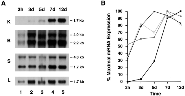
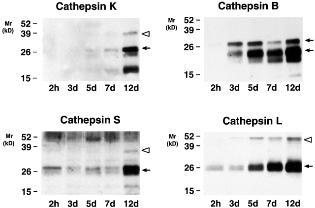
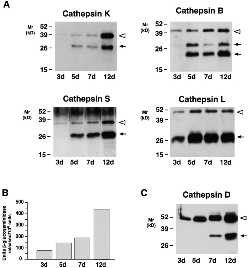
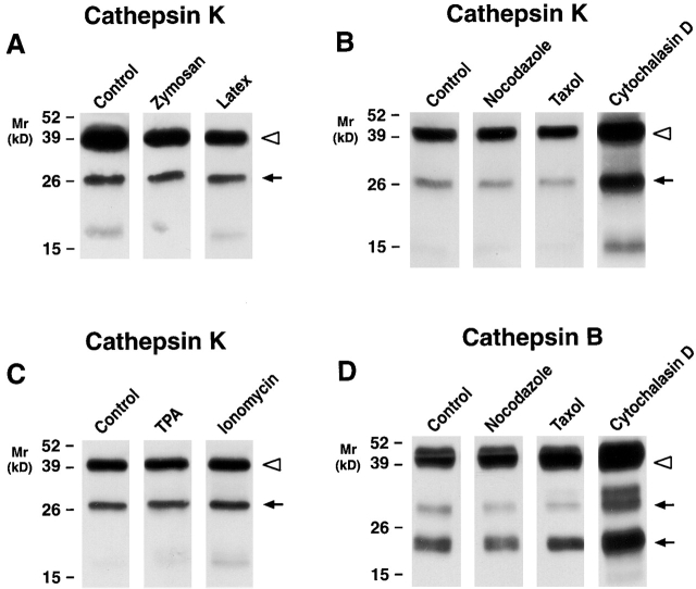
Human macrophages mediate the dissolution of elastic lamina by mobilizing tissue-destructive cysteine proteinases. While macrophage-mediated elastin degradation has been linked to the expression of cathepsins L and S, these cells also express cathepsin K, a new member of the cysteine proteinase family whose elastinolytic potential exceeds that of all known elastases. To determine the relative role of cathepsin K in elastinolysis, monocytes were differentiated under conditions in which they recapitulated a gene expression profile similar to that observed at sites of tissue damage in vivo. After a 12-d culture period, monocyte-derived macrophages (MDMs) expressed cathepsin K in tandem with cathepsins L and S. Though cysteine proteinases are acidophilic and normally confined to the lysosomal network, MDMs secreted cathepsin K extracellularly in concert with cathepsins L and S. Simultaneously, MDMs increased the expression of vacuolar-type H(+)-ATPase components, acidified the pericellular milieu, and maintained extracellular cathepsin K in an active form. MDMs from a cathepsin K-deficient individual, however, retained the ability to express, process, and secrete cathepsins L and S, and displayed normal elastin-degrading activity. Thus, matrix-destructive MDMs exteriorize a complex mix of proteolytic cysteine proteinases, but maintain full elastinolytic potential in the absence of cathepsin K by mobilizing cathepsins L and S.


Figures

Figure 1
Figure 1
The in vitro differentiation of MDMs. (A) Scanning electron micrographs of differentiating monocytes after 2 h, and 3, 5, 7, and 12 d of culture. Bar, 10 μm. (B) Northern blot analysis of osteopontin, gp-39, and chitotriosidase mRNA expression during MDM differentiation. Results are shown from an experiment performed using total RNA pooled from five donors. Equal loading was determined by GAPDH mRNA hybridization (not shown).
Figure 1
Figure 1
The in vitro differentiation of MDMs. (A) Scanning electron micrographs of differentiating monocytes after 2 h, and 3, 5, 7, and 12 d of culture. Bar, 10 μm. (B) Northern blot analysis of osteopontin, gp-39, and chitotriosidase mRNA expression during MDM differentiation. Results are shown from an experiment performed using total RNA pooled from five donors. Equal loading was determined by GAPDH mRNA hybridization (not shown).
Figure 2
Figure 2
Northern blot analysis of cysteine proteinase expression during MDM differentiation. (A) Total RNA was extracted at the indicated time points and hybridizations were performed using probes for cathepsins K, B, S, and L. Results are shown from an experiment performed using total RNA pooled from five donors. (B) The levels of mRNA expression of each cathepsin mRNA were compared with those of GAPDH mRNA at the indicated time points. Data are presented as percentage of maximal expression of each cathepsin mRNA: cathepsin K (▪), cathepsin B (▴), cathepsin S (•), and cathepsin L (□).
Figure 3
Figure 3
Intracellular cysteine proteinase expression during MDM differentiation. Cell lysates (10 μg/lane) were harvested at 2 h, and 3, 5, 7, and 12 d, and immunoblotted with anti–cathepsin K, B, S, and L Abs. The pro- (white arrowheads) and mature (arrows) forms of the enzymes are highlighted. Mature cathepsin B is expressed as a single and two chain active form (M r ∼31 and ∼25 kD, respectively; see reference 7). Results are shown from a single representative experiment of three performed.
Figure 6
Figure 6
Detection of acidic microenvironments in MDM–elastin cocultures. 12-d-old MDMs were either incubated alone (A) or with 2 mg elastin (B) and examined by scanning electron microscopy. (C and D) Cells were incubated with elastin for 24 h and stained with either 0.1% ruthenium red or 0.4 mg/ml cationized ferritin, respectively, and processed for TEM. Micrographs were taken at ×300, ×750, ×10,200, and ×10,100, respectively, with asterisks overlying elastin and arrows pointing to ruthenium red– or ferritin-stained plasma membrane. MDMs incubated alone (E) or with elastin for ∼16 h (F, G, and H) were treated with 10 μg/ml acridine orange and the cells were viewed by confocal laser microscopy. Acidic environments fluoresce orange-red while green fluorescence represents nonspecific association with nucleic acids and unacidified elastin particles (reference 22). The solid white lines outline MDMs; the dotted white lines outline elastin particles in each confocal section. The fluorescence observed surrounding the elastin particles (F) was completely quenched by trypan blue (G). (H) MDMs pretreated with 5 μm bafilomycin A1 (4 h at 37°C) did not acidify cellular compartments as assessed by acridine orange fluorescence. (Inset) Lysates from ∼3 × 10⁴ 12-d-old MDMs (lane 1) were immunoblotted with an mAb (3.2 Fl) to the 73-kD catalytic subunit of the vacuolar-type H+-ATPase. Bovine brain extract (lane 2) served as a positive control.
Figure 6
Figure 6
Detection of acidic microenvironments in MDM–elastin cocultures. 12-d-old MDMs were either incubated alone (A) or with 2 mg elastin (B) and examined by scanning electron microscopy. (C and D) Cells were incubated with elastin for 24 h and stained with either 0.1% ruthenium red or 0.4 mg/ml cationized ferritin, respectively, and processed for TEM. Micrographs were taken at ×300, ×750, ×10,200, and ×10,100, respectively, with asterisks overlying elastin and arrows pointing to ruthenium red– or ferritin-stained plasma membrane. MDMs incubated alone (E) or with elastin for ∼16 h (F, G, and H) were treated with 10 μg/ml acridine orange and the cells were viewed by confocal laser microscopy. Acidic environments fluoresce orange-red while green fluorescence represents nonspecific association with nucleic acids and unacidified elastin particles (reference 22). The solid white lines outline MDMs; the dotted white lines outline elastin particles in each confocal section. The fluorescence observed surrounding the elastin particles (F) was completely quenched by trypan blue (G). (H) MDMs pretreated with 5 μm bafilomycin A1 (4 h at 37°C) did not acidify cellular compartments as assessed by acridine orange fluorescence. (Inset) Lysates from ∼3 × 10⁴ 12-d-old MDMs (lane 1) were immunoblotted with an mAb (3.2 Fl) to the 73-kD catalytic subunit of the vacuolar-type H+-ATPase. Bovine brain extract (lane 2) served as a positive control.
Figure 4
Figure 4
The secretory phenotype of MDMs. (A) Western blot analysis of cathepsins K, B, S, and L secreted by MDMs at the indicated time points. (B) Supernatants collected as above were analyzed for release of β-glucosaminidase or (C) cathepsin D. In A and C, each lane was loaded with 10 μg total protein and the pro- (white arrowheads) and mature (arrows) forms of the enzymes are indicated. Results are shown from a single representative experiment of three performed.
Figure 5
Figure 5
Regulation of cathepsin K secretion in MDMs. 12-d-old MDMs were incubated for 48 h in serum-free media alone or in the presence of (A) zymosan (0.25 mg/ml) or latex beads (1:10 dilution of a 10% suspension), (B) nocodazole (5 μM), taxol (4 μM), or cytochalasin D (5 μM), or (C) TPA (50 ng/ml) or ionomycin (1 M). Cathepsin K secretion into the cell-free supernatants was assessed by Western blot analysis in A, B, and C, and cathepsin B in D. The pro- (white arrowheads) and mature (arrows) forms of the enzymes are indicated. Results are shown from a single representative experiment of three performed.
Figure 7
Figure 7
Vacuolar-type H+-ATPase component expression in MDMs. Total RNA was extracted from monocytes or MDMs at the indicated times and components B (□), a (⋄), E (○), and F (▵) quantified by cDNA microarray analysis. Results are shown as the mean fold increase in mRNA content relative to that expressed by freshly isolated monocytes from a single representative experiment of three performed.
Figure 8
Figure 8
Identification of extracellular cathepsin K–cystatin C complexes in MDM cultures. (A) 12-d-old MDMs were incubated for 48 h in serum-free media (lanes 1–4) or serum-free media supplemented with human cystatin C (100 μg/ml; lanes 5–8). Cystatin C or cathepsin K–cystatin complexes were immunoprecipitated using rabbit preimmune sera (lanes 1, 3, 5, and 7) or a specific anti–cystatin C Ab (lanes 2, 4, 6, and 8). Immunoprecipitates were then resolved by SDS-PAGE, transblotted, and probed with an anti–cathepsin K mAb. In lanes 2 and 6, mature cathepsin K (black arrowhead) was identified as a complex with endogenous or exogenous cystatin C, respectively. When MDMs were incubated with 100 μM E-64 during the 48-h culture period (lanes 3, 4, 7, and 8), secreted cathepsin K was inactivated and did not form complexes with cystatin C. Nonspecific bands are observed as a consequence of a spurious cross-reaction of the secondary Abs with the rabbit polyclonal antisera used to immunoprecipitate cystatin C. Results are shown as a single representative experiment of two performed. (B and C) 12-d-old MDMs were incubated for 48 h in serum-free media alone or in the presence of E-64 (100 μM), bafilomycin A1 (10 μM), or folimycin (50 nM) and the supernatants were examined for cathepsin K (B) or cathepsin B (C) secretion. The pro- (white arrowheads) and mature (arrows) forms of the enzymes are indicated. Results are shown as a single representative experiment of three performed.
Figure 9
Figure 9
The cysteine proteinase expression and elastinolytic activity of normal versus pyknodysostotic MDMs. (A) Western blot analysis of cathepsin K (lanes 1–4), cathepsin B (lanes 5–8), cathepsin S (lanes 9–12), and cathepsin L (lanes 13–16) expression in 12-d-old MDMs from a normal control (lanes 1, 3, 5, 7, 9, 11, 13, and 15) or a pyknodysostotic donor (lanes 2, 4, 6, 8, 10, 12, 14, and 16). Cell lysates (lanes 1, 2, 5, 6, 9, 10, 13, and 14) or cell-free supernatants (lanes 3, 4, 7, 8, 11, 12, 15, and 16) (all 10 μg/lane) were immunoblotted with the respective Abs. The pro- (white arrowheads) and mature (arrows) forms of cathepsins K, B, S, and L are identified as shown. (B) MDM-mediated 3H-elastin degradation. The elastinolytic activity of cells derived from a healthy donor (lanes 1 and 2), an unaffected heterozygous individual (lanes 3 and 4), or an affected subject (lanes 5 and 6) was evaluated in the absence (lanes 1, 3, and 5) or presence (lanes 2, 4, and 6) of 100 μM E-64. Results are shown from a single experiment.

Similar articles

Cited by

References

    1. Carmeliet P., Moons L., Lijnen R., Baes M., Lemaitre V., Tipping P., Drew A., Eckhout Y., Shapiro S., Lupu F., Collen D. Urokinase-generated plasmin activates matrix metalloproteinases during aneurysm formation. Nat. Genet. 1997;17:439–444. - PubMed
    1. Weyand C.M., Wagner A.D., Bjornsson J., Goronzy J.J. Correlation of the topographical arrangement and the functional pattern of tissue-infiltrating macrophages in giant cell arteritis. J. Clin. Invest. 1996;98:1642–1649. - PMC - PubMed
    1. Hautamaki R.D., Kobayashi D.K., Senior R.M., Shapiro S.D. Requirement for macrophage elastase for cigarette smoke-induced emphysema in mice. Science. 1997;277:2002–2004. - PubMed
    1. Sukhova G.K., Shi G.-P., Simon D.I., Chapman H.A., Libby P. Expression of elastolytic cathepsins S and K in human atheroma and regulation of their production in smooth muscle cells. J. Clin. Invest. 1998;102:576–583. - PMC - PubMed
    1. Shi G.-P., Sukhova G.K., Grubb A., Ducharme A., Rhode L.H., Lee R.T., Ridker P.M., Libby P., Chapman H.A. Cystatin C deficiency in human atherosclerosis and aortic aneurysms. J. Clin. Invest. 1999;104:1191–1197. - PMC - PubMed

Publication types

MeSH terms