. 2000 Sep 18;192(6):899-905.
doi: 10.1084/jem.192.6.899.

CC chemokine receptor 2 is critical for induction of experimental autoimmune encephalomyelitis

Affiliations

CC chemokine receptor 2 is critical for induction of experimental autoimmune encephalomyelitis

B T Fife et al. J Exp Med. .

Abstract

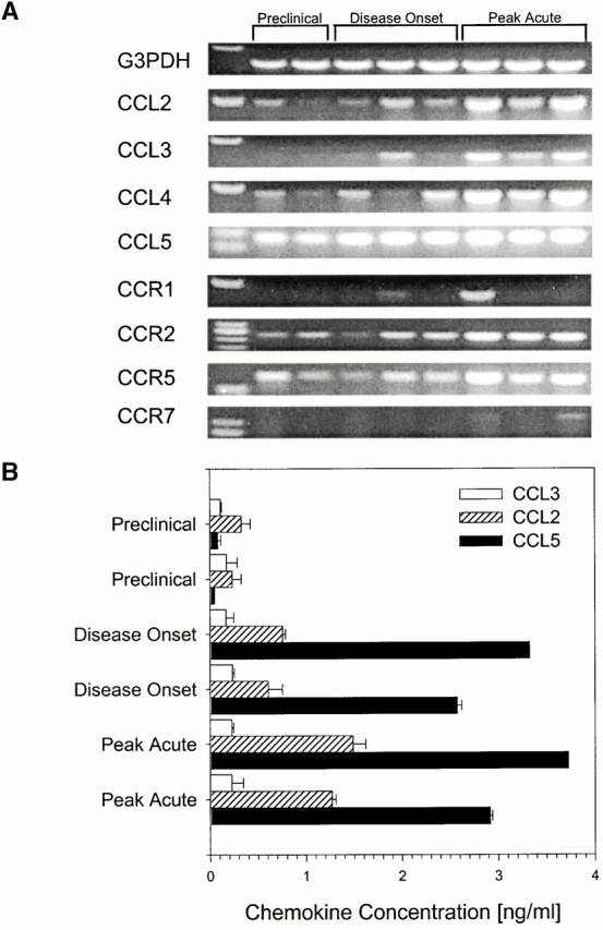
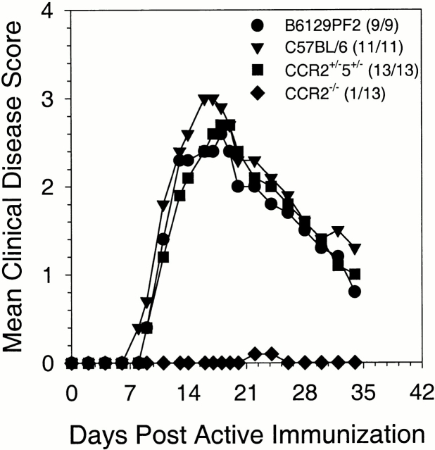
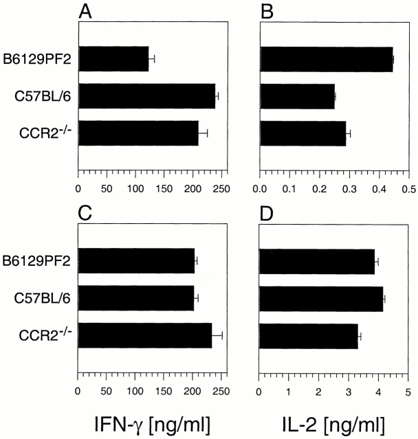
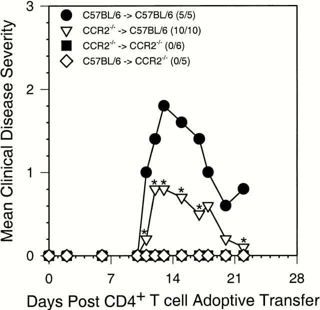
Experimental autoimmune encephalomyelitis (EAE) is a CD4(+) T lymphocyte-mediated disease of the central nervous system (CNS) characterized by mononuclear cell infiltration, demyelination, and paralysis. We previously demonstrated a role for chemokines in acute and relapsing EAE pathogenesis. Presently, we investigated the role of CC chemokine receptor 2 (CCR2) in acute EAE. CCR2(-/-) mice did not develop clinical EAE or CNS histopathology, and showed a significant reduction in T cell- and CNS-infiltrating CD45(high)F4/80(+) monocyte subpopulations. Peripheral lymphocytes from CCR2(-/-) mice produced comparable levels of interferon-gamma (IFN-gamma) and interleukin (IL)-2 in response to antigen-specific restimulation when compared with control mice. Adoptively transferred myelin oligodendrocyte glycoprotein 35-55-specific T cells lacking expression of CCR2 were able to induce EAE, whereas CCR2(-/-) recipients of wild-type T cells failed to develop disease. These results suggest that CCR2 expression on host-derived mononuclear cells is critical for disease induction.


Figures

Figure 1
Figure 1
CNS chemokine and chemokine receptor expression. (A) CCL2, CCL4, CCL5, and chemokine receptor mRNA was detected in CNS tissue samples using RT–PCR at all three time points examined after EAE induction. (B) CNS chemokine protein expression for CCL2, CCL3, CCL4, and CCL5 was quantitated from tissue samples using specific ELISA. The results from two representative mice from each time point are shown. In all cases the data shown are representative of two independent experiments.
Figure 2
Figure 2
CCR2-deficient mice do not develop clinical EAE. Mice were monitored for development of clinical disease. The data are expressed as the mean clinical disease score for all mice in each group as a function of days after immunization with disease incidence indicated in parentheses. Both mean clinical disease severity and disease incidence were significantly decreased in CCR2−/− animals relative to C57BL/6, B6129PF2, and CCR2+/−5+/− controls (P < 0.001) over the entire course of the experiment. The data shown are representative of four independent experiments.
Figure 3
Figure 3
Lymphocytes from CCR2−/− mice produce proinflammatory cytokines upon antigen restimulation. B6129PF2, C57BL/6, and CCR2−/− mice were monitored for the development of clinical EAE. At the time B6129PF2 mice showed peak acute clinical disease, draining LN (A and B) and splenic cells (C and D) pooled from six representative mice for each group were restimulated with the immunizing peptide in vitro. Cultured supernatant was harvested at 48 h and measured for the production of IL-2 and IFN-γ by specific ELISA. CCR2−/− lymphocytes produced comparable levels of IFN-γ and IL-2 to control C57BL/6 lymphocytes in response to specific peptide challenge. The data shown are representative of three independent experiments.
Figure 4
Figure 4
CCR2−/− recipients do not develop clinical EAE. Mice were monitored for the development of clinical disease after the adoptive transfer of MOG35–55-reactivated CD4+ T cells. The data are expressed as the mean clinical disease score for all mice in each group as a function of days after adoptive transfer with disease incidence in parentheses. Both mean clinical disease severity and disease incidence were significantly decreased in both CCR2−/− recipient groups compared with the C57BL/6 wild-type recipient group receiving CD4+ T cells from wild-type C57BL/6 donors by the Student's t test and X2 analysis over the entire course of the experiment. The C57BL/6 recipient mice receiving CCR2−/− CD4+ T cells was significantly reduced when compared with the wild-type C57BL/6 to C57BL/6 recipient group at the days marked (*; P < 0.05). The data shown are representative of two independent experiments.

References

    1. Hohlfeld R. Biotechnological agents for the immunotherapy of multiple sclerosis. Principles, problems, and perspectives. Brain. 1997;120:865–916. - PubMed
    1. Krakowski M.L., Owens T. The central nervous system environment controls effector CD4+ T cell cytokine profile in experimental allergic encephalomyelitis. Eur. J. Immunol. 1997;27:2840–2847. - PubMed
    1. Hickey W.F., Hsu B.L., Kimura H. T-lymphocyte entry into the central nervous system. J. Neurosci. Res. 1991;28:254–260. - PubMed
    1. Cross A.H., Cannella B., Brosnan C.F., Raine C.S. Homing to central nervous system vasculature by antigen-specific lymphocytes. I. Localization of 14C-labeled cells during acute, chronic, and relapsing experimental allergic encephalomyelitis. Lab. Invest. 1990;63:162–170. - PubMed
    1. Yednock T.A., Cannon C., Fritz L.C., Sanchez-Madrid F., Steinman L., Karin N. Prevention of experimental autoimmune encephalomyelitis by antibodies against α4β1 integrin. Nature. 1992;356:63–66. - PubMed

Publication types

MeSH terms