

doi: 10.1101/gad.827300.
p53-independent functions of the p19(ARF) tumor suppressor
Affiliations
- PMID: 10995391
- PMCID: PMC316930
- DOI: 10.1101/gad.827300
Item in Clipboard
p53-independent functions of the p19(ARF) tumor suppressor
Genes Dev.
.
Abstract
The p19(ARF) tumor suppressor antagonizes Mdm2 to induce p53-dependent cell cycle arrest. Individual TKO (triple knock out) mice nullizygous for ARF, p53, and Mdm2 develop multiple tumors at a frequency greater than those observed in animals lacking both p53 and Mdm2 or p53 alone, demonstrating that p19(ARF) can act independently of the Mdm2-p53 axis in tumor surveillance. Reintroduction of ARF into TKO mouse embryo fibroblasts (MEFs), but not into those lacking both p53 and ARF, arrested the cell division cycle in the G1 phase. Inhibition of the retinoblastoma protein had no effect on the ability of ARF to arrest TKO MEFs. Thus, in the absence of Mdm2, p19(ARF) interacts with other targets to inhibit cell proliferation.
Figures
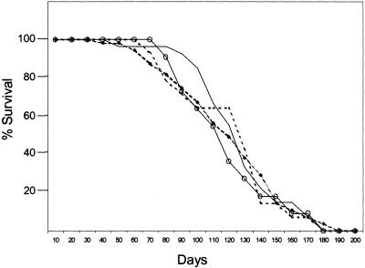
Survival of mice lacking ARF, p53, and/or Mdm2. p53/Mdm2-null (○), ARF/p53-null (dotted line), ARF/p53-null Mdm2 +/- (●), and ARF/p53/Mdm2-null (solid line) mice were observed for 33 weeks. Tumors arising in ARF/p53-null and ARF/p53/Mdm2-null animals are listed in Table 1.
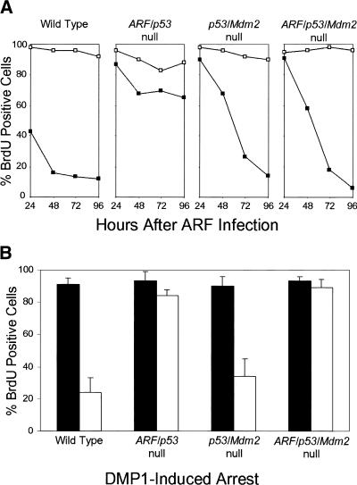
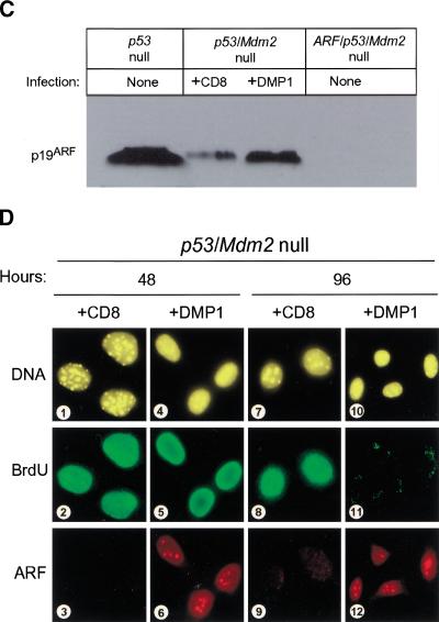
ARF and DMP1 arrest mouse embryo fibroblasts (MEFs) lacking both p53 and Mdm2. (A) Primary MEFs from mice with the indicated genotypes (top) were infected with retroviruses encoding CD8 (□) or p19ARF (▪). Cells labeled with 5-bromodeoxyuridine (BrdU) were analyzed by indirect immunofluorescence (green) and simultaneously scored for p19ARF expression (red) using 3 coverslips and >100 cells per coverslip in 4 independent experiments. (B) Primary MEFs of indicated genotypes (bottom) were infected with retroviruses encoding CD8 (dark bars) or DMP1 (open bars). Cells labeled for 24 hours with BrdU were stained 96 hours postinfection (green) and scored simultaneously for DMP1 expression (red). Error bars indicate standard deviations. (C) Expression of p19ARF protein in MEFs (3T3 strains, each at passage 9) of the indicated genotypes (top). Cells null for both p53 and Mdm2 (middle two lanes) were infected with retroviruses encoding either CD8 or DMP1 as indicated and assayed 96 hours later for ARF protein by direct immunoblotting. Equal quantities of protein (150 μg) were loaded per lane. (D) p53/Mdm2-null MEFs infected with retroviruses encoding CD8 (panels 1–3, 7–9) or DMP1 (panels 4–6, 10–12) were analyzed by immunofluorescence for BrdU incorporation and ARF induction 48 and 96 hours after infection.
ARF and DMP1 arrest mouse embryo fibroblasts (MEFs) lacking both p53 and Mdm2. (A) Primary MEFs from mice with the indicated genotypes (top) were infected with retroviruses encoding CD8 (□) or p19ARF (▪). Cells labeled with 5-bromodeoxyuridine (BrdU) were analyzed by indirect immunofluorescence (green) and simultaneously scored for p19ARF expression (red) using 3 coverslips and >100 cells per coverslip in 4 independent experiments. (B) Primary MEFs of indicated genotypes (bottom) were infected with retroviruses encoding CD8 (dark bars) or DMP1 (open bars). Cells labeled for 24 hours with BrdU were stained 96 hours postinfection (green) and scored simultaneously for DMP1 expression (red). Error bars indicate standard deviations. (C) Expression of p19ARF protein in MEFs (3T3 strains, each at passage 9) of the indicated genotypes (top). Cells null for both p53 and Mdm2 (middle two lanes) were infected with retroviruses encoding either CD8 or DMP1 as indicated and assayed 96 hours later for ARF protein by direct immunoblotting. Equal quantities of protein (150 μg) were loaded per lane. (D) p53/Mdm2-null MEFs infected with retroviruses encoding CD8 (panels 1–3, 7–9) or DMP1 (panels 4–6, 10–12) were analyzed by immunofluorescence for BrdU incorporation and ARF induction 48 and 96 hours after infection.
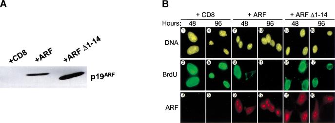
p53-Independent arrest requires the conserved ARF amino-terminal domain. (A) Triple knock out (TKO) mouse embryo fibroblasts (MEFs) infected with retroviruses encoding CD8, ARF, or ARF Δ1–14 were lysed 96 hours after infection, and separated proteins were blotted with affinity-purified polyclonal rabbit antibody to p19ARF. (B) TKO MEFs infected with retroviruses encoding CD8 (panels 1–6), ARF (panels 7–12), or ARF Δ1–14 (panels 13–18) were labeled with 5-bromodeoxyuridine (BrdU) for 24 hours and analyzed 48 and 96 hours postinfection for incorporated BrdU (green) and p19ARF expression (red). DNA was visualized with Hoechst yellow S769121 dye. (C) TKO MEFs (5 × 10⁴) were infected with CD8, ARF, and ARF Δ1–14 retroviruses, stained with propidium iodide 96 hours after infection, and subjected to fluorescence-activated cell sorting to determine DNA content. (D) TKO MEFs were seeded at 300 cells per dish and infected with retroviruses encoding CD8, ARF, or ARF Δ1–14. Colonies were stained 12 days post-infection.
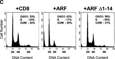
p53-Independent arrest requires the conserved ARF amino-terminal domain. (A) Triple knock out (TKO) mouse embryo fibroblasts (MEFs) infected with retroviruses encoding CD8, ARF, or ARF Δ1–14 were lysed 96 hours after infection, and separated proteins were blotted with affinity-purified polyclonal rabbit antibody to p19ARF. (B) TKO MEFs infected with retroviruses encoding CD8 (panels 1–6), ARF (panels 7–12), or ARF Δ1–14 (panels 13–18) were labeled with 5-bromodeoxyuridine (BrdU) for 24 hours and analyzed 48 and 96 hours postinfection for incorporated BrdU (green) and p19ARF expression (red). DNA was visualized with Hoechst yellow S769121 dye. (C) TKO MEFs (5 × 10⁴) were infected with CD8, ARF, and ARF Δ1–14 retroviruses, stained with propidium iodide 96 hours after infection, and subjected to fluorescence-activated cell sorting to determine DNA content. (D) TKO MEFs were seeded at 300 cells per dish and infected with retroviruses encoding CD8, ARF, or ARF Δ1–14. Colonies were stained 12 days post-infection.
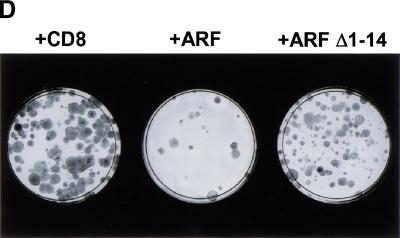
p53-Independent arrest requires the conserved ARF amino-terminal domain. (A) Triple knock out (TKO) mouse embryo fibroblasts (MEFs) infected with retroviruses encoding CD8, ARF, or ARF Δ1–14 were lysed 96 hours after infection, and separated proteins were blotted with affinity-purified polyclonal rabbit antibody to p19ARF. (B) TKO MEFs infected with retroviruses encoding CD8 (panels 1–6), ARF (panels 7–12), or ARF Δ1–14 (panels 13–18) were labeled with 5-bromodeoxyuridine (BrdU) for 24 hours and analyzed 48 and 96 hours postinfection for incorporated BrdU (green) and p19ARF expression (red). DNA was visualized with Hoechst yellow S769121 dye. (C) TKO MEFs (5 × 10⁴) were infected with CD8, ARF, and ARF Δ1–14 retroviruses, stained with propidium iodide 96 hours after infection, and subjected to fluorescence-activated cell sorting to determine DNA content. (D) TKO MEFs were seeded at 300 cells per dish and infected with retroviruses encoding CD8, ARF, or ARF Δ1–14. Colonies were stained 12 days post-infection.
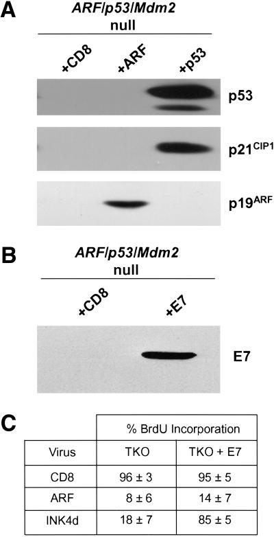
ARF-induced arrest of triple knock out (TKO) cells does not depend on p21Cip1 and is not overridden by HPV E7. (A) TKO mouse embryo fibroblasts (MEFs) infected with retroviruses encoding CD8 or ARF (96 hours postinfection) or wild-type p53 (48 hours postinfection) were lysed, and separated proteins were blotted with antibodies to p53, p21Cip1, and ARF. (B) TKO MEFs infected with retroviruses encoding E7 or CD8 were lysed 48 hours after infection, and separated proteins were blotted with antibody to E7. (C) TKO MEFs infected with MSCV-GFP-IRES or MSCV-GFP-IRES-E7 retroviruses were re-plated 48 hours later and infected with retroviruses encoding CD8, ARF, or p19INK4d. Infected cells were analyzed 96 hours postinfection using an FITC filter to detect GFP-IRES-E7 infected cells (green), immunofluorescence to score 5-bromodeoxyuridine incorporation (red), and antibody to p19ARF followed by Alexa 350 stain to visualize nucleolar ARF (blue). Alternatively, p19INK4d was detected using a rabbit anti-p19INK4d antibody followed by biotinylated anti-rabbit immunoglobulin and streptavidin Texas red to ensure >90% infection efficiency of TKO and E7-containing TKO cells.
References
-
- Bates S, Phillips AC, Clarke PA, Stott F, Peters G, Ludwig RL, Vousden KH. p14ARF links the tumor suppressors pRB and p53. Nature. 1998;395:124–125. - PubMed
-
- Carnero A, Hudson JD, Price CM, Beach DH. p16INK4a and p19ARF act in overlapping pathways in cellular immortalization. Nat Cell Biol. 2000;2:148–155. - PubMed
-
- Donehower L, Harvey AM, Slagle BL, McArthur MJ, Montgomery CA, Jr, Butel JS, Bradley A. Mice deficient for p53 are developmentally normal but susceptible to spontaneous tumours. Nature. 1992;356:215–221. - PubMed
Publication types
MeSH terms
Substances
Grants and funding
LinkOut - more resources
Full Text Sources
Other Literature Sources
Molecular Biology Databases
Research Materials
Miscellaneous