. 2000 Sep 15;14(18):2377-91.

HFR1 encodes an atypical bHLH protein that acts in phytochrome A signal transduction

Affiliations

HFR1 encodes an atypical bHLH protein that acts in phytochrome A signal transduction

C D Fairchild et al. Genes Dev. .

Abstract

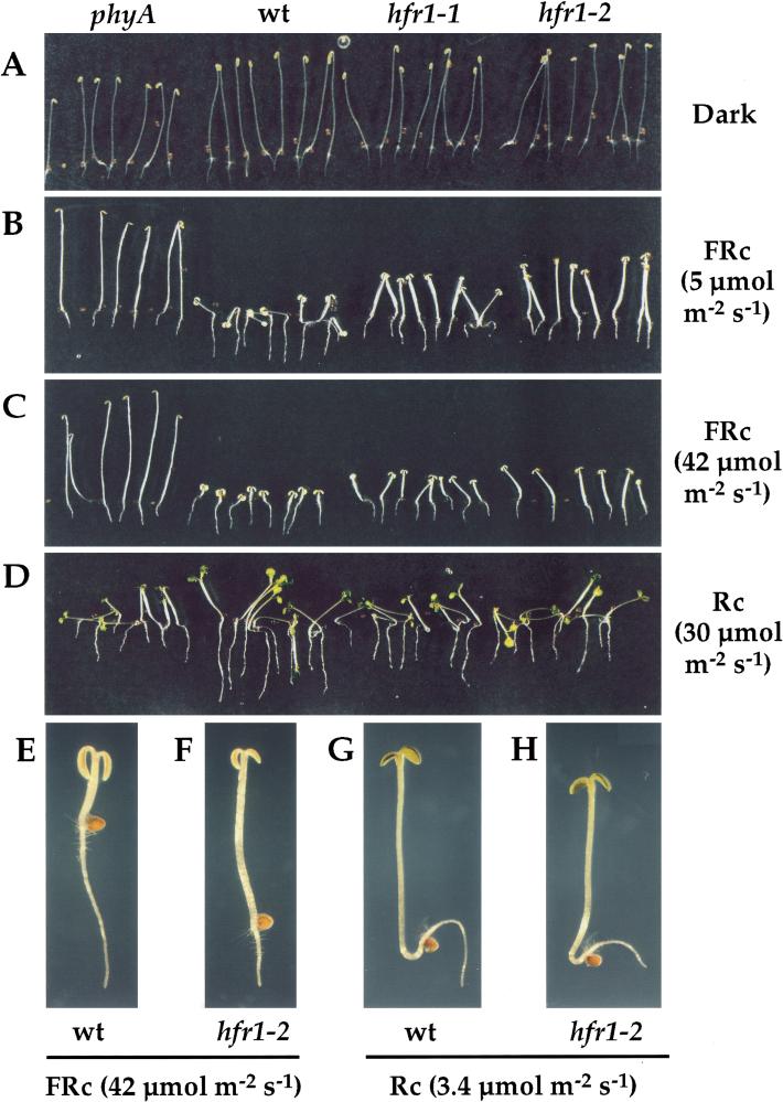
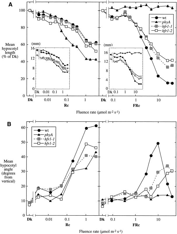
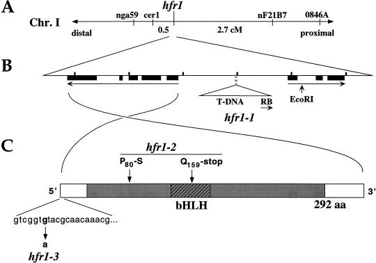
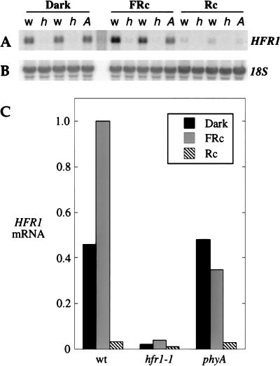
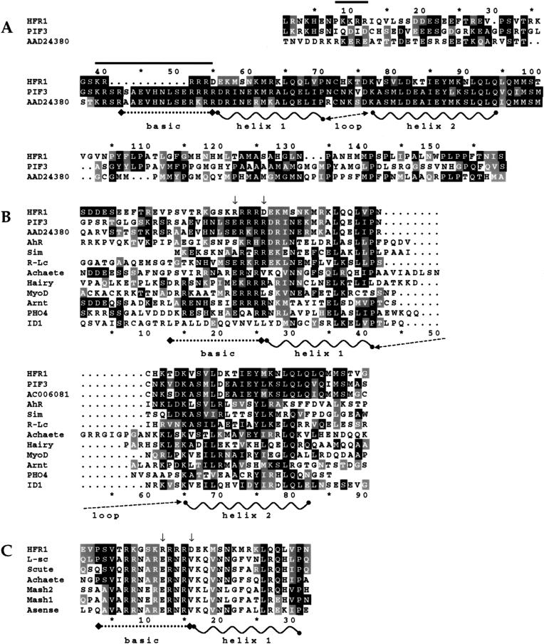
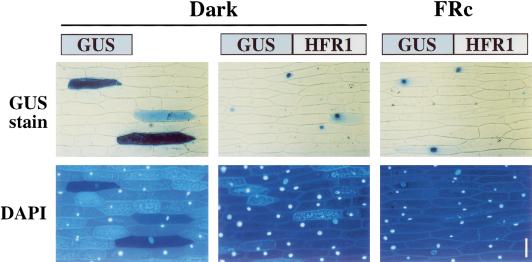
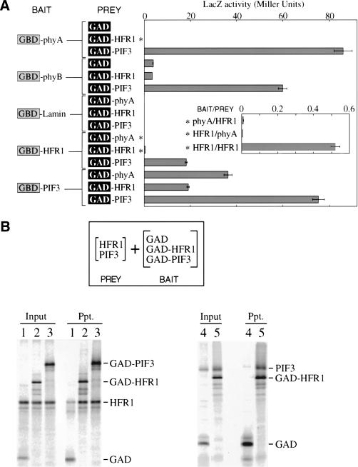
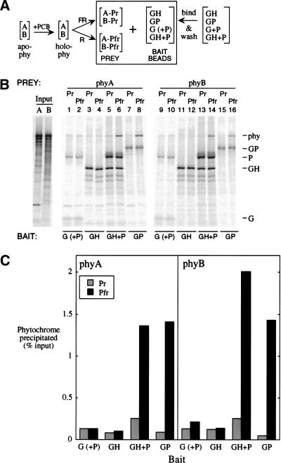
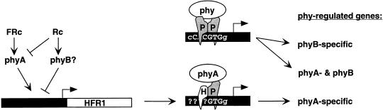
Phytochromes are informational photoreceptors through which plants adapt their growth and development to prevailing light conditions. These adaptations are effected primarily through phytochrome regulation of gene expression by mechanisms that remain unclear. We describe a new mutant, hfr1 (long hypocotyl in far-red), that exhibits a reduction in seedling responsiveness specifically to continuous far-red light (FRc), thereby suggesting a locus likely to be involved in phytochrome A (phyA) signal transduction. Using an insertionally tagged allele, we cloned the HFR1 gene and subsequently confirmed its identity with additional alleles derived from a directed genetic screen. HFR1 encodes a nuclear protein with strong similarity to the bHLH family of DNA-binding proteins but with an atypical basic region. In contrast to PIF3, a related bHLH protein previously shown to bind phyB, HFR1 did not bind either phyA or B. However, HFR1 did bind PIF3, suggesting heterodimerization, and both the HFR1/PIF3 complex and PIF3 homodimer bound preferentially to the Pfr form of both phytochromes. Thus, HFR1 may function to modulate phyA signaling via heterodimerization with PIF3. HFR1 mRNA is 30-fold more abundant in FRc than in continuous red light, suggesting a potential mechanistic basis for the specificity of HFR1 to phyA signaling.


Figures

Figure 1
Figure 1
Visible defects in hfr1 seedling photomorphogenesis. Col-5 wild-type (wt) and mutant (phyA-211 [Reed et al. 1994], hfr1-1 and hfr1-2) seedlings were grown for 4 d in complete darkness or in various light conditions on vertically oriented agar surfaces. They were then photographed without rearrangement. Seedlings grown in (A) darkness, (B) moderate FRc, (C) strong FRc, (D) Rc. (E–H) Representative wt (E,G) and hfr1-2 (F,H) seedlings grown in strong FRc (E,F) or in Rc (G,H). In (EH), the root/hypocotyl junction is roughly marked by the empty seed coats.
Figure 2
Figure 2
Hypocotyl responses of hfr1 mutants over a range of Rc and FRc fluence rates. (A) Mean hypocotyl lengths expressed for each line as a percent of the respective value for seedlings grown in darkness (Dk); (insets) the same data expressed as lengths in millimeters. Bars represent the standard error of the mean; where not visible, the bars are eclipsed by a symbol. (B) Mean hypocotyl angle from vertical, a measure of hypocotyl negative gravitropism. An angle of 0° represents maximal negative gravitropism.
Figure 3
Figure 3
Molecular cloning of the HFR1 locus and identification of lesions in the hfr1 mutants. (A) Genetic linkage of hfr1 to markers at the top end of Chromosome I. (B) Diagram of the 6-kb region surrounding the HFR1 locus that was sequenced in the present study (corresponding to BAC T6A9 coordinates 60330–66530). The length between tick marks is 1 kb. Transcribed regions flanking the hfr1-1 T-DNA insertion are represented by long arrows (exons, solid boxes). The RB and EcoRI site shown are the termini of the flanking region initially isolated from hfr1-1 by PCR walk. In (A,B), centromere distal (and the top of Chromosome I) is to the left side, centromere proximal to the right. (C) Schematic diagram of the mature HFR1 transcript and lesions found in the hfr1-2 and hfr1-3 alleles. The shaded region indicates the predicted coding region for the HFR1 protein.
Figure 4
Figure 4
HFR1 is a light-regulated gene with strongly reduced expression in the hfr1-1 mutant. Northern blot analysis of total RNA from 3-d-old seedlings grown under the indicated light conditions. (A) HFR1 probe; w, wild-type; h, hfr1-1; A, phyA-211. Duplicates of wild-type and hfr1-1 represent separate RNA preparations. (B) 18S rRNA reprobe of the membrane from (A). (C) HFR1 mRNA levels adjusted for 18S signal and expressed relative to the wild-type FRc value. Wild-type and hfr1-1 values are averages of two samples.
Figure 5
Figure 5
Sequence comparison of HFR1 to other bHLH proteins. Identical amino acid residues are shaded black; similar residues are shaded grey. (A) Alignment of HFR1 (amino acid residues 105–248) and its closest known homologs, the Arabidopsis protein PIF3 (GenBank accession no. AF100166) and predicted protein AAD24380, over the region of significant sequence similarity. Solid bars indicate prospective monopartite nuclear localization signals in HFR1. (B) Alignment of a more restricted region of HFR1 (amino acid residues 122–194) to a broader set of representative members of the bHLH protein family from various organisms. Arrows indicate key positions where HFR1 is dissimilar to most (perhaps all) known bHLH proteins. GenBank accession nos.: AhR, P30561; Sim, P05709; R-Lc, P13526; Achaete, P10083; Hairy, P14003; MyoD, CAA40000; Arnt, P41739; PHO4, P07270; ID1, P20067. (C) Similarity of the basic region of the HFR1 bHLH domain (amino acid residues 132–153) to the basic regions of the Achaete/Scute bHLH subfamily. In this alignment, only amino acid residues with similarity to HFR1 are shaded. GenBank accession nos.: L-sc, P09774; Scute, P10084; MASH2, P19360; MASH1, P19359; Asense, P09775.
Figure 6
Figure 6
HFR1 protein is constitutively nuclear localized when transiently expressed in onion epidermal cells. Onion epidermal peels were bombarded with constructs for expression of either the GUS reporter only or a GUS-HFR1 chimera. The peels were then incubated in darkness or FRc for 17 h before staining. In the top row, blue color results from GUS activity; below, the fluorescence from DAPI stain shows the position of the nucleus in each cell. Bar, 100 μm.
Figure 7
Figure 7
HFR1 protein can bind the phytochrome-interactor PIF3. (A) Two-hybrid assays for interaction in yeast between HFR1, PIF3, phyA C-terminal half, or phyB C-terminal half, along with negative controls nuclear lamin and unfused GAL4 activation domain (GAD). GBD refers to the GAL4 DNA-binding domain. Asterisks mark assay results that are also shown in an expanded view in the inset. Bars represent the standard error of the mean. (B) In vitro binding of HFR1 to PIF3. Shown are autoradiograms of SDS-PAGE separated proteins from immunoprecipitations. The GAL4 activation domain (GAD) was used as an epitope tag in bait constructs in fusion with PIF3 (GAD-PIF3) and HFR1 (GAD-HFR1). Prey HFR1 (HFR1) and PIF3 (PIF3) were expressed without the epitope tag. Bait proteins were immunoprecipitated using antibody to GAD immobilized on beads. Input lanes were loaded with half the fraction of each binding reaction that was loaded for washed precipitate lanes (Ppt.). (Lanes 1–3) precipitation of HFR1 by GAD control (1), GAD-HFR1 (2), and GAD-PIF3 (3). (Lanes 4,5) precipitation of PIF3 by GAD control (4) and GAD-HFR1 (5).
Figure 8
Figure 8
Both phyA and phyB bind as Pfr in vitro to both the PIF3 homodimer and a HFR1/PIF3 complex. (A) PhyA or phyB apoproteins were expressed separately in vitro (apo-phy), and combined with chromophore (PCB) to make spectrally active holoprotein (holo-phy); portions of each phytochrome solution were then irradiated with either FR or R to form predominantly Pr (A-Pr; B-Pr) or Pfr (A-Pfr; B-Pfr) forms, respectively. Bait beads were prepared by binding GAD-tagged proteins (G; alone or coexpressed with untagged PIF3 [P]), GAD-HFR1 (GH), or GAD-PIF3 (GP) to MAb-Protein A-beads, and then washing to remove loosely bound protein. Bait proteins were labeled at 2% of the specific activity of the phytochromes. (B) Autoradiograms of proteins separated by SDS-PAGE. Input phyA (A) and phyB (B) lanes were loaded with one-fifth the fraction of each binding reaction that was loaded for washed precipitates. (C) Quantitation of the phytochrome precipitated in (B).
Figure 9
Figure 9
A model for the role of HFR1 in phytochrome signaling. FRc, acting through phyA, enhances HFR1 transcription and Rc, acting through phyB or another phytochrome, suppresses HFR1 transcription. Phytochromes in their Pfr form (phy) translocate to the nucleus, where they are recruited to target gene promoters in genomic DNA (thick black bars, with recognition sequences overlaid and with arrows representing transcription initiation sites) by PIF3 (P). Whereas the PIF3 homodimer recognizes a G-box and potentially regulates gene expression in response to Rc or FRc, the heterodimer of PIF3 and HFR1 (H), formed predominantly in FRc, recognizes a distinct sequence in phyA-specific gene targets.

References

    1. Abe H, Yamaguchi-Shinozaki K, Urao T, Iwasaki T, Hosokawa D, Shinozaki K. Role of ArabidopsisMYC and MYB homologs in drought- and abscisic acid–regulated gene expression. Plant Cell. 1997;9:1859–1868. - PMC - PubMed
    1. Atchley WR, Terhalle W, Dress A. Positional dependence, cliques, and predictive motifs in the bHLH protein domain. J Mol Evol. 1999;48:501–516. - PubMed
    1. Barnes SA, Nishizawa NK, Quaggio RB, Whitelam GC, Chua NH. Far-red light blocks greening of arabidopsis seedlings via a phytochrome A–mediated change in plastid development. Plant Cell. 1996;8:601–615. - PMC - PubMed
    1. Bell CJ, Ecker JR. Assignment of 30 microsatellite loci to the linkage map of Arabidopsis. Genomics. 1994;19:137–144. - PubMed
    1. Bolle C, Koncz C, Chua NH. PAT1, a new member of the GRAS family, is involved in phytochrome A signal transduction. Genes & Dev. 2000;14:1269–1278. - PMC - PubMed

Publication types

MeSH terms