

Apoptosis in the skeletal muscle of patients with heart failure: investigation of clinical and biochemical changes
- PMID: 10995417
- PMCID: PMC1729437
- DOI: 10.1136/heart.84.4.431
Apoptosis in the skeletal muscle of patients with heart failure: investigation of clinical and biochemical changes
Abstract
Objective: To investigate the contribution of apoptosis in the development of the skeletal myopathy in chronic heart failure.
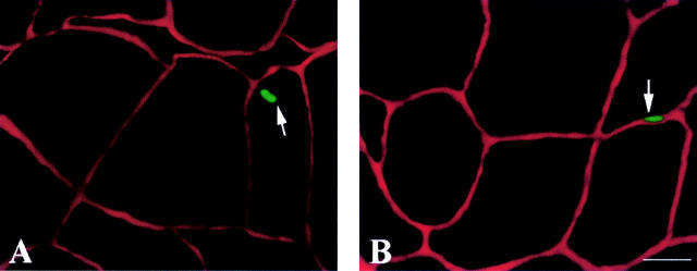
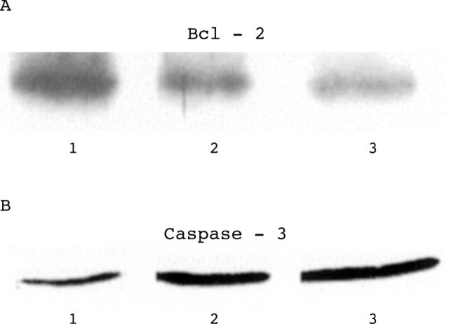
Design: The electrophoretic pattern of myosin heavy chains (MHC), fibre cross sectional area, number of in situ nick end labelling (TUNEL) positive apoptotic myocyte nuclei, and the tissue levels of caspase-3, Bcl-2, and ubiquitin were determined in biopsies taken from the vastus lateralis muscle. The study involved nine patients with severe chronic heart failure caused by ischaemic heart disease and hibernating myocardium and five controls.
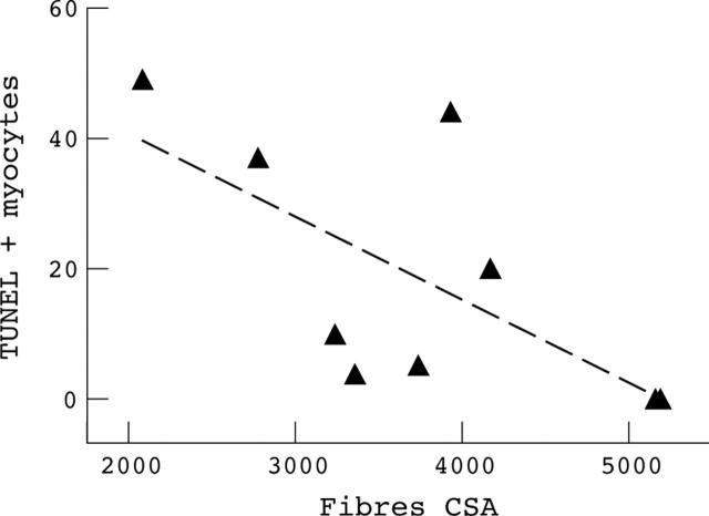
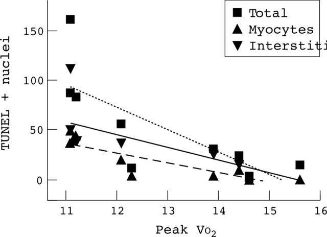
Results: In chronic heart failure patients the vastus lateralis showed a significant increase of MHC(2a) and MHC(2b) and a greater degree of fibre atrophy, as demonstrated by the decreased cross sectional area. There was also an increased number of TUNEL positive apoptotic myocyte nuclei. Tissue concentrations of Bcl-2 were decreased, while those of caspase-3 and ubiquitin were increased. Peak oxygen consumption (VO(2)) was negatively correlated with the number of TUNEL positive nuclei and the fibre cross sectional area. There was a correlation between the number of apoptotic nuclei and the fibre cross sectional area, but no correlation between myosin heavy chains and number of apoptotic nuclei.
Conclusions: Myocyte apoptosis occurs in the skeletal muscle of patients with chronic heart failure, and its magnitude is associated with the severity of exercise capacity limitation and the degree of muscle atrophy. Muscle atrophy contributes to the limitation of exercise capacity, together with the increased synthesis of fast, more fatiguable myosin heavy chains.
Figures
Comment in
-
Apoptosis: a pivotal event or an epiphenomenon in the pathophysiology of heart failure?Heart. 2000 Oct;84(4):355-6. doi: 10.1136/heart.84.4.355. Heart. 2000. PMID: 10995395 Free PMC article. No abstract available.
-
Reduction in Fas/APO-1 plasma concentrations correlates with improvement in left ventricular function in patients with idiopathic dilated cardiomyopathy treated with pentoxifylline.Heart. 2000 Oct;84(4):438-9. doi: 10.1136/heart.84.4.438. Heart. 2000. PMID: 10995419 Free PMC article. Clinical Trial. No abstract available.
References
Publication types
MeSH terms
Substances
LinkOut - more resources
Full Text Sources
Other Literature Sources
Medical
Research Materials