. 2000 Sep 18;150(6):1223-32.
doi: 10.1083/jcb.150.6.1223.

The reduction of chromosome number in meiosis is determined by properties built into the chromosomes

Affiliations

The reduction of chromosome number in meiosis is determined by properties built into the chromosomes

L V Paliulis et al. J Cell Biol. .

Abstract

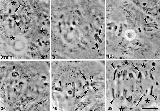
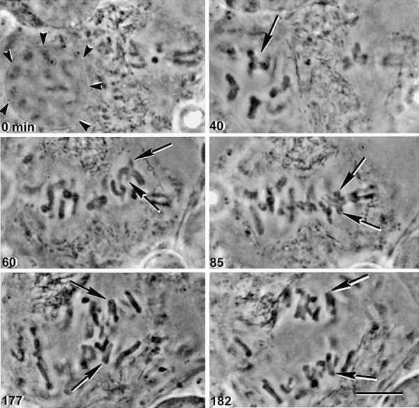
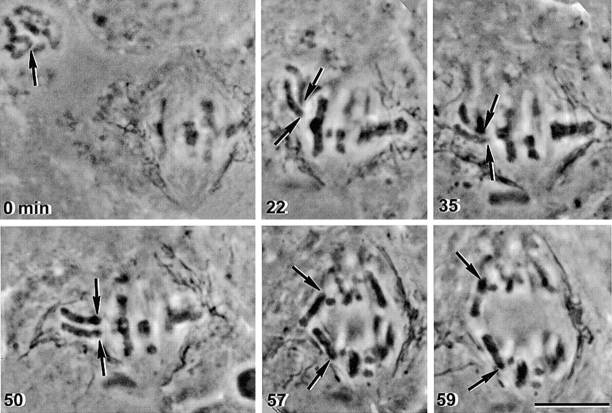
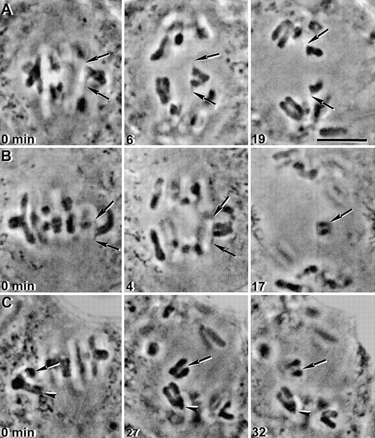
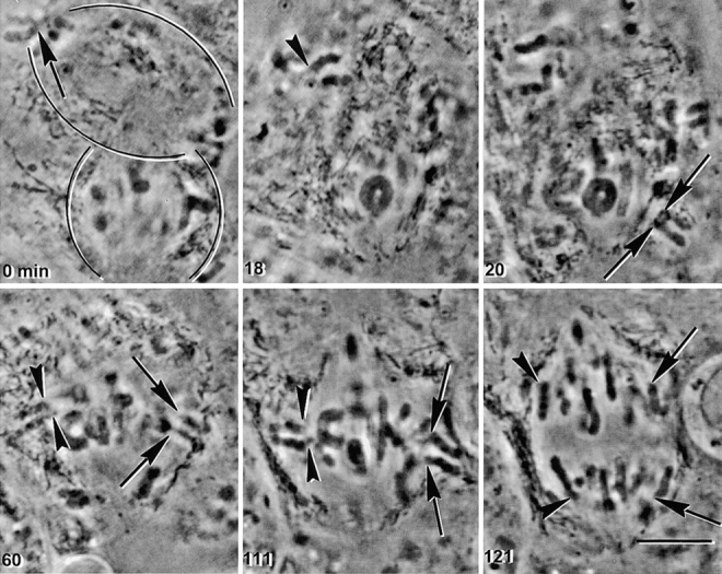
In meiosis I, two chromatids move to each spindle pole. Then, in meiosis II, the two are distributed, one to each future gamete. This requires that meiosis I chromosomes attach to the spindle differently than meiosis II chromosomes and that they regulate chromosome cohesion differently. We investigated whether the information that dictates the division type of the chromosome comes from the whole cell, the spindle, or the chromosome itself. Also, we determined when chromosomes can switch from meiosis I behavior to meiosis II behavior. We used a micromanipulation needle to fuse grasshopper spermatocytes in meiosis I to spermatocytes in meiosis II, and to move chromosomes from one spindle to the other. Chromosomes placed on spindles of a different meiotic division always behaved as they would have on their native spindle; e.g., a meiosis I chromosome attached to a meiosis II spindle in its normal fashion and sister chromatids moved together to the same spindle pole. We also showed that meiosis I chromosomes become competent meiosis II chromosomes in anaphase of meiosis I, but not before. The patterns for attachment to the spindle and regulation of cohesion are built into the chromosome itself. These results suggest that regulation of chromosome cohesion may be linked to differences in the arrangement of kinetochores in the two meiotic divisions.


Figures

Figure 1
Figure 1
Kinetochore attachment to the spindle and chromosome cohesion in mitosis and meiosis. Mitosis, mitotic chromosomes consist of two sister chromatids. Kinetochore arrangement: one kinetochore faces one pole while its sister kinetochore faces the opposite pole and they move to opposite poles in anaphase. Chromosome cohesion: in metaphase, sister chromatids are held together by cohesion along chromosome arms and between centromeres. In anaphase both centromere and arm cohesion are released. Meiosis I, Bivalents consist of two homologous chromosomes, each of which is composed of a pair of sister chromatids. Kinetochore arrangement: both sister kinetochores face the same spindle pole, while the homologous pair of sister kinetochores faces the opposite spindle pole. Therefore, in anaphase I, both sister chromatids of one homologue move toward the same pole. Chromosome cohesion: sister chromatids in meiosis I are held together by cohesion between centromeres and between arms of sister chromatids. The bivalent is held together by both recombination and arm cohesion. Recombination sites are at the junction between black and grey chromosome segments. In anaphase of meiosis I, arm cohesion is released and half-bivalents separate. (Meiosis II) Each meiosis II chromosome consists of a pair of sister chromatids. Kinetochore arrangement: one kinetochore faces one pole, while its sister kinetochore faces the opposite pole. Consequently, sister kinetochores separate from one another in anaphase of meiosis II. Chromosome cohesion: in early stages of meiosis II, only the cohesion between centromeres remains; it is removed in anaphase II.
Figure 2
Figure 2
Determinants for the pattern of chromosome attachment to the spindle and release of chromosome cohesion are built into the chromosome. A metaphase I grasshopper spermatocyte was fused to a metaphase II spermatocyte. Spindle poles are indicated by asterisks, manipulated meiosis I chromosomes by straight arrows, unmanipulated meiosis I chromosomes by curved arrows, manipulated meiosis II chromosomes by filled arrowheads, and unmanipulated meiosis II chromosomes by open arrowheads. The fused cell contains two spindles. A bivalent was detached from the meiosis I spindle and placed near the meiosis II spindle (0 and 8 min, straight arrows). The bivalent attached to the meiosis II spindle with a pair of sister kinetochores facing each pole (48 min, straight arrows). Pairs of sister chromatids segregated to each pole (69 min, straight arrows). Unmanipulated bivalents on the meiosis I spindle had a pair of sister kinetochores facing each pole (48 min, curved arrows). In anaphase in unmanipulated bivalents, pairs of sister chromatids separated from one another (69 min, curved arrows). A meiosis II chromosome (12 min, filled arrowhead) was detached from the meiosis II spindle and placed near the meiosis I spindle (36 min, filled arrowhead). The meiosis II chromosome attached to the meiosis I spindle with a single sister kinetochore facing each pole (48 min, filled arrowhead), and single sister chromatids moved to opposite poles in anaphase (69 min, filled arrowheads). Unmanipulated meiosis II chromosomes attached with a single sister kinetochore facing each pole (48 min, open arrowheads) and moved to opposite poles in anaphase (69 min, open arrowheads). Bar, 10 μm.
Figure 3
Figure 3
The way a meiosis I chromosome attaches to the spindle and releases cohesion does not depend on its initial spindle attachment. A late-prophase I spermatocyte and a metaphase II spermatocyte were fused (0 min). The prophase nuclear envelope was still present (0 min, arrowheads). After nuclear envelope breakdown, a bivalent that had not yet attached to the meiosis I spindle (40 min, arrow) was placed near the meiosis II spindle (60 min, arrows). The manipulated bivalent attached to the meiosis II spindle (85 min, arrows). Pairs of sister chromatids segregated to opposite poles in anaphase. The upper pair is more clearly visible in the 177 min image, while the lower pair is more clearly visible in the 182 min image. Bar, 10 μm.
Figure 4
Figure 4
The way a meiosis II chromosome attaches to the spindle and releases cohesion does not depend on its initial spindle attachment. A prophase II spermatocyte and a metaphase I spermatocyte were fused (0 min). After nuclear envelope breakdown, a meiosis II chromosome that had not yet attached to the meiosis II spindle (0 min, arrow) moved near the meiosis I spindle (22 min, arrows). It attached with one chromatid facing each pole (22, 35, and 50 min, arrows), and single sister chromatids segregated to opposite poles in anaphase (57 and 59 min, arrows). Bar, 10 μm.
Figure 5
Figure 5
Bivalents can be induced to attach to the spindle with a single sister kinetochore facing each pole, but they neither attach nor separate in the normal meiosis II manner. Bivalents in unfused spermatocytes were micromanipulated. (A) One pair of sister kinetochores of the bivalent was induced to attach to opposite spindle poles (0 min, arrows). The chromosome remained, with stretched-out kinetochores, at the equator of the spindle after anaphase onset (6 min, arrows). 13 min after anaphase onset, the sister chromatids started to slowly separate from one another (19 min), moving towards the poles to which they were attached. (B) Another example of a bivalent in which one pair of sister kinetochores was induced to attach to opposite poles (0 min, arrows). In anaphase I, sister kinetochores were greatly stretched towards their spindle poles, but the sister chromatids did not separate from one another (4 and 17 min, arrows). (C) One pair of sister kinetochores of the bivalent was induced to attach to opposite poles (0 min, arrow), while the other pair of sister kinetochores attached to the same pole (0 min, arrowhead). In the pair that did attach to opposite poles, the sister chromatids did not separate from one another (27 and 32 min, arrows). The other pair of sister kinetochores attached to the lower spindle pole (0 and 27 min, arrowheads) and moved together to that pole in anaphase (27 and 32 min, arrowheads). Bar, 10 μm.
Figure 6
Figure 6
Chromosomes acquire meiosis II properties after anaphase I. A spermatocyte in anaphase I was fused to a spermatocyte in metaphase I (0 min). The spindles are outlined: anaphase I above, metaphase I below. Two different anaphase chromosomes were studied in this experiment, one indicated by an arrow and the other by an arrowhead. The chromosomes were detached from the anaphase I spindle (0 min, arrow; 18 min, arrowhead) and placed near the metaphase I spindle (20 min, arrows; 60 min, arrowheads). The chromosomes attached to the metaphase I spindle, with a sister kinetochore facing each pole (20 and 60 min, arrows; 60 min, arrowheads). The manipulated meiosis I chromosomes behaved just like meiosis II chromosomes when the cell entered anaphase, sending a single chromatid to each pole (111 and 121 min, arrowheads and arrows). Bar, 10 μm.

References

    1. Bickel S.E., Moore D.P., Lai C., Orr-Weaver T.L. Genetic interactions between mei-S332 and ord in the control of sister-chromatid cohesion. Genetics. 1998;150:1467–1476. - PMC - PubMed
    1. Bickel S.E., Orr-Weaver T.L. Holding chromatids together to ensure they go their separate ways. Bioessays. 1996;18:293–300. - PubMed
    1. Gimenez-Abian J.F., Clarke D.J., Garcia de la Vega C., Gimenez-Martin G. The role of sister chromatid cohesiveness and structure in meiotic behaviour. Chromosoma. 1997;106:422–434. - PubMed
    1. Goldstein L.S. Kinetochore structure and its role in chromosome orientation during the first meiotic division in male D. melanogaster . Cell. 1981;25:591–602. - PubMed
    1. Kerrebrock A.W., Miyazaki W.Y., Birnby D., Orr-Weaver T.L. The Drosophila mei-s332 gene promotes sister-chromatid cohesion in meiosis following kinetochore differentiation. Genetics. 1992;130:827–841. - PMC - PubMed

Publication types