. 2000 Sep 18;150(6):1283-98.
doi: 10.1083/jcb.150.6.1283.

Desmin cytoskeleton linked to muscle mitochondrial distribution and respiratory function

Affiliations

Desmin cytoskeleton linked to muscle mitochondrial distribution and respiratory function

D J Milner et al. J Cell Biol. .

Abstract

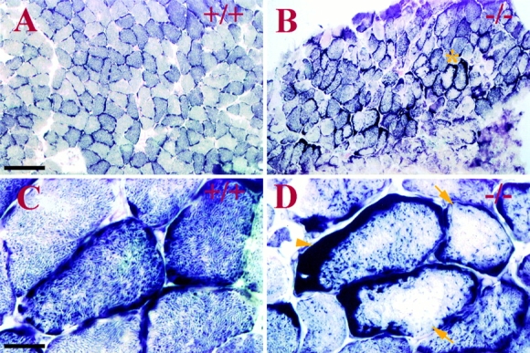
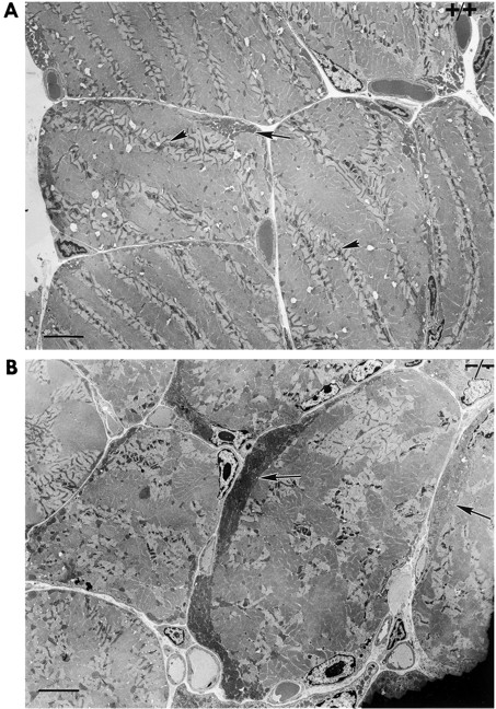
Ultrastructural studies have previously suggested potential association of intermediate filaments (IFs) with mitochondria. Thus, we have investigated mitochondrial distribution and function in muscle lacking the IF protein desmin. Immunostaining of skeletal muscle tissue sections, as well as histochemical staining for the mitochondrial marker enzymes cytochrome C oxidase and succinate dehydrogenase, demonstrate abnormal accumulation of subsarcolemmal clumps of mitochondria in predominantly slow twitch skeletal muscle of desmin-null mice. Ultrastructural observation of desmin-null cardiac muscle demonstrates in addition to clumping, extensive mitochondrial proliferation in a significant fraction of the myocytes, particularly after work overload. These alterations are frequently associated with swelling and degeneration of the mitochondrial matrix. Mitochondrial abnormalities can be detected very early, before other structural defects become obvious. To investigate related changes in mitochondrial function, we have analyzed ADP-stimulated respiration of isolated muscle mitochondria, and ADP-stimulated mitochondrial respiration in situ using saponin skinned muscle fibers. The in vitro maximal rates of respiration in isolated cardiac mitochondria from desmin-null and wild-type mice were similar. However, mitochondrial respiration in situ is significantly altered in desmin-null muscle. Both the maximal rate of ADP-stimulated oxygen consumption and the dissociation constant (K(m)) for ADP are significantly reduced in desmin-null cardiac and soleus muscle compared with controls. Respiratory parameters for desmin-null fast twitch gastrocnemius muscle were unaffected. Additionally, respiratory measurements in the presence of creatine indicate that coupling of creatine kinase and the adenine translocator is lost in desmin-null soleus muscle. This coupling is unaffected in cardiac muscle from desmin-null animals. All of these studies indicate that desmin IFs play a significant role in mitochondrial positioning and respiratory function in cardiac and skeletal muscle.


Figures

Figure 1
Figure 1
Alteration of mitochondrial distribution in desmin-null soleus skeletal muscle. SDH histochemical staining (A–D) demonstrates alteration of mitochondrial distribution in desmin-null (−/−) soleus muscle. In sections of normal (+/+) 10-mo-old mouse soleus muscle (A), a muscle consisting mostly of highly oxidative slow twitch fibers, a checkerboard pattern of intensely staining fibers is observed, with a variable amount of fibers staining with less intensity. The staining is mostly granular throughout the fibers, with slight enhancement of subsarcolemmal staining in a few fiber sections (C). In soleus sections from corresponding desmin-null animals (B, same scale as A), this discernible checkerboard pattern is partially lost. Large clumps of subsarcolemmal staining appear in a majority of the fibers (arrowhead), and the interior of fibers frequently show reduced SDH staining (arrows) (C and D, higher magnification view of A and B accordingly).
Figure 2
Figure 2
Alteration of mitochondrial distribution occurs in other muscles. SDH histochemical staining (A–F) demonstrates alteration of mitochondrial distribution in the red gastrocnemius (A and E, left), an area comprised mostly of slow twitch fibers. Control wild-type muscle (+/+; A) shows granular staining with some slight subsarcolemmal clumping, whereas subsarcolemmal clumps are pronounced in desmin null (−/−) sections (B, arrowheads), and some fibers appear to have lost mitochondrial staining from the fiber interior altogether (B, arrows). Sections of fast twitch skeletal muscle of the gastrocnemius (+/+ and −/− in C and D, respectively; and A and E, right) do not show any changes in SDH staining patterns. When young (1 mo) mouse muscle is stained for SDH, some slight enhancement of clumping is seen in desmin-null sections (F, arrowheads), and the staining of the interior of the fibers is slightly reduced compared with wild-type (E) fibers.
Figure 3
Figure 3
Ultrastructural observation of mitochondrial distribution in skeletal muscle. Electron microscopic analysis of oblique sections from soleus skeletal muscle confirm the change in mitochondrial distribution in desmin null (−/−) skeletal muscle demonstrated by SDH staining in Fig. 1 and Fig. 2. Wild-type (+/+, A and C) soleus muscle displays regularly arrayed mitochondria (arrowheads) throughout the interior of the fiber visible at the level of the lighter color I band (I in C) and interspersed between the myofibrils (arrowheads). Only slight subsarcolemmal clumping can be observed (arrow). A desmin-null fiber (B) showing increased subsarcolemmal clumping (arrow) and decrease in intermyofibrillar mitochondria. (D) A severe case of mitochondrial (M) clumping in a desmin-null fiber (arrow). Note the internal nucleus (n) and the lack of intermyofibrillar mitochondria compared with the wild-type section (C). Bars, 5 μm.
Figure 3
Figure 3
Ultrastructural observation of mitochondrial distribution in skeletal muscle. Electron microscopic analysis of oblique sections from soleus skeletal muscle confirm the change in mitochondrial distribution in desmin null (−/−) skeletal muscle demonstrated by SDH staining in Fig. 1 and Fig. 2. Wild-type (+/+, A and C) soleus muscle displays regularly arrayed mitochondria (arrowheads) throughout the interior of the fiber visible at the level of the lighter color I band (I in C) and interspersed between the myofibrils (arrowheads). Only slight subsarcolemmal clumping can be observed (arrow). A desmin-null fiber (B) showing increased subsarcolemmal clumping (arrow) and decrease in intermyofibrillar mitochondria. (D) A severe case of mitochondrial (M) clumping in a desmin-null fiber (arrow). Note the internal nucleus (n) and the lack of intermyofibrillar mitochondria compared with the wild-type section (C). Bars, 5 μm.
Figure 4
Figure 4
Ultrastructural observations of mitochondrial alterations in cardiac muscle. (A) Wild type (+/+) cardiac muscle showing normal cardiomyocyte ultrastructural appearance with laterally aligned myofibrils and strands of mitochondria (arrow) between the myofibrils. (B) Desmin-null (−/−) cardiac muscle demonstrating proliferation and swelling of mitochondria (arrow) between myofibrils and subsarcolemmal clumping of mitochondria (m). (C) Desmin-null cardiac muscle from exercised mice exhibiting severe mitochondrial swelling, including mitochondria with broken cristae (arrow). An area of severe mitochondrial proliferation is also observed (p). (D) Desmin-null cardiac muscle showing severe architectural disruption, large empty spaces surrounded by mitochondria, and mitochondria sequestered within a membrane-limited structure (arrowhead). (E) High magnification view of area in D (asterisk) showing mitochondria enclosed in a membrane-limited structure (arrowhead) within a myocyte. Note the mitochondria display normal condensed morphology. Bars: (A–D) 5 μm; (E) 1 μm.
Figure 4
Figure 4
Ultrastructural observations of mitochondrial alterations in cardiac muscle. (A) Wild type (+/+) cardiac muscle showing normal cardiomyocyte ultrastructural appearance with laterally aligned myofibrils and strands of mitochondria (arrow) between the myofibrils. (B) Desmin-null (−/−) cardiac muscle demonstrating proliferation and swelling of mitochondria (arrow) between myofibrils and subsarcolemmal clumping of mitochondria (m). (C) Desmin-null cardiac muscle from exercised mice exhibiting severe mitochondrial swelling, including mitochondria with broken cristae (arrow). An area of severe mitochondrial proliferation is also observed (p). (D) Desmin-null cardiac muscle showing severe architectural disruption, large empty spaces surrounded by mitochondria, and mitochondria sequestered within a membrane-limited structure (arrowhead). (E) High magnification view of area in D (asterisk) showing mitochondria enclosed in a membrane-limited structure (arrowhead) within a myocyte. Note the mitochondria display normal condensed morphology. Bars: (A–D) 5 μm; (E) 1 μm.
Figure 5
Figure 5
Western blot analysis of total protein extracts from myocardial tissue of desmin null (Des−/−) and wild-type (Des+/+) mice for the mitochondrial proteins porin and cytochrome oxidase (COXI). The membranes were probed with the corresponding antibodies. The lower porin band is a commonly obtained porin degradation product. A: twice the total protein of B.
Figure 6
Figure 6
Mitochondrial changes begin before other ultrastructural changes in young desmin-null mice. Sections of cardiac muscle from 2-wk-old desmin-null (−/−) animals (B–E) display mitochondrial changes similar to but less severe than those seen in adults, including occasional clumping and slight increase in mitochondria number (C, arrow), swelling, and degeneration (D and E, arrows) compared with wild-type (+/+) sections (A). Other ultrastructural changes seen in adult desmin-null animals are not observed in 2-wk-old desmin-null sections, as myofibrils (arrow) and intercalated disks (arrowhead) appear normal (B). Also note the large membrane-limited structure (C, asterisk) inside a cardiomyocyte that is similar to the structures seen in adult null cardiomyocytes (Fig. 4 D). M, mitochondria. Bars: (A–C) 5 μm; (D and E) 1 μm.
Figure 6
Figure 6
Mitochondrial changes begin before other ultrastructural changes in young desmin-null mice. Sections of cardiac muscle from 2-wk-old desmin-null (−/−) animals (B–E) display mitochondrial changes similar to but less severe than those seen in adults, including occasional clumping and slight increase in mitochondria number (C, arrow), swelling, and degeneration (D and E, arrows) compared with wild-type (+/+) sections (A). Other ultrastructural changes seen in adult desmin-null animals are not observed in 2-wk-old desmin-null sections, as myofibrils (arrow) and intercalated disks (arrowhead) appear normal (B). Also note the large membrane-limited structure (C, asterisk) inside a cardiomyocyte that is similar to the structures seen in adult null cardiomyocytes (Fig. 4 D). M, mitochondria. Bars: (A–C) 5 μm; (D and E) 1 μm.

References

    1. Almahbobi G., Williams L.J., Han X.G., Hall P.F. Binding of lipid droplets and mitochondria to intermediate filaments in rat Leydig cells. J. Reprod. Fertil. 1993;98:209–217. - PubMed
    1. Bereiter-Hahn J., Voth M. Dynamics of mitochondria in living cellsshape changes, dislocations, fusion, and fission of mitochondria. Microsc. Res. Tech. 1994;27:198–219. - PubMed
    1. Bilak S.R., Sernett S.W., Bilak M.M., Bellin R.M., Stromer M.H., Huiatt T.W., Robson R.M. Properties of the novel intermediate filament protein synemin and its identification in mammalian muscle. Arch. Biochem. Biophys. 1998;355:63–76. - PubMed
    1. Capetanaki Y., Milner D. Desmin cytoskeleton in muscle integrity and function. Subcell. Biochem. 1998;31:463–495. - PubMed
    1. Capetanaki Y., Ngai J., Lazarides E. Characterization and regulation in the expression of a gene encoding for the intermediate filament protein desmin. Proc. Natl. Acad. Sci. USA. 1984;81:6909–6912. - PMC - PubMed

Publication types

MeSH terms