. 2000 Sep 18;150(6):1349-60.
doi: 10.1083/jcb.150.6.1349.

Cell to cell communication in response to mechanical stress via bilateral release of ATP and UTP in polarized epithelia

Affiliations

Cell to cell communication in response to mechanical stress via bilateral release of ATP and UTP in polarized epithelia

L Homolya et al. J Cell Biol. .

Abstract

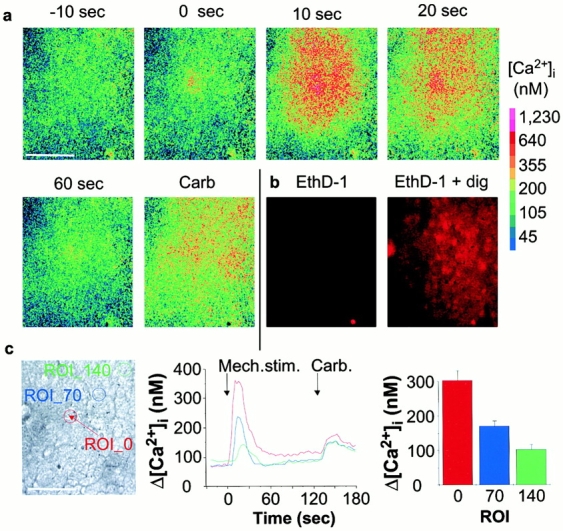
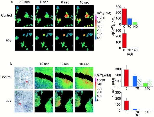
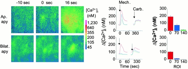
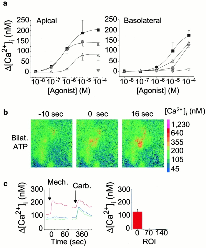
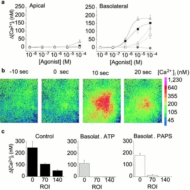
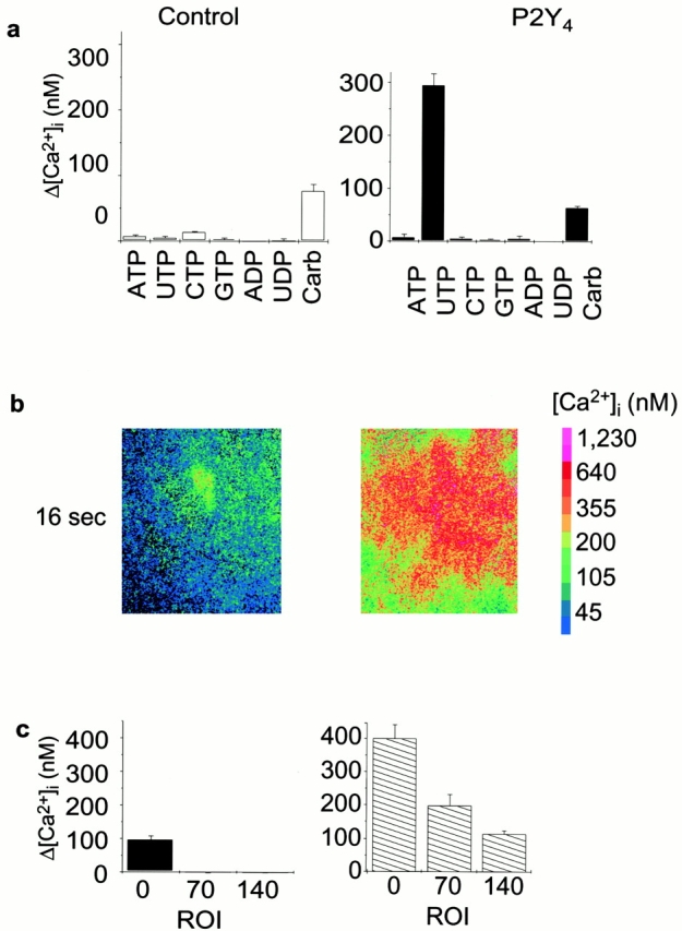
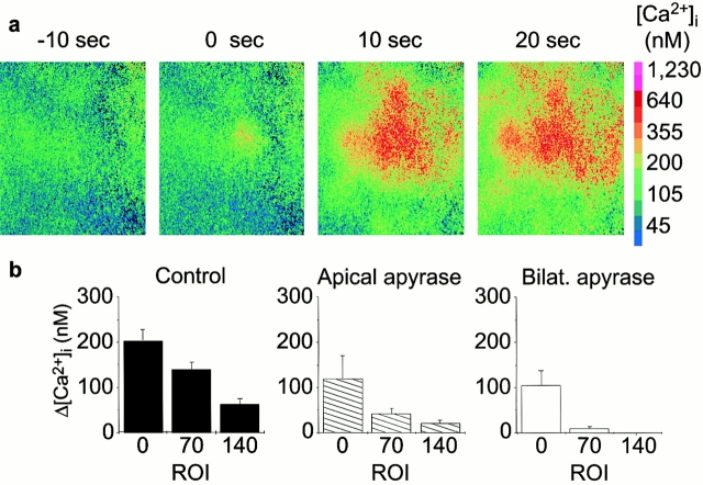
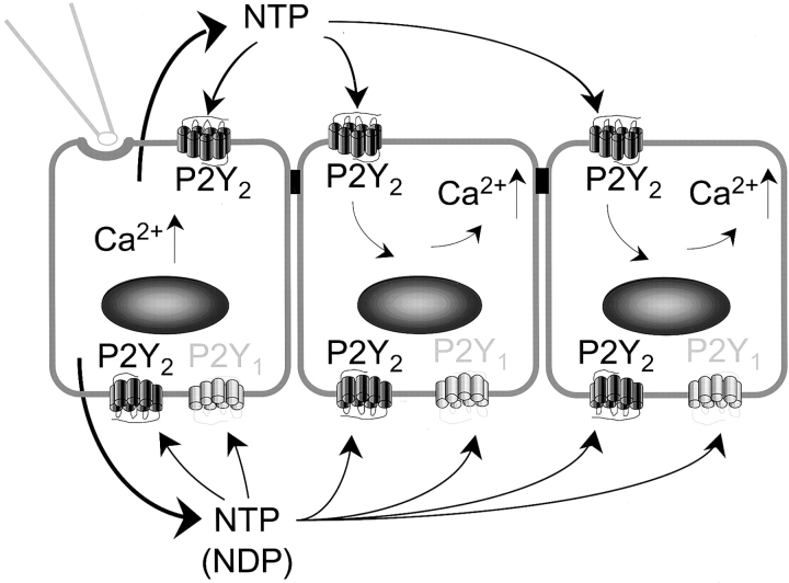
Airway epithelia are positioned at the interface between the body and the environment, and generate complex signaling responses to inhaled toxins and other stresses. Luminal mechanical stimulation of airway epithelial cells produces a propagating wave of elevated intracellular Ca(2+) that coordinates components of the integrated epithelial stress response. In polarized airway epithelia, this response has been attributed to IP(3) permeation through gap junctions. Using a combination of approaches, including enzymes that destroy extracellular nucleotides, purinergic receptor desensitization, and airway cells deficient in purinoceptors, we demonstrated that Ca(2+) waves induced by luminal mechanical stimulation in polarized airway epithelia were initiated by the release of the 5' nucleotides, ATP and UTP, across both apical and basolateral membranes. The nucleotides released into the extracellular compartment interacted with purinoceptors at both membranes to trigger Ca(2+) mobilization. Physiologically, apical membrane nucleotide-release coordinates airway mucociliary clearance responses (mucin and salt, water secretion, increased ciliary beat frequency), whereas basolateral release constitutes a paracrine mechanism by which mechanical stresses signal adjacent cells not only within the epithelium, but other cell types (nerves, inflammatory cells) in the submucosa. Nucleotide-release ipsilateral and contralateral to the surface stimulated constitutes a unique mechanism by which epithelia coordinate local and distant airway defense responses to mechanical stimuli.


Figures

Figure 1
Figure 1
Propagation of Ca2+ signals in polarized cultures of normal mouse nasal epithelial cells. a, Ca2+ waves: a single cell in a confluent, Fura-2 loaded culture grown on a permeable membrane support was mechanically stimulated with a micropipette. This stimulation resulted in a radially propagating Ca2+ wave. The numbers above the pseudocolor images indicate the time in seconds relative to the stimulus. The first image in which an elevation in Ca2+ level was observed is considered as time 0. Subsequent exposure to 1 mM carbachol (Carb) induced a second Ca2+ response in the cells (including the stimulated one), showing that the cells remain viable and responsive. Color bar shows the calibration for [Ca2+]i. Bar, 100 μm. b, Cell viability: viability of the cells after mechanical stimulation was also tested by the addition of 10 mM ethidium-homodimer (EthD-1). Only one cell (distinct from the mechanically stimulated one) showed dye uptake after a 5-min incubation. As control for the viability assay, all the cells were lysed with 80 M digitonin in the continuous presence of the dye (EthD-1 + dig). c, Quantitation of Ca2+ waves: Ca2+ concentrations were quantitated in three circular regions of interest 70 μm apart (ROI_0, ROI_70 and ROI_140) shown in the bright-field image (left). An arrow indicates the stimulated cell. Bar, 100 μm. Time course of [Ca2+]i for the three regions of interest in the same experiment is shown in the central panel. The color of each tracing corresponds to the color of circles denoting each ROI shown in the bright-field image on the left. The right panel depicts the mean change in Ca2+ levels (± SEM) for the ROI_0, ROI_70 and ROI_140 at 8, 12, and 16 s after the stimulus, respectively (n = 20).
Figure 2
Figure 2
Cell–cell contact is not required for the mechanically induced Ca2+ wave in mouse nasal epithelial cells. a, Propagating Ca2+ waves in not-contacting cells. Mechanical stimulation of a single cell in a nonpolarized culture grown on a glass coverslip resulted in a Ca2+ wave propagation (top). Elevation in Ca2+ level was observed in cells that were not in physical contact with the stimulated cell. Apyrase (10 U/ml) abolished the spread of Ca2+ wave (bottom). Low intensity pixels (<32) of the background were omitted on the pseudocolor images. Bar graph on the right depicts summary data for multiple experiments (n = 4 and 6). For other details, see Fig. 1 c. b, The Ca2+ wave propagates over a physical gap: a confluent culture of mouse epithelial cells grown on permeable membrane support was gently scraped with a micropipette 1 h before the experiment to produce a gap in the epithelial sheet. A single cell close to the scrape (marked with a red arrow on the bright-field image on the left) was mechanically stimulated in the absence (top) and presence (bottom) of apyrase (10 U/ml, grade V). The Ca2+ wave propagated over the gap (top) indicating that the signal transmission mechanism involves extracellular substance(s). The mediator was sensitive to apyrase (bottom). Bar, 100 μm. To avoid artificial background noise effects of low intensity fields, the gap was masked on the basis of the bright-field image. The mean changes in Ca2+ level (± SEM) are shown in the bar graph in the right panels (n = 4 and 4, respectively). The solid bars refer to the ROIs on the stimulated side, whereas hatched bars refer to the ROIs on the opposite side of the gap. No significant differences between the two sides were found in the spread of the Ca2+ wave.
Figure 3
Figure 3
Inhibition of intercellular Ca2+ waves with extracellular nucleotidase activity. Pseudocolor images on the left demonstrate the intracellular Ca2+ levels at designated time points in representative experiments. In the center, time courses of [Ca2+]i in these experiments are shown. Summary data for multiple experiments are shown on the right (n = 6 and 13). Apical addition of apyrase (10 U/ml) attenuated, but did not block, the mechanically induced intercellular Ca2+ wave (top), whereas bilateral addition of apyrase completely prevented the Ca2+ spread (bottom). Subsequent carbachol-induced Ca2+ responses were not affected as compared with control experiments shown in Fig. 1 c (middle).
Figure 4
Figure 4
Participation of P2-receptors in intercellular Ca2+ waves. a, Functional expression of P2Y-receptors in polarized mouse epithelial cells. Nucleotide-induced Ca2+ responses to apical versus basolateral additions of ATP (squares), UTP (circles), ADP (upright triangles), and UDP (inverted triangles) were measured. Basal to peak changes in [Ca2+]i levels (mean ± SEM) are plotted (n = 3–6/point). b, Inhibition of intercellular Ca2+ wave by desensitization of P2-receptors. Bilateral pretreatment with ATP (300 μM) completely prevented the propagation of Ca2+ signals without affecting the carbachol-induced Ca2+ response. c, Quantitative analysis of the inhibition of intercellular Ca2+ waves: Time course of [Ca2+]i for the three regions of interest in the same experiment shown (left) and mean change in Ca2+ levels (± SEM) for several similar experiments (n = 15) (right). For details see legend of Fig. 1 c.
Figure 5
Figure 5
Propagating Ca2+ waves in P2Y2-R (−/−) airway epithelial cells. a, Characterization of polarized cultures of nasal cells isolated from P2Y2-receptor gene targeted (−/−) mice for residual expression of P2Y-receptors. Nucleotide-induced Ca2+ responses to ATP (squares), UTP (circles), ADP (upright triangles), and UDP (inverted triangles) were measured (n = 3–6/point). The effects of ATP (open squares) and ADP (open up triangles) were also assessed in the presence of 100 μM PAPS, a P2Y1-receptor antagonist (Boyer et al. 1996). The pharmacological profiles indicate that the residual P2-receptor (most likely P2Y1-R) is expressed solely in the basolateral membrane. b, Mechanically induced intercellular Ca2+ wave in P2Y2-R (−/−) cells in a representative experiment. The residual Ca2+ propagation in P2Y2-R (−/−) cells suggests basolateral release of nucleotides interacting with P2Y1-receptors in the basolateral membrane. For details see Fig. 1 a. c, Inhibition of Ca2+ propagation in P2Y2-R (−/−) cells. The magnitude of the Ca2+ waves was smaller in P2Y2-R (−/−) cells as compared with wild-type cells (Fig. 1 c, right). Basolateral pretreatment with ATP (hatched bars) or PAPS (open bars) completely abolished the intercellular Ca2+ wave as compared with P2Y2-R (−/−) cells without treatment (solid bars; n = 11, 6, and 9, respectively). For details for quantitative analysis see legend of Fig. 1 c.
Figure 6
Figure 6
Participation of released UTP in the mechanically induced Ca2+ wave propagation. a, Characterization of a mouse P2Y2-R (−/−) nasal cell line expressing the human P2Y4-receptor. Ca2+ responses to nucleotides (100 μM) and carbachol (1 mM) were measured in P2Y2-R (−/−) cells transduced with the Hygror-only vector (Control) and with the human P2Y4-receptor (P2Y4) (n = 3–4/point). Control [P2Y2(−/−)] cells grown on glass coverslips showed no substantial Ca2+ responses to nucleotides but to carbachol. In contrast, UTP stimulated significant Ca2+ responses in P2Y4-R transduced cells. No other nucleotide stimulated substantial Ca2+ response in the P2Y4-receptor expressing cells. b, Mechanically induced intercellular Ca2+ wave in polarized culture of P2Y4-R transduced cells in a representative experiment. Pseudocolor images demonstrate the intracellular Ca2+ levels in the empty vector-transduced P2Y2(−/−) cells (left) and the P2Y4-R transduced P2Y2(−/−) cells (right) 16 s after local mechanical stimulation in the continuous presence of 100 μM PAPS in the basolateral bath. c, Quantitative analysis of Ca2+ wave propagation in control and P2Y4-R transduced cells. Summary data for experiments similar to that shown in Fig. 6 b. Mechanical stimulation of a single cell did not elicit Ca2+ wave in the P2Y2(−/−) cells transduced with the Hygror-only vector, when 100 μM PAPS was present in the basolateral bath, whereas an extensive Ca2+ wave was observed in P2Y4-R transduced cells (n = 3 and 3, respectively). For details for quantitative analysis see legend of Fig. 1 c.
Figure 7
Figure 7
Intercellular Ca2+ waves in polarized human airway epithelial cells. a, A representative experiment. Local mechanical stimulation elicited cell to cell propagation of Ca2+ signals in well-differentiated human bronchial epithelial cell cultures. b, Block of Ca2+ wave propagation with extracellular nucleotidase activity. The intercellular Ca2+ waves were partially inhibited by apical addition of apyrase (hatched bars; n = 5) as compared with control (solid bars; n = 3). Bilateral treatment with apyrase completely abolished the spread of Ca2+ signals (open bars; n = 10).
Figure 8
Figure 8
A proposed model for intercellular Ca2+ waves in airway epithelia. Mechanical stimulation on the apical surface of a polarized airway epithelial cell induces ipsilateral and contralateral release of nucleotides that activates the adjacent cells via P2Y2-receptors on the apical membrane and via both P2Y1- and P2Y2-receptors on the basolateral membrane.

References

    1. Abraham E.H., Okunieff P., Scala S., Vos P., Oosterveld M.J.S., Chen A.Y., Shrivastav B., Guidotti G. Cystic fibrosis transmembrane conductance regulator and adenosine triphosphate. Science. 1997;275:1324–1325. - PubMed
    1. Boitano S., Dirksen E.R., Sanderson M.J. Intercellular propagation of calcium waves mediated by inositol trisphosphate. Science. 1992;258:292–295. - PubMed
    1. Boitano S., Sanderson M.J., Dirksen E.R. A role for Ca2+-conducting ion channels in mechanically-induced signal transduction of airway epithelial cells. J. Cell Sci. 1994;107:3037–3044. - PubMed
    1. Boitano S., Dirksen E.R., Evans W.H. Sequence-specific antibodies to connexins block intercellular calcium signaling through gap junctions. Cell Calcium. 1998;23:1–9. - PubMed
    1. Boyer J.L., Romero-Avila T., Schachter J.B., Harden T.K. Identification of competitive agonists of the P2Y1-receptor. Mol. Pharmacol. 1996;50:1323–1329. - PubMed

Publication types

MeSH terms