. 2000 Sep 18;150(6):1385-98.
doi: 10.1083/jcb.150.6.1385.

Absence of alpha-syntrophin leads to structurally aberrant neuromuscular synapses deficient in utrophin

Affiliations

Absence of alpha-syntrophin leads to structurally aberrant neuromuscular synapses deficient in utrophin

M E Adams et al. J Cell Biol. .

Abstract

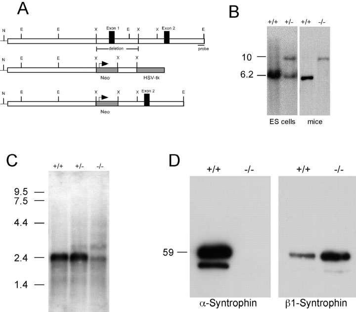
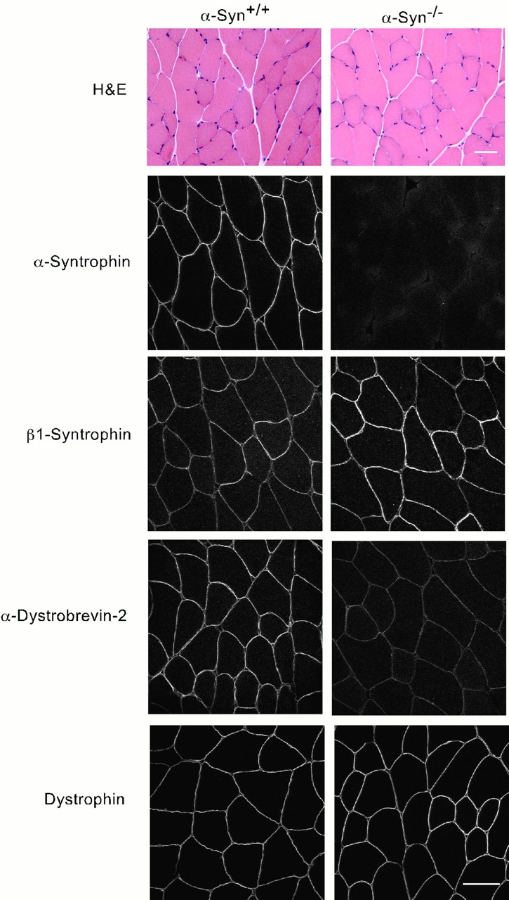
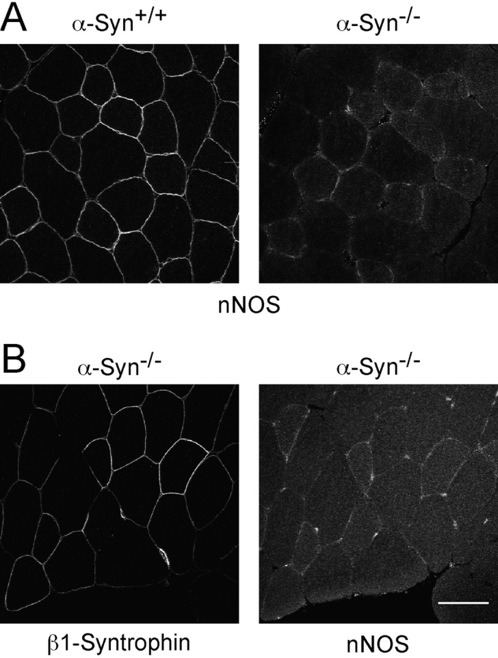
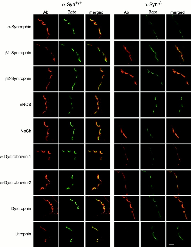
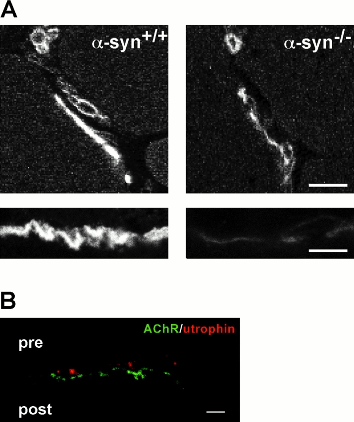
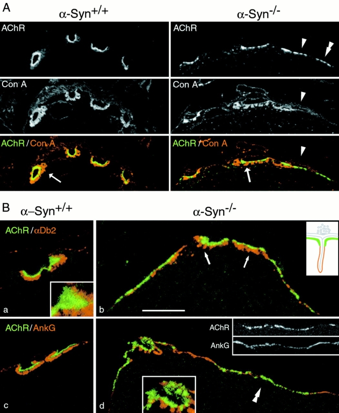
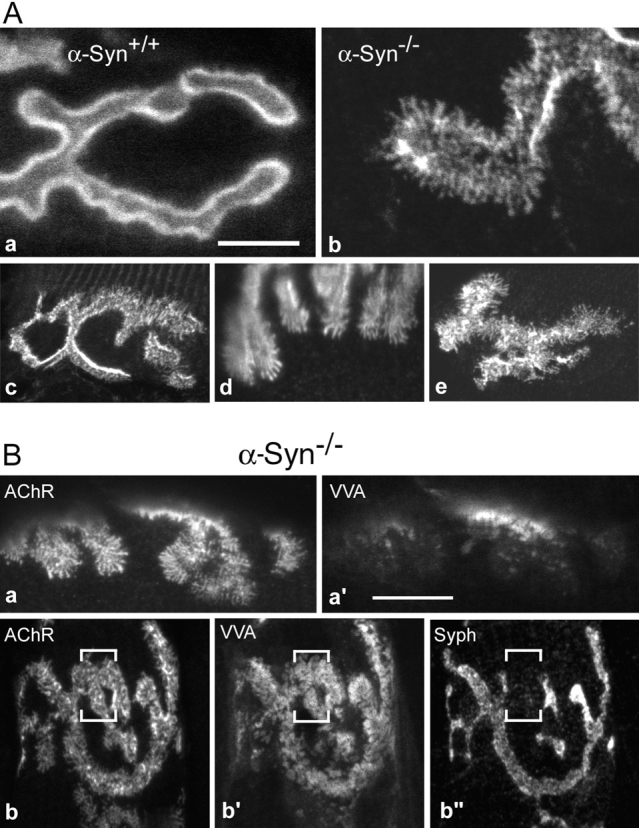
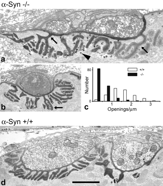
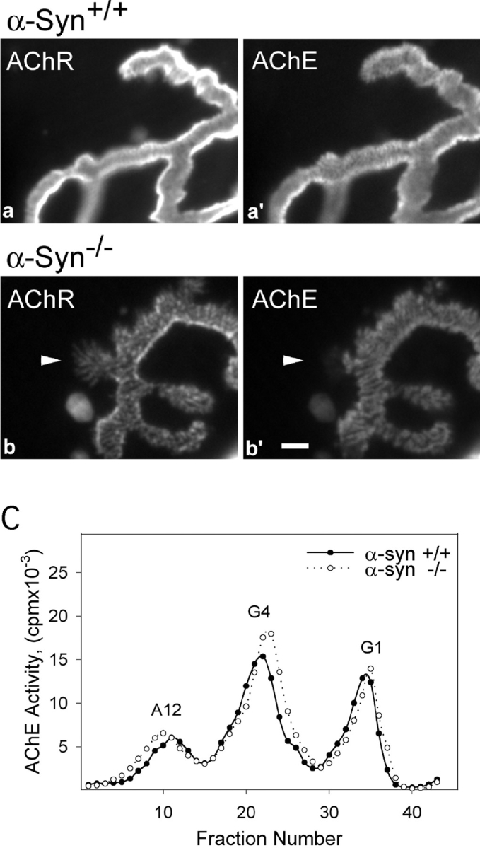
The syntrophins are a family of structurally related proteins that contain multiple protein interaction motifs. Syntrophins associate directly with dystrophin, the product of the Duchenne muscular dystrophy locus, and its homologues. We have generated alpha-syntrophin null mice by targeted gene disruption to test the function of this association. The alpha-Syn(-/)- mice show no evidence of myopathy, despite reduced levels of alpha-dystrobrevin-2. Neuronal nitric oxide synthase, a component of the dystrophin protein complex, is absent from the sarcolemma of the alpha-Syn(-/)- mice, even where other syntrophin isoforms are present. alpha-Syn(-/)- neuromuscular junctions have undetectable levels of postsynaptic utrophin and reduced levels of acetylcholine receptor and acetylcholinesterase. The mutant junctions have shallow nerve gutters, abnormal distributions of acetylcholine receptors, and postjunctional folds that are generally less organized and have fewer openings to the synaptic cleft than controls. Thus, alpha-syntrophin has an important role in synapse formation and in the organization of utrophin, acetylcholine receptor, and acetylcholinesterase at the neuromuscular synapse.


Figures

Figure 1
Figure 1
Generation and characterization of α-syntrophin null mice. A, A targeting vector was constructed using a 7.4-kb Not I (N)/XbaI (X) restriction fragment long arm and a 1.5-kb XbaI fragment short arm. Homologous recombination resulted in the deletion of 2.8-kb of the α-syntrophin gene including all of exon 1. B, Southern blot analysis was performed using genomic DNA (isolated from ES cells or from mice) digested with EcoRI (E). Hybridization using the 500-bp probe shown in A, detected a 6.2-kb wild-type band and a 10-kb recombinant band. C, RNA blot analysis of poly A+ RNA from the mice indicated. The positions of RNA standards are indicated. D, Immunoblot of muscle proteins partially purified with mAb 1351 and detected with α-syntrophin and β1-syntrophin isoform specific antibodies. The position of a 59-kD protein standard is indicated on the left.
Figure 2
Figure 2
Analysis of mouse quadriceps muscle. Hematoxylin and eosin (H&E, top) stained quadriceps muscle from wild-type (α-Syn+/+) and α-syntrophin null (α-Syn−/−) mice. The bottom panels show immunofluorescence microscopy using the indicated antibody. Bar, 50 μm.
Figure 3
Figure 3
nNOS distribution in wild-type and α-syntrophin null mouse quadriceps muscle. A, Immunofluorescent labeling of nNOS shows sarcolemmal staining in wild-type that is lost in the α-syntrophin null muscle. B, Immunofluorescence of serial sections of α-syntrophin null muscle shows nNOS is lost from β1-syntrophin containing fibers. Bar, 50 μm.
Figure 4
Figure 4
Distribution of syntrophins and associated proteins at the NMJ. Mouse quadriceps muscle sections were double-labeled with the indicated antibody and bodipy-labeled Bgtx. The two images were combined (merged) to show the relative positions of the indicated proteins and AChRs. All wild-type (α-Syn+/+) and α-syntrophin null (α-Syn−/−) images were collected under identical conditions. Bar, 5 μm.
Figure 5
Figure 5
Loss of utrophin from the α-Syn−/− postsynaptic membrane. A, Mouse quadriceps (8-μm sections) were labeled with antiutrophin antibody. NMJs are intensely labeled in α-Syn+/+ mice and weakly labeled in α-Syn−/− mice, whereas blood vessels are labeled with similar intensity. Bars, 10μm. B, High resolution images of 150-nm sections show that α-Syn−/− NMJ utrophin labeling is largely presynaptic. Bar, 2 μm.
Figure 6
Figure 6
Distribution of AChR in α-Syn−/− and α -Syn+/+ NMJs. Sternomastoid NMJs from littermate α-Syn−/− and α-Syn+/+ mice were labeled as indicated for AChR, concanavalin A receptors (basal lamina and other extracellular matrix materials), and postsynaptic/sarcolemmal proteins. Inset drawing in B, b, is a schematic of normal junctional folds with the utrophin/AChR-rich membrane in green, the sodium channel/ankyrin-G/dystrophin-rich membrane in red. A, Nerve terminals are unlabeled areas bounded by synaptic gutters and the overlying matrix. Arrow shows a contact with sparse junctional folds. A similar contact lies to the left, and a contact apparently without folds, to the right (arrowhead). AChR is organized in clusters at all three contacts, and clusters of AChR extend beyond nerve-muscle contacts (double arrowhead). B, α-Dystrobrevin-2 was present mainly in the troughs of the junctional folds in α-Syn−/− NMJs (b, arrows), as in wild-type mice (a; see also Peters et al. 1998). Ankyrin G appeared to be present exclusively in the troughs in α-Syn−/− NMJs (d), as in the wild type (c; Flucher and Daniels 1989). Synaptic AChR fields were smooth and continuous in the α-Syn+/+ NMJs (a, inset), but broken into clusters in the α-Syn−/− NMJs (d, color inset). Perisynaptically, these proteins interdigitated with clusters of AChR (grayscale insets). Microscope settings were separately optimized for each image. Bar: (A, α-Syn+/+) 10.6 μm; (A, α-Syn−/−) 8.9 μm; (B, a) 6.3 μm; (B, a, inset) 2.2 μm; (B, b) 6.2 μm; (B, c) 7.2 μm; (B, d) 7.7 μm, (B, d, color inset) 4.0 μm; (B, d, grayscale insets) 10.3 μm.
Figure 7
Figure 7
Global distribution of AChR, folds, and synaptic vesicles within α-Syn−/− NMJs. A, Sternomastoid NMJs from wild-type and α-Syn−/− mice were labeled for AChR and imaged en face. In wild-type NMJs (a), AChR was distributed smoothly throughout the synaptic gutter and the gutter edges appeared bright. In α-Syn−/− NMJs (b–e), AChR in the gutters was distributed in streaks and clusters (b–e) and could even be absent in places (b). The gutter edges often showed little additional intensity, indicating shallow gutters. Most α-Syn−/− NMJs contained lines of AChR extending beyond the gutters (b–e). Some NMJs contained areas of near normal appearance (c), whereas others were abnormal throughout (e). Bar: (a) 5.0 μm; (b) 4.8 μm; (c) 14.7 μm; (d) 11.4 μm; (e) 13.1 μm. B, Sternomastoid NMJs from α-Syn−/− mice were double- or triple-labeled as indicated. Regions of highly altered AChR distribution (a) could be largely devoid of VVA-B4 labeling (a′), suggesting absence of junctional folds. In other NMJs, virtually the entire AChR field (b) was well labeled by VVA-B4 (b′). Regions that gave strong signals for both AChR (b) and junctional folds (b′), which suggests maturity, could nevertheless be devoid of synaptophysin labeling (b′′), suggesting absence of a functional nerve terminal. Bar: (a and a′) 12.1 μm; (b and b′′) 16.1 μm.
Figure 8
Figure 8
Ultrastructural analysis of α-Syn−/− NMJs. Nerve-muscle contacts in null (a and b) and wild-type (d) sternomastoid muscles treated with tannic acid to highlight synaptic clefts and junctional folds (synaptic basal lamina). Folds in α-Syn−/− NMJs varied from near normal in appearance (straight, oriented toward the membrane; b) to moderately deranged (curved elements, short folds, folds parallel to the membrane; a). The number of junctional fold openings to the synaptic cleft was reduced in α-Syn−/− NMJs. Arrows in a and b indicate the only folds that open to the cleft in the views shown. In contrast, 12 such folds are apparent in the view of the wild-type NMJ (d). Pooled data for junctional fold openings per micrometer of presynaptic membrane in wild-type and null NMJs are shown in c. The small vesicle-like structures (arrowhead in a), which appear to be caveoli budding from the junctional folds, were plentiful in all NMJs, although few are present in the α-Syn+/+ image shown here (d). Bar: (a) 1.1 μm; (b) 1.5 μm; (d) 1.4 μm.
Figure 9
Figure 9
AChE in wild-type and α-Syn−/− NMJs. Sternomastoid NMJs from wild-type and α-Syn−/− mice were double-labeled for AChR and AChE, and imaged en face under identical microscope settings. The intensities of both labels are substantially reduced in the null NMJs (b and b′) compared with wild-type (a and a′). The AChE distribution in null NMJs shows most of the alterations found in the AChR distributions, except that AChE does not extend into the thin fingers of AChR (arrowhead in b and b′). Bar, 2 μm. C, The muscles from α-Syn−/− mice show no apparent differences in the synthesis and assembly of AChE. The AChE from sternomastoid muscles of α-Syn+/+ and α-Syn−/− mice was extracted and analyzed by velocity sedimentation. G1, Monomeric AChE; G4, tetrameric AChE; A12, synaptic form of AChE consisting of three tetramers attached to a collagen-like tail.

Similar articles

Cited by

References

    1. Adams M.E., Butler M.H., Dwyer T.M., Peters M.F., Murnane A.A., Froehner S.C. Two forms of mouse syntrophin, a 58 kd dystrophin-associated protein, differ in primary structure and tissue distribution. Neuron. 1993;11:531–540. - PubMed
    1. Adams M.E., Dwyer T.M., Dowler L.L., White R.A., Froehner S.C. Mouse alpha 1- and beta 2-syntrophin gene structure, chromosome localization, and homology with a discs large domain. J. Biol. Chem. 1995;270:25859–25865. - PubMed
    1. Ahn A.H., Kunkel L.M. Syntrophin binds to an alternatively spliced exon of dystrophin. J. Cell Biol. 1995;128:363–371. - PMC - PubMed
    1. Ahn A.H., Freener C.A., Gussoni E., Yoshida M., Ozawa E., Kunkel L.M. The three human syntrophin genes are expressed in diverse tissues, have distinct chromosomal locations, and each bind to dystrophin and its relatives. J. Biol. Chem. 1996;271:2724–2730. - PubMed
    1. Bewick G.S., Nicholson L.V.B., Young C., O'Donnell E., Slater C.R. Different distributions of dystrophin and related proteins at nerve-muscle junctions. Neuro. Report. 1992;3:857–860. - PubMed

Publication types

MeSH terms