. 2000 Sep 18;150(6):1445-60.
doi: 10.1083/jcb.150.6.1445.

Expression and function of alpha(v)beta(3) and alpha(v)beta(5) integrins in the developing pancreas: roles in the adhesion and migration of putative endocrine progenitor cells

Affiliations

Expression and function of alpha(v)beta(3) and alpha(v)beta(5) integrins in the developing pancreas: roles in the adhesion and migration of putative endocrine progenitor cells

V Cirulli et al. J Cell Biol. .

Abstract

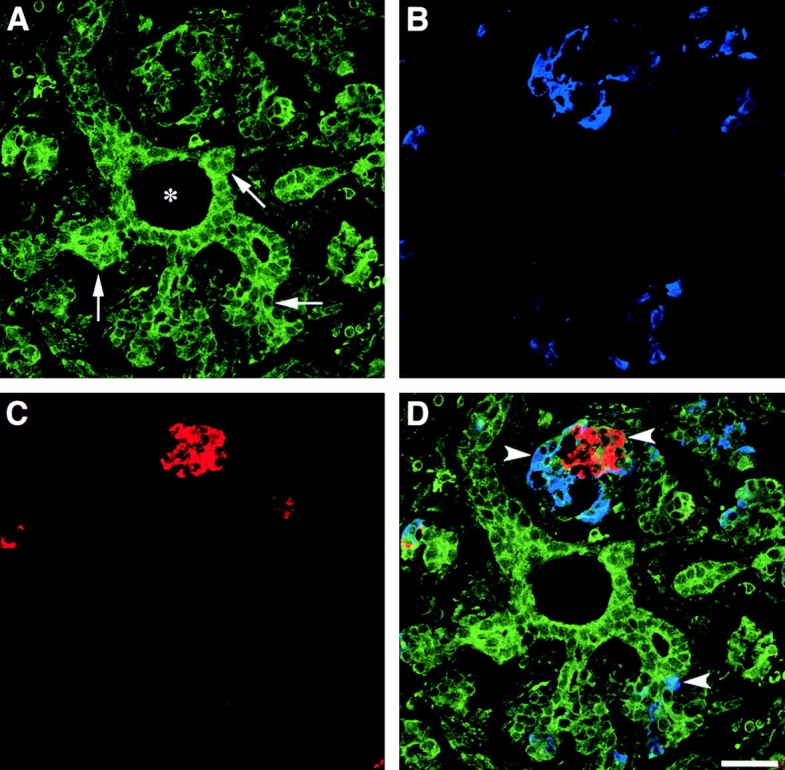
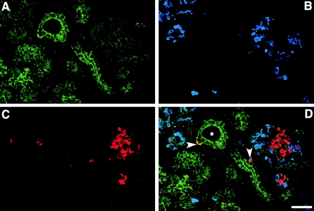
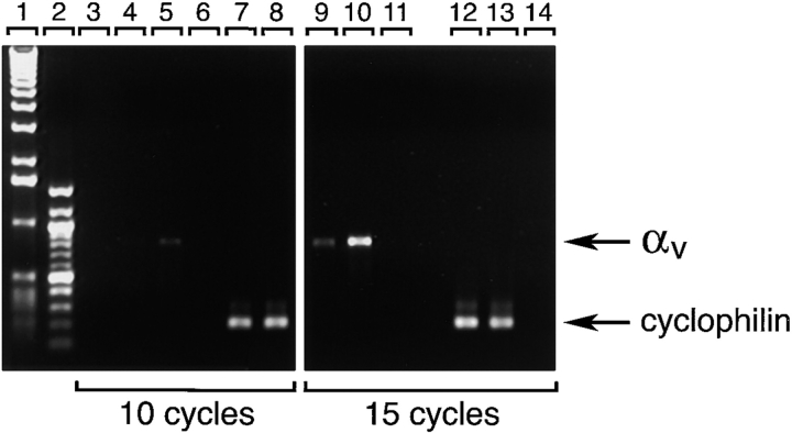
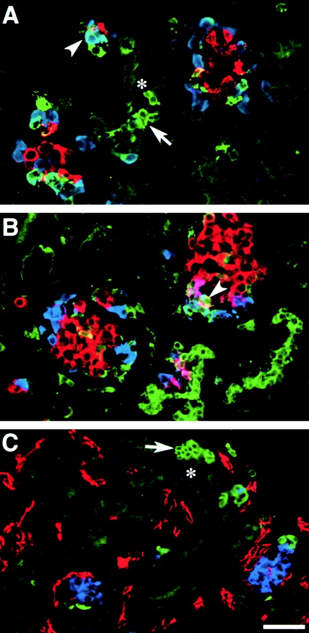
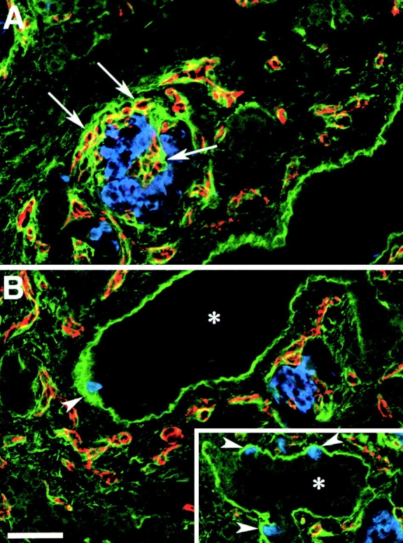
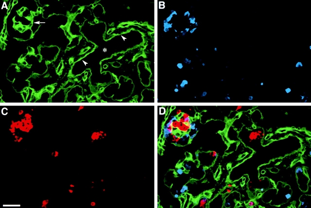
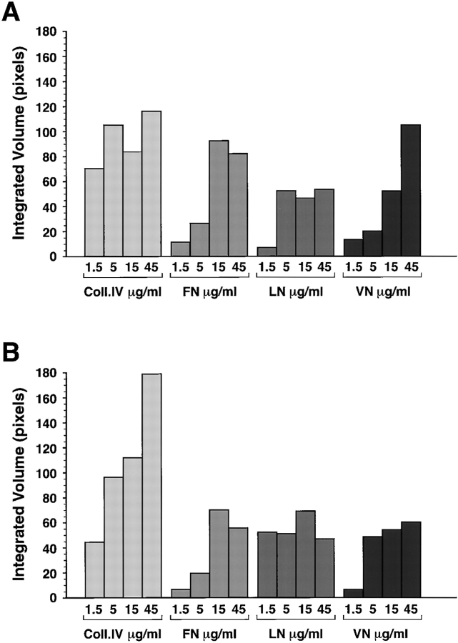
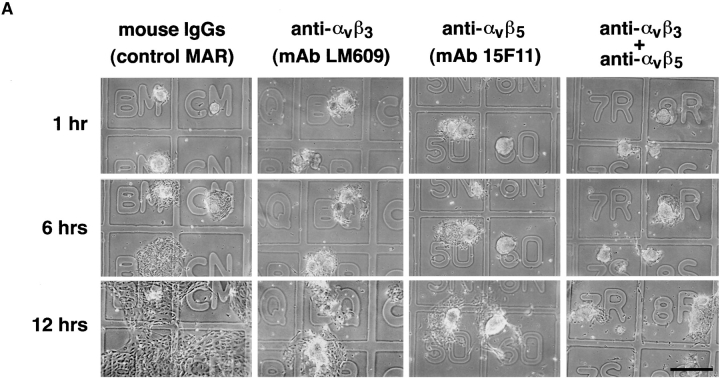
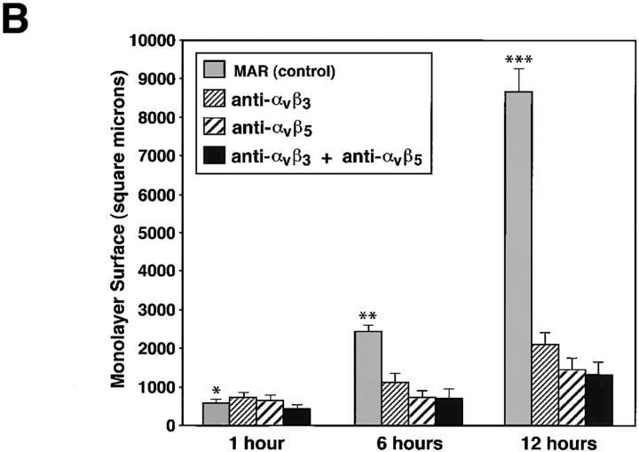
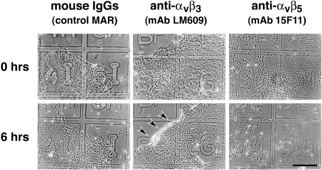
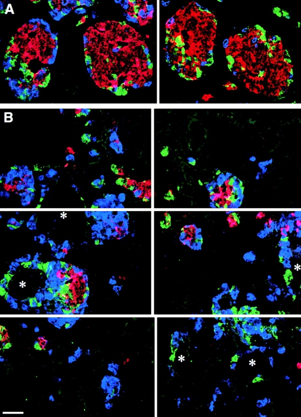
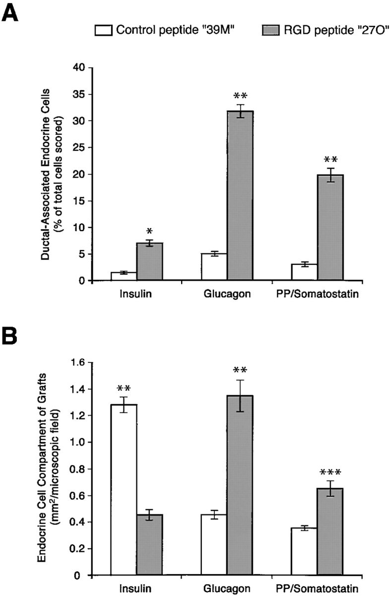
Cell-cell and cell-matrix interactions play a critical role in tissue morphogenesis and in homeostasis of adult tissues. The integrin family of adhesion receptors regulates cellular interactions with the extracellular matrix, which provides three-dimensional information for tissue organization. It is currently thought that pancreatic islet cells develop from undifferentiated progenitors residing within the ductal epithelium of the fetal pancreas. This process involves cell budding from the duct, migration into the surrounding mesenchyme, differentiation, and clustering into the highly organized islet of Langerhans. Here we report that alpha(v)beta(3) and alpha(v)beta(5), two integrins known to coordinate epithelial cell adhesion and movement, are expressed in pancreatic ductal cells and clusters of undifferentiated cells emerging from the ductal epithelium. We show that expression and function of alpha(v)beta(3) and alpha(v)beta(5) integrins are developmentally regulated during pancreatic islet ontogeny, and mediate adhesion and migration of putative endocrine progenitor cells both in vitro and in vivo in a model of pancreatic islet development. Moreover, we demonstrate the expression of fibronectin and collagen IV in the basal membrane of pancreatic ducts and of cell clusters budding from the ductal epithelium. Conversely, expression of vitronectin marks a population of epithelial cells adjacent to, or emerging from, pancreatic ducts. Thus, these data provide the first evidence for the contribution of integrins alpha(v)beta(3) and alpha(v)beta(5) and their ligands to morphogenetic events in the human endocrine pancreas.


Figures

Figure 1
Figure 1
Expression of αvβ3 in the developing human pancreas. αvβ3-specific immune reactivity (A, green) highlights the pancreatic ductal epithelium and clusters of cells branching from the ducts (arrows). Note the αvβ3-specific staining marking the regions of cell–cell and cell–matrix contacts. A number of developing endocrine cells budding from the ductal epithelium are identified by the insulin- (C, red) and glucagon-specific (B, blue) immune reactivities. The three fluorescence patterns are merged in D, demonstrating colocalization of hormone-positive cells with αvβ3 expression (arrowheads). We consistently observed that the brightness of αvβ3-specific immune reactivity is decreased in developing islet clusters when compared with the levels of αvβ3 expression in ductal cells. Bar, 50 μm.
Figure 3
Figure 3
Identification of αvβ5 in the developing human pancreas. Confocal immune fluorescence on cryostat sections stained for αvβ5 (A, green), insulin (C, red), and glucagon (B, blue). The three fluorophore spectra are merged in D. Bright levels of αvβ5-specific immune reactivity are detected in pancreatic ducts (asterisk), whereas significantly lower levels characterize endocrine cells organized in larger islet-like clusters. However, single endocrine cells adjacent to, or emerging from, the ductal epithelium retain significant higher levels of αvβ5 expression (D; arrowheads). Bar, 100 μm.
Figure 2
Figure 2
Decreased transcription of the αv integrin subunit in endocrine differentiated cells. Total RNA isolated from fetal and adult human pancreatic islets was reversed transcribed to generate cDNAs and amplify αv-specific sequences. Products of PCR reactions obtained at 10 and 15 cycles using αv -specific and cyclophilin-specific primers are shown. Lanes 1 and 2, 1-Kb and 100-bp ladder, respectively. Lanes 3, 6, 11, and 14 were loaded with PCR reactions performed using no template DNA (water); lanes 4, 7, 9, and 12 were loaded with reactions performed using cDNA from adult pancreatic islets; and lanes 5, 8, 10, and 13 were loaded with reactions performed using cDNA from fetal pancreatic islets. Significantly higher levels of αv-specific PCR product (824 bp) are detected in samples of fetal pancreatic islets as compared with samples of adult islets. Representative of three independent determinations using three independent tissue donors.
Figure 5
Figure 5
VN expression pattern in the developing human pancreas. A and B show representative fields recorded by confocal microscopy from cryostat sections stained for VN (green), glucagon (blue), and insulin (red). C shows staining for VN (green), PECAM-1 (red), and insulin (blue). Note the strong VN-specific immune reactivity identifying groups of epithelial cells emerging from, or adjacent to, ductal structures (asterisk; A and C, arrows). Some VN-positive cells coexpress glucagon (A, arrowhead) or insulin (B, arrowhead), suggesting a cell lineage relationship of VN-positive cells with putative endocrine progenitors. Staining for PECAM-1 (C, red) shows that none of the VN-positive cells are endothelial in nature. Bar, 100 μm.
Figure 4
Figure 4
FN expression pattern in the developing human pancreas. Confocal images of two adjacent microscopic fields (A and B) from cryostat sections stained for FN (green), insulin (blue), and PECAM-1 (red). A strong FN-specific immune reactivity (green) highlights the basal membrane of ducts (asterisk) and of insulin-positive cells (blue) emerging from the ductal epithelium (arrowheads, B and inset). The PECAM-1-specific staining (red), identifying blood vessels, reveals that the basal membrane of endothelial cells, infiltrating developing islet clusters, is also marked by a bright FN-specific staining (arrows). Bar, 100 μm.
Figure 6
Figure 6
Identification of Coll-IV in the developing human pancreas. Three-color confocal immune fluorescence on cryostat sections of human fetal pancreas. A strong Coll-IV-specific immune reactivity (A, green) identifies basal membranes (A, arrowheads) of cells in the ducts (asterisk), and in clusters of developing islet cells, identified by staining for insulin (C, red) and glucagon (B, blue). The three fluorophore spectra are combined in D. Bar, 100 μm.
Figure 7
Figure 7
Adhesion of fetal and adult pancreatic epithelial cells to purified ECM proteins. Adhesion to Coll-IV is constitutively high in both fetal (A) and adult cells (B), although adult cells consistently demonstrated stronger adhesion. Conversely, adhesion to FN and VN increased progressively at the higher concentrations for fetal as compared with adult cells.
Figure 9
Figure 9
Integrins αvβ3 and αvβ5 mediate migration of fetal pancreatic epithelial cells. Time course adhesion and migration of fetal pancreatic epithelial cells on 804G matrix–coated coverslips marked by an alpha-numerical grid to identify identical microscopic fields over time (A). Note that cell migration is inhibited by anti-αvβ3 and anti-αvβ5 function blocking antibodies. Quantitative morphometric analysis performed on multiple microscopic fields (n = 50 fields/condition) from four independent experiments revealed that blocking antibodies produced >75% overall inhibition of monolayer development in these assays (B). Bar, 150 μm. *n.s.; **P < 0.05; ***P < 0.0001.
Figure 9
Figure 9
Integrins αvβ3 and αvβ5 mediate migration of fetal pancreatic epithelial cells. Time course adhesion and migration of fetal pancreatic epithelial cells on 804G matrix–coated coverslips marked by an alpha-numerical grid to identify identical microscopic fields over time (A). Note that cell migration is inhibited by anti-αvβ3 and anti-αvβ5 function blocking antibodies. Quantitative morphometric analysis performed on multiple microscopic fields (n = 50 fields/condition) from four independent experiments revealed that blocking antibodies produced >75% overall inhibition of monolayer development in these assays (B). Bar, 150 μm. *n.s.; **P < 0.05; ***P < 0.0001.
Figure 8
Figure 8
Subcellular localization of αvβ3 and αvβ5 integrins. Representative confocal microscopic fields showing distinctive patterns for αvβ3 (A) and αvβ5 (B) fluorescence (green) colocalizing with F-actin filaments (red fluorescence) in fetal pancreatic epithelial cells. While αvβ3-specific immune reactivity is mainly confined at the very tips of F-actin filaments (A, arrows), fluorescence specific for αvβ5 extends back along the F-actin filaments identifying a larger number of focal adhesion contacts in the basal pole of adherent cells. Bar, 2.5 μm.
Figure 10
Figure 10
Integrins αvβ3 and αvβ5 mediate attachment of fetal pancreatic epithelial cells. Established cell monolayers were challenged with anti-αvβ3 and anti-αvβ5 function blocking antibodies (25 μg/ml) to assess the role of these two integrin receptors in the maintenance of cell monolayer integrity. After 6 h of incubation in the presence of anti-αvβ3, mAb cells from the periphery of monolayers started to detach and roll back into three-dimensional clusters (lower middle panel, arrows). Conversely, incubation with the anti-αvβ5 mAb resulted in the almost complete detachment of cell monolayers from the matrix (lower right panel). Results shown are representative of four independent experiments. Bar, 150 μm.
Figure 11
Figure 11
Migration of developing islet cells from the ductal epithelium is perturbed by cyclic RGD peptide analogues. A shows two representative adjacent microscopic fields of pancreatic grafts from animals treated with the control 39M peptide. Large and well organized islet clusters are evident with the majority of insulin-positive cells (red) located in the center, surrounded by cells that are positive for glucagon (blue) and somatostatin/pancreatic polypeptide (green). In contrast, analysis of the pancreatic grafts harvested from mice treated with the RGD peptide inhibitor 27O reveals a profoundly perturbed organization and development of islet cell clusters (B, reconstruction of six adjacent microscopic fields). Most islet cells appear closely associated with ductal structures (asterisk), often completely or nearly surrounding ducts. Bar, 100 μm.
Figure 12
Figure 12
Morphometric changes in islet development by cyclic RGD peptide analogues. Human pancreatic grafts harvested from mice treated with the 27O RGD peptide analogue show an increased frequency of islet cells associated with ductal elements (A). Thus, the frequency of ductal-associated islet cells is increased 4.5-fold for insulin-producing cells, 6.5-fold for glucagon-producing cells, and 6.6-fold for somatostatin/pancreatic polypeptide–producing cells. Measurements of the relative surface area per microscopic field calculated for each islet cell type (B) reveal a 65% decrease of insulin-producing cells, paralleled by a 193% increase of glucagon-producing cells, and an 85% increase of somatostatin/pancreatic polypeptide–producing cells. *P < 0.001; **P < 0.0001; ***P < 0.05.

Similar articles

Cited by

References

    1. Alpert S., Hanahan D., Tietelman G. Hybrid insulin gene reveal a developmental lineage for pancreatic endocrine cell and imply a relationship with neurons. Cell. 1988;53:295–308. - PubMed
    1. Bauer G.E., Balsamo J., Lilien J. Cadherin-mediated adhesion in pancreatic islet cells is modulated by a cell surface N-acetylgalactosaminylphosphotransferase. J. Cell Sci. 1992;103:1235–1241. - PubMed
    1. Beattie G.M., Levine F., Mally M.I., Otonkoski T., O'Brien J.S., Salomon D.R., Hayek A. Acid β-galactosidasea developmentally regulated marker of endocrine cell precursors in the human fetal pancreas. J. Clin. Endocrinol. Metab. 1994;78:1232–1240. - PubMed
    1. Beattie G.M., Rubin J.S., Mally M.I., Otonkoski T., Hayek A. Regulation of proliferation and differentiation of human fetal pancreatic islet cells by extracellular matrix, hepatocyte growth factor and cell-cell contact. Diabetes. 1996;45:1223–1228. - PubMed
    1. Beattie G.M., Cirulli V., Lopez A.D., Hayek A. Ex vivo expansion of human pancreatic endocrine cells. J. Clin. Endocrinol. Metab. 1997;82:1852–1856. - PubMed

Publication types

MeSH terms